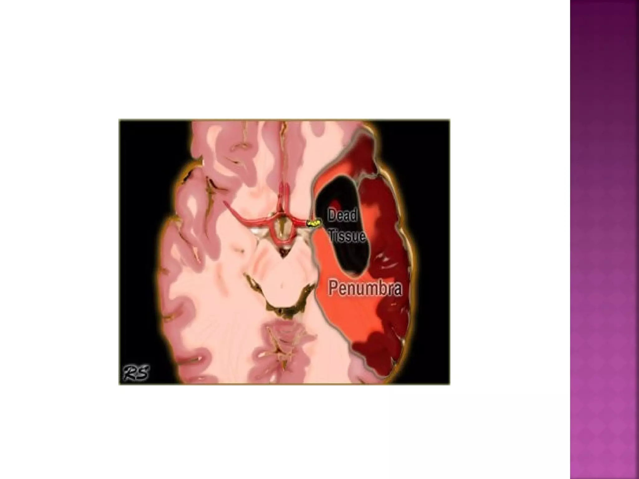
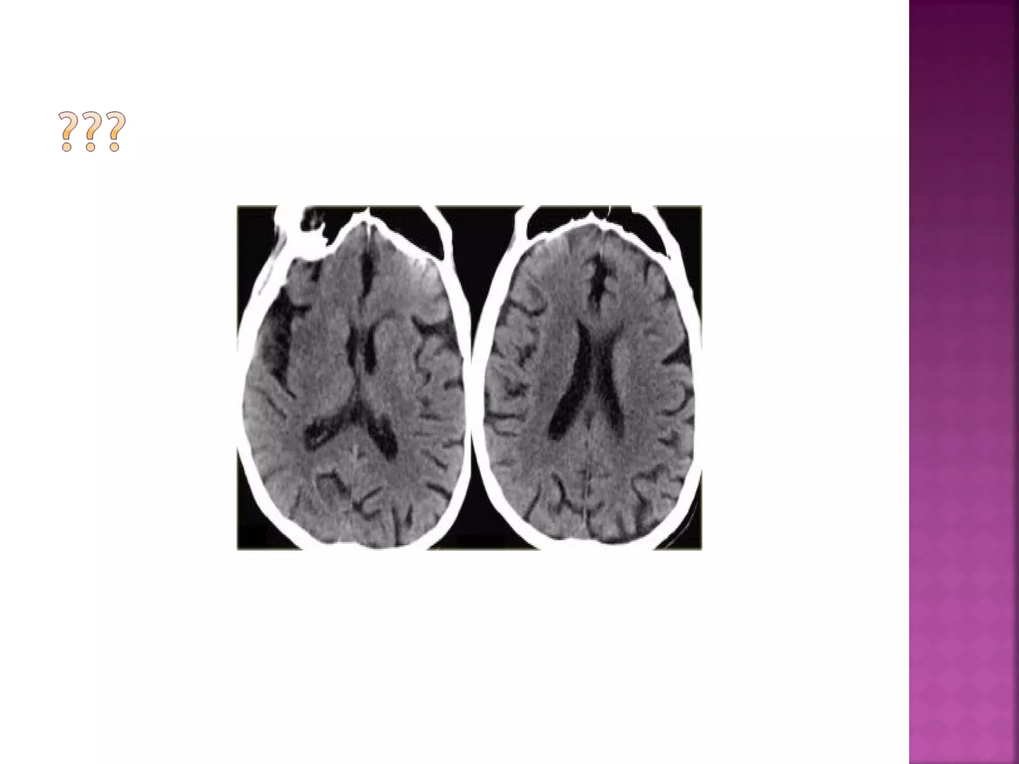
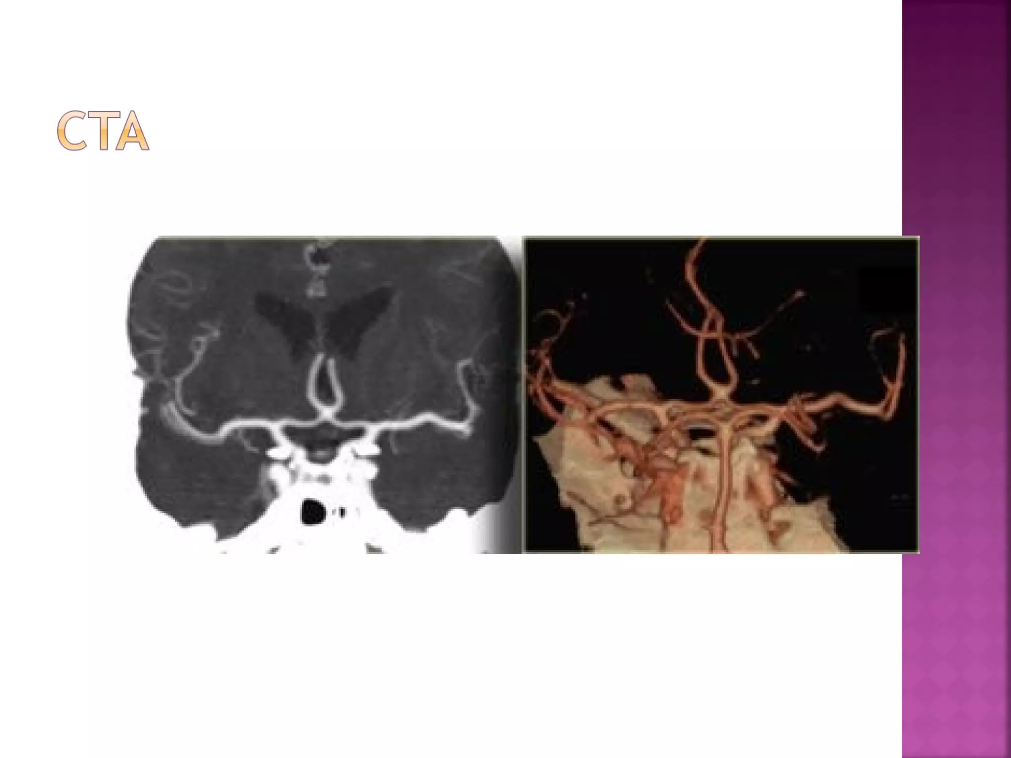
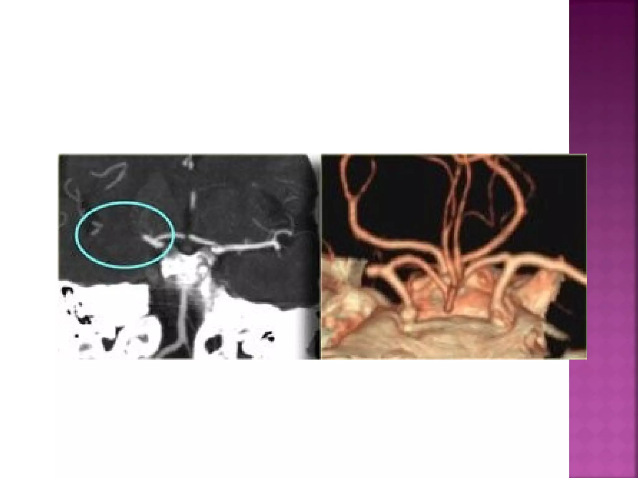
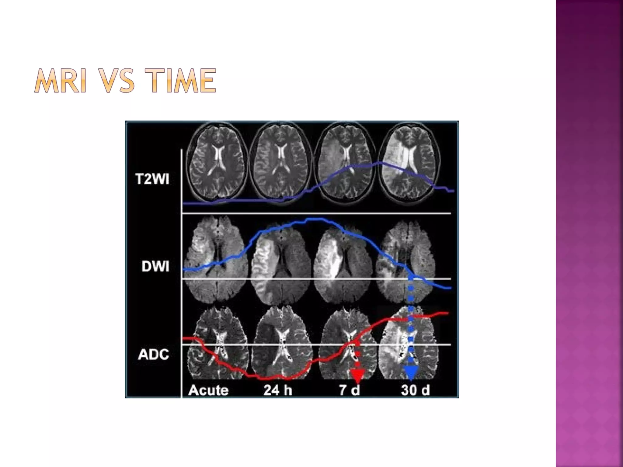
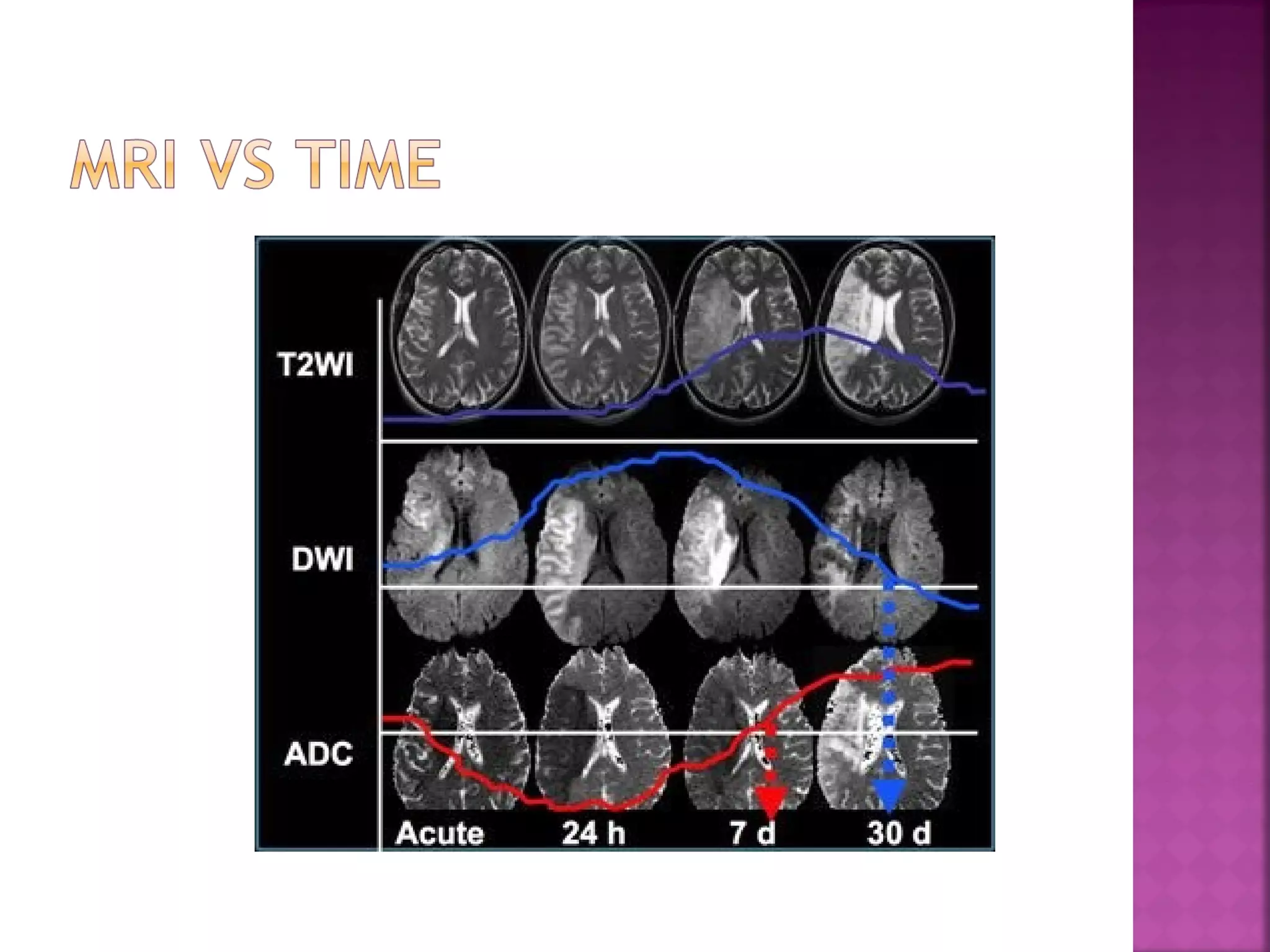
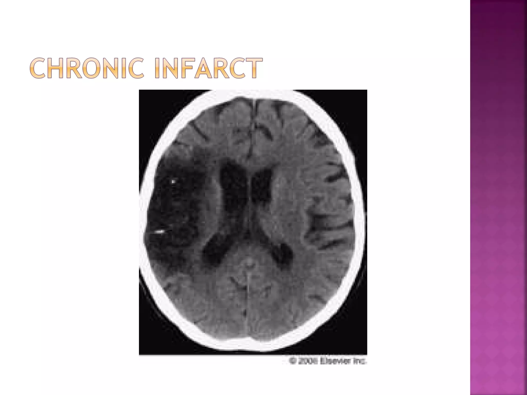
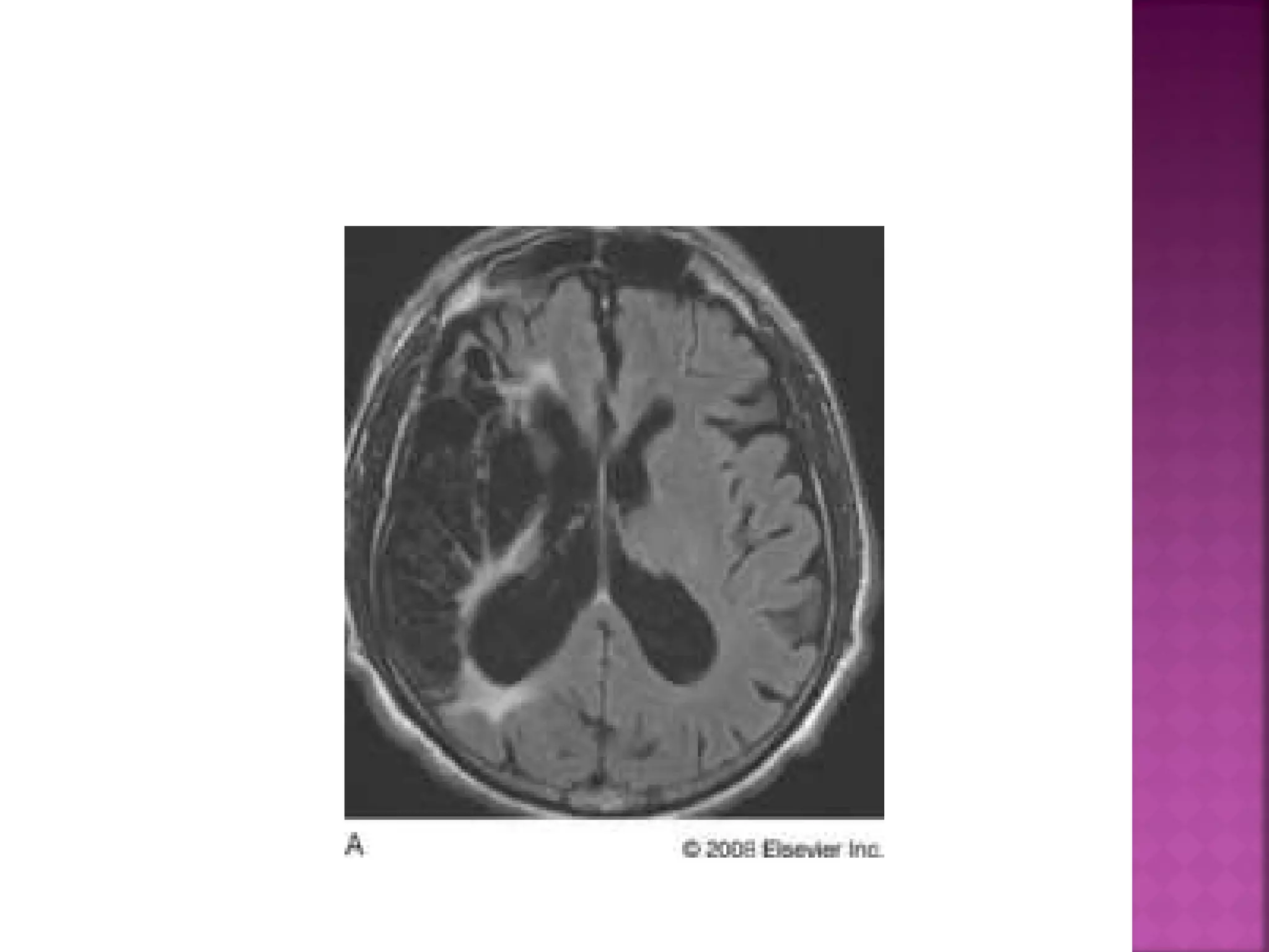
BY. DR NADIA
The goal of imaging in a patient with acute
stroke is:
 Exclude hemorrhage
 Differentiate between irreversibly affected
brain tissue and reversibly impaired tissue
(dead tissue versus tissue at risk)
 Identify stenosis or occlusion of major extraand intracranial arteries


CT has the advantage of being available 24
hours a day and is the gold standard for
hemorrhage.
Hemorrhage on MR images can be quite
confusing.
On CT 60% of infarcts are seen within 3-6 hrs
and virtually all are seen in 24 hours.


The diagnosis is infarction, because of the
location (vascular territory of the middle
cerebral artery (MCA) and because of the
involvement of gray and white matter, which
is also very typical for infarction.


Obscuration of the lentiform nucleus, also
called blurred basal ganglia, is an important
sign of infarction.
It is seen in middle cerebral artery infarction
and is one of the earliest and most
frequently seen signs.
The basal ganglia are almost always involved
in MCA-infarction.


This refers to hypodensity and swelling of the
insular cortex.
It is a very indicative and subtle early CTsign of infarction in the territory of the
middle cerebral artery.
It has to be differentiated from herpes
encephalitis.


This is a result of thrombus or embolus in the
MCA.


On T2WI and FLAIR infarction is seen as high
SI.
These sequences detect 80% of infarctions
before 24 hours.
They may be negative up to 2-4 hours postictus!


High signal on conventional MR-sequences is
comparable to hypodensity on CT.
It is the result of irreversible injury with cell
death.
So hyperintensity means BAD news: dead
brain.


DWI is the most sensitive sequence for stroke
imaging.
DWI is sensitive to restriction of Brownian
motion of extracellular water due to
imbalance caused by cytotoxic edema.
Normally water protons have the ability to
diffuse extracellularly and loose signal.
High intensity on DWI indicates restriction of
the ability of water protons to diffuse
extracellularly.




In the acute phase T2WI will be normal, but
in time the infarcted area will become
hyperintense.
The hyperintensity on T2WI reaches its
maximum between 7 and 30 days. After this
it starts to fade




DWI is already positive in the acute phase
and then becomes more bright with a
maximum at 7 days.
DWI in brain infarction will be positive for
approximately for 3 weeks after onset (in
spinal cord infarction DWI is only positive for
one week!).


ADC will be of low signal intensity with a
maximum at 24 hours and then will increase
in signal intensity and finally becomes bright
in the chronic stage.
THANK YOU

Brain ischemia

  • 1.
  • 3.
    The goal ofimaging in a patient with acute stroke is:  Exclude hemorrhage  Differentiate between irreversibly affected brain tissue and reversibly impaired tissue (dead tissue versus tissue at risk)  Identify stenosis or occlusion of major extraand intracranial arteries
  • 4.
     CT has theadvantage of being available 24 hours a day and is the gold standard for hemorrhage. Hemorrhage on MR images can be quite confusing. On CT 60% of infarcts are seen within 3-6 hrs and virtually all are seen in 24 hours.
  • 7.
     The diagnosis isinfarction, because of the location (vascular territory of the middle cerebral artery (MCA) and because of the involvement of gray and white matter, which is also very typical for infarction.
  • 9.
     Obscuration of thelentiform nucleus, also called blurred basal ganglia, is an important sign of infarction. It is seen in middle cerebral artery infarction and is one of the earliest and most frequently seen signs. The basal ganglia are almost always involved in MCA-infarction.
  • 11.
     This refers tohypodensity and swelling of the insular cortex. It is a very indicative and subtle early CTsign of infarction in the territory of the middle cerebral artery. It has to be differentiated from herpes encephalitis.
  • 15.
     This is aresult of thrombus or embolus in the MCA.
  • 20.
     On T2WI andFLAIR infarction is seen as high SI. These sequences detect 80% of infarctions before 24 hours. They may be negative up to 2-4 hours postictus!
  • 21.
     High signal onconventional MR-sequences is comparable to hypodensity on CT. It is the result of irreversible injury with cell death. So hyperintensity means BAD news: dead brain.
  • 23.
     DWI is themost sensitive sequence for stroke imaging. DWI is sensitive to restriction of Brownian motion of extracellular water due to imbalance caused by cytotoxic edema. Normally water protons have the ability to diffuse extracellularly and loose signal. High intensity on DWI indicates restriction of the ability of water protons to diffuse extracellularly.
  • 29.
      In the acutephase T2WI will be normal, but in time the infarcted area will become hyperintense. The hyperintensity on T2WI reaches its maximum between 7 and 30 days. After this it starts to fade
  • 31.
      DWI is alreadypositive in the acute phase and then becomes more bright with a maximum at 7 days. DWI in brain infarction will be positive for approximately for 3 weeks after onset (in spinal cord infarction DWI is only positive for one week!).
  • 33.
     ADC will beof low signal intensity with a maximum at 24 hours and then will increase in signal intensity and finally becomes bright in the chronic stage.
  • 43.