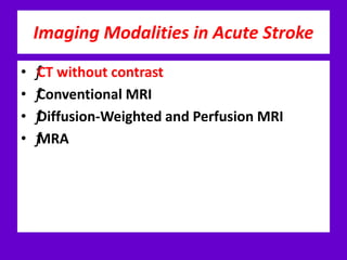
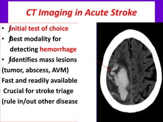
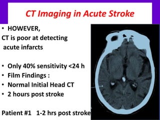
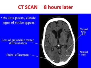
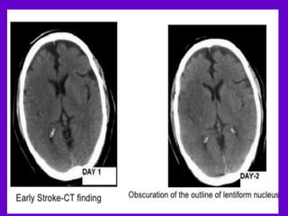
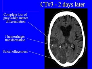
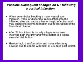
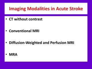
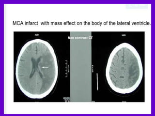
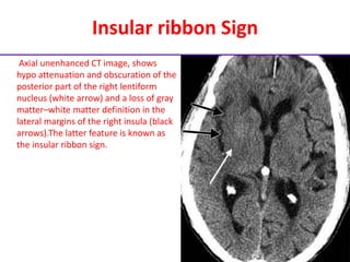
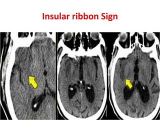
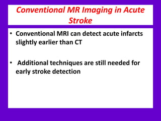
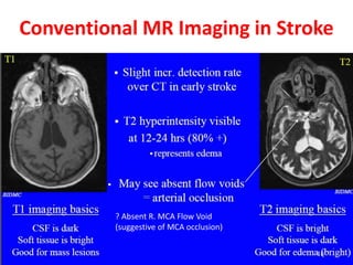
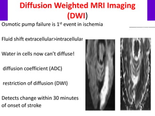
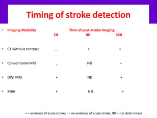
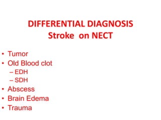
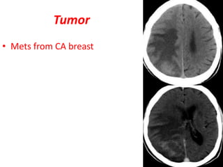
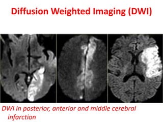
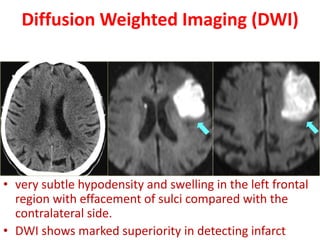
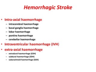
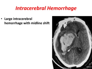
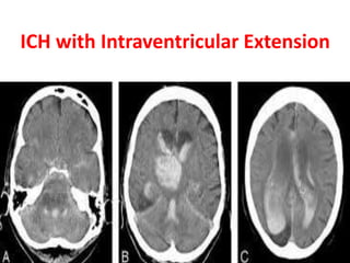
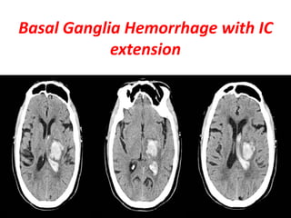
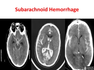
CT SCAN , MRI IN STROCKE
• DR. SAAD ALYOUSEF
QATAR RED CRESCENT- AL HEMAILA CENTRE
• SYRIAN BOARD /DIGNOSTIC RADIOLOGY
Stroke - Definition and Statistics
• ƒAcute, vascular injury to CNS
• <24 hrs = TIA
• >24 hrs = stroke (CVA)
• ƒAffects 600,000 people/ yr
• (that is 1 stroke per minute!)
• ƒIs #3 cause of mortality in adults
• Is #1 cause of disability
Types of Stroke
• ƒHemorrhagic (20%)
• ƒusually hypertensive hemorrhage
• ƒIschemic (80%)
• ƒ Thrombotic (40%)
• ƒintracerebral atherosclerosis
• ƒ Embolic (60%)
• ƒCardiac embolus (thrombus, tumor, septic embolus)
• ƒartery-to-artery (mainly carotid thrombus)
• ƒParadoxical embolus (thrombus, fat, air)
Risk Factors for Stroke
• ƒAtherosclerosis risk factors
• ƒFamily history of CVA, TIA, or MI
• ƒHypertension
• ƒSmoking
• ƒDiabetes
• ƒHypercholesterolemia
• ƒPrevious CVA, TIA, or MI
• ƒAtrial fibrillation
Differential Diagnosis
• ƒMany CNS diseases can mimic ischemic
• stroke
• ƒHemorrhage
• ƒMass lesion (tumor, abscess, AVM)
• ƒSeizure (Todd’s paralysis)
• ƒHemiplegic migraine
• ƒMS flare
• ƒVenous infarct
• Goals of Imaging in Acute Stroke
• 1. Rule in or out other disease processes
• 2. Define location, extent and age of infarct
Cerebrovascular Anatomy
Imaging Modalities in Acute Stroke
• ƒCT without contrast
• ƒConventional MRI
• ƒDiffusion-Weighted and Perfusion MRI
• ƒMRA
CT Imaging in Acute Stroke
• ƒInitial test of choice
• ƒBest modality for
detecting hemorrhage
• ƒIdentifies mass lesions
(tumor, abscess, AVM)
Fast and readily available
Crucial for stroke triage
(rule in/out other diseases)
CT Imaging in Acute Stroke
• HOWEVER,
CT is poor at detecting
acute infarcts
• Only 40% sensitivity <24 h
• Film Findings :
• Normal Initial Head CT
• 2 hours post stroke
Patient #1 1-2 hrs post stroke
CT SCAN 8 hours later
• Therefore, other imaging
modalities are used to detect
strokes < 6 hours!
Imaging Modalities in Acute Stroke
• CT without contrast
• Conventional MRI
• Diffusion-Weighted and Perfusion MRI
• MRA
CT SCAN EARLY SIGN
• Hypo attenuating brain tissue
• Obscuration of lentiform nucleus
• Dense MCA sign
• Insular ribbon sign
• Loss of sulcal effacement
Hypo attenuating brain tissue
• MCA infarction: on CT an
area of hypo attenuation
appearing within six
hours is highly specific
for irreversible ischemic
brain damage
Obscuration lentiform nucleus
Axial unenhanced CT image
shows hypo attenuation and
obscuration of the left
lentiform nucleus (arrows),
which, because of acute
ischemia in the lenticulostriate
distribution, appears
abnormal in comparison with
the right lentiform nucleus.
Obscuration lentiform nucleus
Axial unenhanced CT images show obscuration of the right lentiform nucleus (arrow in b). This feature is less
visible with the routine brain imaging window used for a (window width, 80 HU; center, 35 HU)
than with the narrower window used for b (window width, 10 HU; center, 28 HU).
Insular ribbon Sign
Axial unenhanced CT image, shows
hypo attenuation and obscuration of the
posterior part of the right lentiform
nucleus (white arrow) and a loss of gray
matter–white matter definition in the
lateral margins of the right insula (black
arrows).The latter feature is known as
the insular ribbon sign.
Insular ribbon Sign
Conventional MR Imaging in Acute
Stroke
• Conventional MRI can detect acute infarcts
slightly earlier than CT
• Additional techniques are still needed for
early stroke detection
Conventional MR Imaging in Stroke
? Absent R. MCA Flow Void
(suggestive of MCA occlusion)
Diffusion Weighted MRI Imaging
(DWI)
Osmotic pump failure is 1st event in ischemia
Fluid shift extracellular>intracellular
Water in cells now can’t diffuse!
diffusion coefficient (ADC)
restriction of diffusion (DWI)
Detects change within 30 minutes
of onset of stroke
Timing of stroke detection
• Imaging Modality Time of post-stroke imaging
2H 8H 30H
• CT without contrast _ + +
• Conventional MRI _ ND +
• DWI MRI + ND +
• MRA + ND +
+ = evidence of acute stroke; - = no evidence of acute stroke; ND = not determined
DIFFERENTIAL DIAGNOSIS
Stroke on NECT
• Tumor
• Old Blood clot
– EDH
– SDH
• Abscess
• Brain Edema
• Trauma
Tumor
• Mets from CA breast
Chronic Hemorrhage
• CT axial- left chronic frontoparietal
subdural hematoma, hypo intense
area.
• Subacute hemorrhage
Abcess
• NECT
• CECT
Brain Edema
Traumatic Contusion
CTA
• Insular ribbon sign in
right insular cortex
• CTA disclose
thrombus in rt. MCA
CT Perfusion (CTP)
• With CT and MR imaging we can get a good
impression of the area that is infracted.
• but we cannot preclude a large ischemic penumbra
(tissue at risk).
With perfusion studies we monitor the first pass of
an iodinated contrast agent bolus through the
cerebral vasculature.
Perfusion will tell us which area is at risk.
Approximately 26% of patients will require a
perfusion study to come to the proper diagnosis.
MRI
• On PD/T2WI and FLAIR infarction is seen as high SI.
• These sequences detect 80% of infarctions before
24 hours.
• They may be negative up to 2-4 hours post-ictus!
• MR Hperintensity = CT Hypodensity
• T2WI and FLAIR
demonstrating
hyperintensity in
the territory of the
middle cerebral
artery.
• Notice the
involvement of the
lentiform nucleus
and insular cortex.
Diffusion Weighted Imaging (DWI)
• DWI is the most sensitive sequence for stroke
imaging.
• Also called Stroke sequence
DWI in posterior, anterior and middle cerebral
infarction
Diffusion Weighted Imaging (DWI)
• very subtle hypodensity and swelling in the left frontal
region with effacement of sulci compared with the
contralateral side.
• DWI shows marked superiority in detecting infarct
Hemorrhagic Stroke
• Intra-axial haemorrhage
– intracerebral haemorrhage
– basal ganglia haemorrhage
– lobar haemorrhage
– pontine haemorrhage
– cerebellar haemorrhage
• Intraventricular haemorrhage (IVH)
• extra-axial haemorrhage
– extradural haemorrhage (EDH)
– subdural haemorrhage (SDH)
– subarachnoid haemorrhage (SAH)
Intracerebral Hemorrhage
• Large intracerebral
hemorrhage with midline shift
ICH with Intraventricular Extension
Basal Ganglia Hemorrhage with IC
extension
Subarachnoid Hemorrhage
THANX

Imaginginacutestroke 140320043301-phpapp02

  • 1.
    CT SCAN ,MRI IN STROCKE • DR. SAAD ALYOUSEF QATAR RED CRESCENT- AL HEMAILA CENTRE • SYRIAN BOARD /DIGNOSTIC RADIOLOGY
  • 2.
    Stroke - Definitionand Statistics • ƒAcute, vascular injury to CNS • <24 hrs = TIA • >24 hrs = stroke (CVA) • ƒAffects 600,000 people/ yr • (that is 1 stroke per minute!) • ƒIs #3 cause of mortality in adults • Is #1 cause of disability
  • 3.
    Types of Stroke •ƒHemorrhagic (20%) • ƒusually hypertensive hemorrhage • ƒIschemic (80%) • ƒ Thrombotic (40%) • ƒintracerebral atherosclerosis • ƒ Embolic (60%) • ƒCardiac embolus (thrombus, tumor, septic embolus) • ƒartery-to-artery (mainly carotid thrombus) • ƒParadoxical embolus (thrombus, fat, air)
  • 4.
    Risk Factors forStroke • ƒAtherosclerosis risk factors • ƒFamily history of CVA, TIA, or MI • ƒHypertension • ƒSmoking • ƒDiabetes • ƒHypercholesterolemia • ƒPrevious CVA, TIA, or MI • ƒAtrial fibrillation
  • 5.
    Differential Diagnosis • ƒManyCNS diseases can mimic ischemic • stroke • ƒHemorrhage • ƒMass lesion (tumor, abscess, AVM) • ƒSeizure (Todd’s paralysis) • ƒHemiplegic migraine • ƒMS flare • ƒVenous infarct
  • 6.
    • Goals ofImaging in Acute Stroke • 1. Rule in or out other disease processes • 2. Define location, extent and age of infarct
  • 7.
  • 8.
    Imaging Modalities inAcute Stroke • ƒCT without contrast • ƒConventional MRI • ƒDiffusion-Weighted and Perfusion MRI • ƒMRA
  • 9.
    CT Imaging inAcute Stroke • ƒInitial test of choice • ƒBest modality for detecting hemorrhage • ƒIdentifies mass lesions (tumor, abscess, AVM) Fast and readily available Crucial for stroke triage (rule in/out other diseases)
  • 10.
    CT Imaging inAcute Stroke • HOWEVER, CT is poor at detecting acute infarcts • Only 40% sensitivity <24 h • Film Findings : • Normal Initial Head CT • 2 hours post stroke Patient #1 1-2 hrs post stroke
  • 11.
    CT SCAN 8hours later
  • 15.
    • Therefore, otherimaging modalities are used to detect strokes < 6 hours!
  • 16.
    Imaging Modalities inAcute Stroke • CT without contrast • Conventional MRI • Diffusion-Weighted and Perfusion MRI • MRA
  • 18.
    CT SCAN EARLYSIGN • Hypo attenuating brain tissue • Obscuration of lentiform nucleus • Dense MCA sign • Insular ribbon sign • Loss of sulcal effacement
  • 19.
    Hypo attenuating braintissue • MCA infarction: on CT an area of hypo attenuation appearing within six hours is highly specific for irreversible ischemic brain damage
  • 20.
    Obscuration lentiform nucleus Axialunenhanced CT image shows hypo attenuation and obscuration of the left lentiform nucleus (arrows), which, because of acute ischemia in the lenticulostriate distribution, appears abnormal in comparison with the right lentiform nucleus.
  • 21.
    Obscuration lentiform nucleus Axialunenhanced CT images show obscuration of the right lentiform nucleus (arrow in b). This feature is less visible with the routine brain imaging window used for a (window width, 80 HU; center, 35 HU) than with the narrower window used for b (window width, 10 HU; center, 28 HU).
  • 22.
    Insular ribbon Sign Axialunenhanced CT image, shows hypo attenuation and obscuration of the posterior part of the right lentiform nucleus (white arrow) and a loss of gray matter–white matter definition in the lateral margins of the right insula (black arrows).The latter feature is known as the insular ribbon sign.
  • 23.
  • 24.
    Conventional MR Imagingin Acute Stroke • Conventional MRI can detect acute infarcts slightly earlier than CT • Additional techniques are still needed for early stroke detection
  • 25.
    Conventional MR Imagingin Stroke ? Absent R. MCA Flow Void (suggestive of MCA occlusion)
  • 26.
    Diffusion Weighted MRIImaging (DWI) Osmotic pump failure is 1st event in ischemia Fluid shift extracellular>intracellular Water in cells now can’t diffuse! diffusion coefficient (ADC) restriction of diffusion (DWI) Detects change within 30 minutes of onset of stroke
  • 27.
    Timing of strokedetection • Imaging Modality Time of post-stroke imaging 2H 8H 30H • CT without contrast _ + + • Conventional MRI _ ND + • DWI MRI + ND + • MRA + ND + + = evidence of acute stroke; - = no evidence of acute stroke; ND = not determined
  • 28.
    DIFFERENTIAL DIAGNOSIS Stroke onNECT • Tumor • Old Blood clot – EDH – SDH • Abscess • Brain Edema • Trauma
  • 29.
  • 30.
    Chronic Hemorrhage • CTaxial- left chronic frontoparietal subdural hematoma, hypo intense area. • Subacute hemorrhage
  • 31.
  • 32.
  • 33.
  • 34.
    CTA • Insular ribbonsign in right insular cortex • CTA disclose thrombus in rt. MCA
  • 35.
    CT Perfusion (CTP) •With CT and MR imaging we can get a good impression of the area that is infracted. • but we cannot preclude a large ischemic penumbra (tissue at risk). With perfusion studies we monitor the first pass of an iodinated contrast agent bolus through the cerebral vasculature. Perfusion will tell us which area is at risk. Approximately 26% of patients will require a perfusion study to come to the proper diagnosis.
  • 36.
    MRI • On PD/T2WIand FLAIR infarction is seen as high SI. • These sequences detect 80% of infarctions before 24 hours. • They may be negative up to 2-4 hours post-ictus! • MR Hperintensity = CT Hypodensity
  • 37.
    • T2WI andFLAIR demonstrating hyperintensity in the territory of the middle cerebral artery. • Notice the involvement of the lentiform nucleus and insular cortex.
  • 38.
    Diffusion Weighted Imaging(DWI) • DWI is the most sensitive sequence for stroke imaging. • Also called Stroke sequence
  • 39.
    DWI in posterior,anterior and middle cerebral infarction Diffusion Weighted Imaging (DWI)
  • 40.
    Diffusion Weighted Imaging(DWI) • very subtle hypodensity and swelling in the left frontal region with effacement of sulci compared with the contralateral side. • DWI shows marked superiority in detecting infarct
  • 41.
    Hemorrhagic Stroke • Intra-axialhaemorrhage – intracerebral haemorrhage – basal ganglia haemorrhage – lobar haemorrhage – pontine haemorrhage – cerebellar haemorrhage • Intraventricular haemorrhage (IVH) • extra-axial haemorrhage – extradural haemorrhage (EDH) – subdural haemorrhage (SDH) – subarachnoid haemorrhage (SAH)
  • 42.
    Intracerebral Hemorrhage • Largeintracerebral hemorrhage with midline shift
  • 43.
  • 44.
    Basal Ganglia Hemorrhagewith IC extension
  • 45.
  • 46.