Intracranial hemorrhage- shruthi s jayaraj, calicut medical college
•Download as PPTX, PDF•
105 likes•31,314 views


Recommended
More Related Content
What's hot
What's hot (20)
Viewers also liked
Similar to Intracranial hemorrhage- shruthi s jayaraj, calicut medical college
Similar to Intracranial hemorrhage- shruthi s jayaraj, calicut medical college (20)
Recently uploaded
Recently uploaded (20)
Intracranial hemorrhage- shruthi s jayaraj, calicut medical college
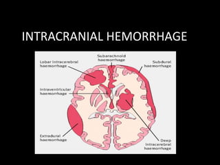
- 2. INTRACRANIAL HEMORRHAGES ARE CLASSIFIED ON THE BASIS OF BOTH • LOCATION • UNDERLYING VASCULAR PATHOLOGY
- 3. DEPENDING ON LOCATION INTRAAXIAL & EXTRA AXIAL
- 5. INTRA AXIAL HEMORRAGE- INTRA PARENCHYMAL - INTRA VENTRICULAR EXTRA AXIAL HEMORRHAGE – EPIDURAL HEMORRHAGE - SUBDURAL HEMORRHAGE - SUBARACHNIOD HEMORRHAGE
- 6. INTRA PARENCHYMAL HEMORRHAGE 1. AETIOLOGY A) B) C) D) E) Hypertension Trauma Cerebral amyloid angiopathy Advanced age Cocaine and methamphetamine use
- 7. F) HEMORRHAGIC DISORDERS G) NEOPLASMS H) VASCULAR MALFORMATIONS
- 8. HYPERTENSIVE INTRAPARENCHYMAL HMRG • SPONTANEOUS RUPTURE OF PENETRATING ARTERIES DEEP IN THE BRAIN • SITES 1. BASAL GANGLIA 2.THALAMUS 3.CEREBELLUM 4.PONS
- 9. • FOCAL DEFICIT EVOLVE OVER 20- 30 MINUTES • DIMINISHING LEVEL OF CONSCIOUSNESS • SIGNS OF RAISED ICP
- 10. C/F
- 11. putamen • • • • C/L hemiparesis Arm & legs gradually weaken Slurred speech Eye deviate away from side of hemiparesis large – brain stem compression
- 12. Thalamic hemorrhage • • • • • c/l hemiparesis Prominent sensory deficit Dominant thalamus – aphasia Non dominant – constructional apraxia Ocular disturbance- extension into upper midbrain
- 13. • • • • Ocular disturbances Deviation of eyes downward & inward Unequal pupils with absence of light reactions Ipsilateral horner’s syndrome Paralysis of vertical gaze,nystagmus
- 14. Pontine hemorrhage • Deep coma with quadriplegia over few minutes • Pin point pupil reacting to light • Impaired reflex horizontal eye movements • Hyperpnoea,hyperhydrosis,hypertension are common
- 15. Cerebellar hemorrhage • • • • Occipital headahe Repeated vomiting Ataxia Dizziness and vertigo may be prominent
- 16. • Paresis of conjugate lateral gaze to the side of hemorrhage • Ipsilateral 6th nerve palsy • Dysphagia,dysarthria
- 17. Cerebellar hmrg… • Later stage – BRAIN STEM COMPRESSION/HYDROCEPHALUS IMMEDIATE EVACUATION CAN BE LIFE SAVING !!
- 18. LOBAR HEMORRHAGE • occipital hemorrhage - hemianopia; • left temporal hemorrhage,-aphasia and delirium; • parietal hemorrhage - hemisensory loss; • frontal hemorrhage,-arm weakness • Focal headache and vomiting can occur
- 24. Cerebral amyloid angiopathy • Elderly • arteriolar degeneration and amyloid deposition • most common cause of lobar hemorrhage in the elderly
- 26. • intracranial hemorrhages associated with IV thrombolysis given for MI • patients who present with multiple hemorrhages (and infarcts) over several months or years • patients with "micro-bleeds" seen on brain MRI sequences sensitive for hemosiderin
- 27. • pathologic demonstration of Congo red staining of amyloid in cerebral vessels • no specific therapy, although antiplatelet and anticoagulating agents are typically avoided.
- 28. • Cocaine and methamphetamine are frequent causes of stroke in young (age <45 years) patients
- 29. Cocaine • enhances sympathetic activity • acute, sometimes severe, hypertension, • and this may lead to hemorrhage
- 30. • Intracranial hemorrhages associated with anticoagulant therapy can occur at any location • evolve slowly, over 24–48 hours
- 31. • hematologic disorders (leukemia, aplastic anemia, thrombocytopenic purpura) • multiple ICHs. • Skin and mucous membrane bleeding offers a diagnostic clue
- 32. • Hemorrhage into a brain tumor may be the first manifestation of neoplasm I. Choriocarcinoma, II. malignant melanoma, III. renal cell carcinoma, and IV. bronchogenic carcinoma are among the most common metastatic tumors associated with ICH
- 33. Other causes • Head injury • Hypertensive encephalopathy • Sepsis
- 36. • BLEEDING • HEADACHE • SEIZURES
- 37. • MRI / Contrast CT / Angiogram
- 38. • Treatment: Surgery / stereotaxic radiation
- 39. Venous anomalies • As a result of anomalous cerebral, cerbellar / brainstem venous drainage • Are functional venous channels • Surgery – risk of venous infarction and hemorrhage
- 40. Capillary telangiectasia • May be associated with Hereditary hemorrhagic telangiectasia / osler rendu weber syndrome
- 41. • Typically : pons, deep cerebral white matter
- 45. Cavernous angioma • tuft of capillary sinusoids within deep hemispheric white matter and brain stem with normal intervening neural structures
- 46. • < 1 cm diameter typically • a/w venous anomalies • Surgical resection reduce seizure risk and bleeding risk
- 48. • connection b/w dural sinus and dural artery • Pulsatile tinnitus / headache • Surgical and endovascular techniques are curative
- 50. • PROGNOSIS & CLINICAL OUTCOME – ICH SCORING SYSTEM
- 53. EMERGENCY MANAGEMENT • Airway managemant • Expansion of hemorrhage and elevated B.P ?? • CURRENT RECOMMENDATION : “ KEEP CEREBRAL PERFUSION PRESSURE ABOVE 60 mm Hg “ ( MAP – ICP )
- 54. • • • • ELEVATED ICP – Tracheal intubation and acute hyperventilation Mannitol administration Elevation of head end of bed CSF drainage
- 55. • Blood pressure lowered with nonvasodilating IV drugs like nicardipine • Cerebellar hematoma > 3 cm – evacuation <1 cm- surgical removal usually unnecessary 1 cm – 3cm : carefully monitored
- 56. • Special attention to platelet count , PT, PTT to identify coagulopathy
- 57. EXTRA AXIAL HEMORRHAGES (EDH, SDH,SAH)
- 58. EDH
- 59. • Most common – tempero parietal region • VESSELS : 1. Anterior & Posterior branches of middle meningeal artery 2. Middle meningeal vein ‘’ lucid interval present ‘’
- 60. Kernohan’s notch effect • EDH – RAISED ICP CONING OF SUPRATENTORIAL CONTENT THROUGH THE TENTORIAL HIATUS SHIFT OF MIDBRAIN TO THE OPPOSITE SIDE – INJURED BY SHARP END OF TENTORIUM CEREBELLI
- 61. CORTICOSPINAL TRACT ON OPPOSITE SIDE BEFORE DECUSSATION GETS INJURED HEMIPARESIS AND PUPILLARY CHANGES ON THE SIDE OF HEMATOMA
- 64. C/F • h/o trauma/ fall…Transient loss of consciousness..lucid interval…regain consciousness • Pupillary changes – hutchinsonian pupil • Features of raised ICP
- 65. • X RAY & CT are diagnostic • Immediate surgical intervention is life saving • Complications – meningitis, post traumatic amnesia,post traumatic epilepsy
- 67. • • • • • Old age, h/o minor trauma No lucid interval,severe primary brain damage LOC immediately – progressive 2 varieties : acute , chronic Chronic – 2 – 4 weeks - chronic subdural hygroma
- 68. Treatment : • Craniotomy and clot evacuation • Antibiotics • Anticonvulsants for 3 years • D/D – ICSOL , Electrolyte imbalance
- 69. SAH • Sponateousnly / trauma Causes : ANEURYSM RUPTURE Hypertensiom AV malformation Blood dyscrasias anticoagulant therapy
- 71. C/F • • • • Features of raised ICP SIGNS OF MENINGEAL IRRITATION CRANIAL NERVES- 3,4,6 Pressure effect on surrounding structures
- 72. management • Medical – adequate rest - analgesics and sedatives for headache -antifibrinolytics prevent rebleeding -dehydrating measures for brain -LP to relieve severe headache Surgery – aneurysm ( clipping of its neck ) / excision of AV malformation after 6-14 days
- 74. Questions????
- 75. THANK YOU