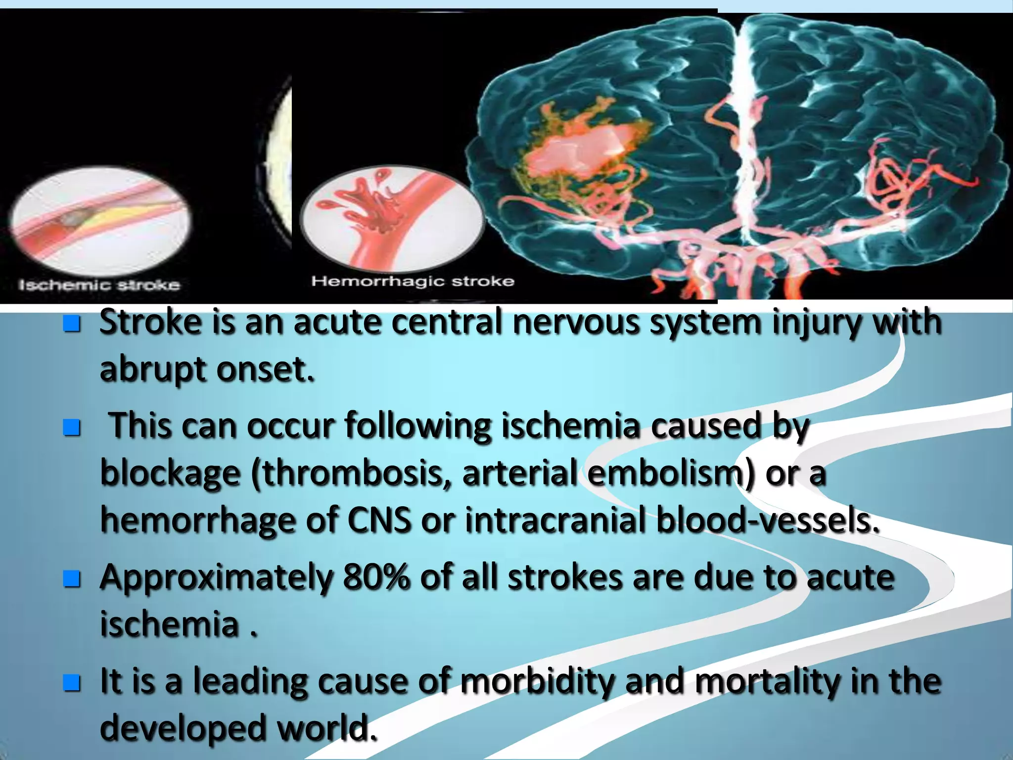
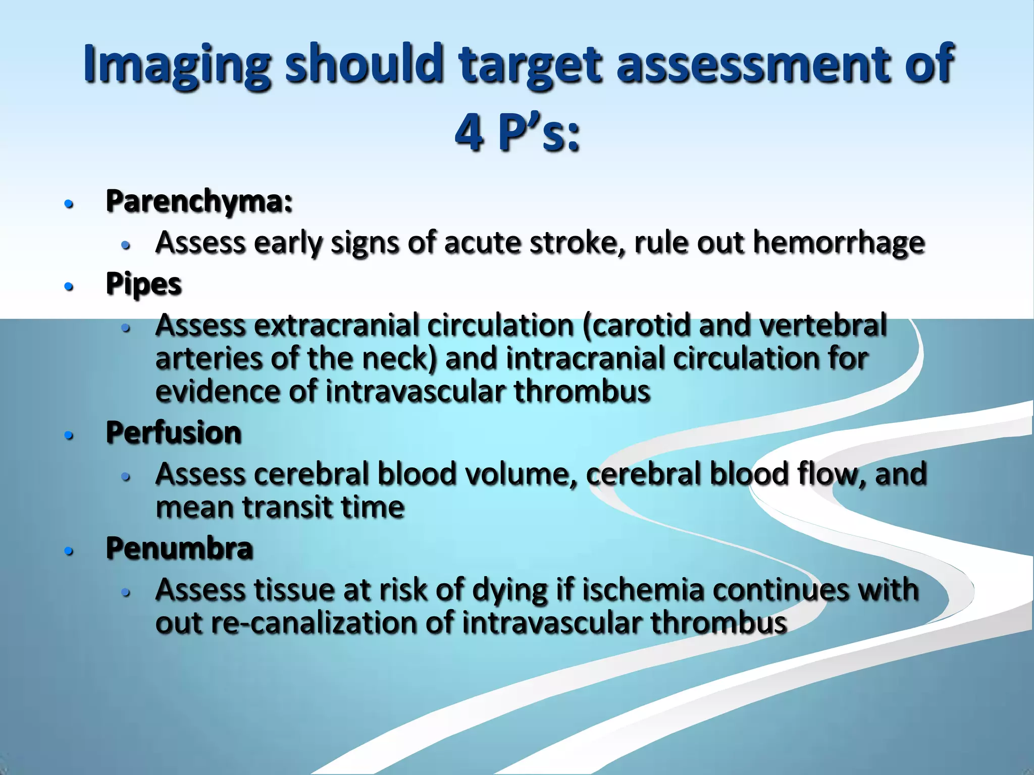
This document discusses various imaging modalities for stroke, focusing on their ability to assess the 4 P's: parenchyma, pipes, perfusion, and penumbra. CT techniques like non-contrast CT, CTA, and CTP can quickly detect hemorrhage, visualize vessels for clots, and assess perfusion/penumbra. MRI techniques like DWI, PWI, MRA provide highly sensitive visualization of acute ischemia and perfusion abnormalities to identify the ischemic core and penumbra. Imaging plays a crucial role in the early diagnosis and management of stroke by establishing the diagnosis, guiding therapy decisions, and identifying salvageable brain tissue.
Its important to recognise the myelination pattern in neonates and infants. This presentation talks about the myelination pattern and imaging of white matter diseases in children.
Radiology Spotters collection by Dr Pradeep. Nice collection Radiology spotters mixed collection ppt made by or collected by Dr. Pradeep, this is a collection of confusing spotter and very important spotter commonly asked in exams, our references is radiopaedia, learning radiology and Aunt Minnie.. Thanks
Magnetic Resonance Angiography and VenographyAnjan Dangal
Introduction to MR Angiography and Venography Procedure of Brain . Includes Indication, MRI protocol, planning and anatomy as well as brief intoduction to physics behind MRA and MRV principle.
A detailed description of ct coronary angiography and calcium scoring with various aspects regarding the preparation, procedure, limitations and a short review regarding post CABG imaging.
Brain CT Anatomy and Basic Interpretation Part ISakher Alkhaderi
Detailed anatomy and Radiological guidelines for radiologist and general physicians to facilitate use of BRAIN CT SCAN in medical diagnosis and emergencies supported by images and scientific data.
CEREBRAL INFARCTS
Pathophysiology
Significantly diminished blood supply to all parts(global ischemia) or selected areas(regional or focal ischemia) of the brain
Focal ischemia- cerebral infarction
Global ischemia-hypoxic ischemic encephalopathy(HIE), hypotensive cerebral infarction
Infarct vs pneumbra
In the central core of the infarct, the severity of hypoperfusion results in irreversible cellular damage
Around this core, there is a region of decreased flow in which either:
The critical flow threshold for cell death has not reached
Or the duration of ischemia has been insufficient to cause irreversible damage.
Current therapies attempt to rescue these ‘at risk’ cells
Goal of imaging
Exclude hemorrhage
Identify the presence of an underlying structural lesion such as tumour , vascular malformation, subdural hematoma that can mimic stroke
Identify stenosis or occlusion of major extra- and intracranial arteries
Differentiate between irreversibly affected brain tissue and reversibly impaired tissue (dead tissue versus tissue at risk)
Imaging modalities
CT
MRI
Diffusion weighted imaging
MRA
MRS
CT angiography
CT perfusion imaging
Perfusion-weighted MR Imaging
Trans cranial doppler
Cerebral angiography
Classification
Hyper acute infarct (<12 hours)
Acute infarct (12 to 48 hours)
Subacute infarct (2 to 14 days)
Chronic infarct (>2 weeks)
Old infarct (> 8 to 10 weeks)
CT-Hyperacute infarct
Normal in 50 – 60%
Hyperdense MCA sign-acute intraluminal thrombus
Obscuration of lentiform nulei
Dot sign-occluded MCA branch in sylvian fissure
Insular ribbon sign –grey white interface loss along the lateral insula
Hyperdense MCA sign
Obscuration of lentiform nuclei
Insular ribbon sign
Insular ribbon sign
MRI –Hyperacute infarct
Absence of normal flow void with intra vascular arterial enhancement
Anatomic changes in T1WI
Sulcal effacement,
Gyral edema,
Loss of grey white interface
Sulcal effacement
CT- Acute infarct
Low density basal ganglia
Sulcal effacement
Wedge shaphed parenchymal hypo density area that involves both grey and white matter
Increasing mass effect
Hemorrhagic transformation may occur -15 to 45% ( basal ganglia and cortex common site) in 24 to 48 hours
Sulcal effacement
MRI –Acute infarct
T2WI-hyperintensity in affected area
Meningeal enhancement adjacent to infarct(12 to 24 hours)
Early parenchymal enhancement
Hemorrhagic transformation becomes evident
MRI –Acute infarct
MRI –Acute infarct
CT – sub acute infarct
NECT
Wedge-shaped area of decreased attenuation involving gray/white matter in typical vascular distribution
Mass effect initially increases, then begins to
diminish by 7-10 days
HT of initially ischemic infarction occurs in 15-20% of MCA occlusions, usually by 48-72 hrs
CECT
Enhancement patterns typically patchy or gyral
May appear as early as 2-3 days after ictus, persisting up to 8-10 weeks
Neuroimaging is the use of various techniques to either directly or indirectly image the structure, function of the nervous system.
Neuroimaging plays a pivotal role in the diagnosis of central nervous system (CNS) disorders.
Main modalities of neuroimaging techniques are CT scan and MRI.
Synthetic Fiber Construction in lab .pptxPavel ( NSTU)
Synthetic fiber production is a fascinating and complex field that blends chemistry, engineering, and environmental science. By understanding these aspects, students can gain a comprehensive view of synthetic fiber production, its impact on society and the environment, and the potential for future innovations. Synthetic fibers play a crucial role in modern society, impacting various aspects of daily life, industry, and the environment. ynthetic fibers are integral to modern life, offering a range of benefits from cost-effectiveness and versatility to innovative applications and performance characteristics. While they pose environmental challenges, ongoing research and development aim to create more sustainable and eco-friendly alternatives. Understanding the importance of synthetic fibers helps in appreciating their role in the economy, industry, and daily life, while also emphasizing the need for sustainable practices and innovation.
2024.06.01 Introducing a competency framework for languag learning materials ...Sandy Millin
http://sandymillin.wordpress.com/iateflwebinar2024
Published classroom materials form the basis of syllabuses, drive teacher professional development, and have a potentially huge influence on learners, teachers and education systems. All teachers also create their own materials, whether a few sentences on a blackboard, a highly-structured fully-realised online course, or anything in between. Despite this, the knowledge and skills needed to create effective language learning materials are rarely part of teacher training, and are mostly learnt by trial and error.
Knowledge and skills frameworks, generally called competency frameworks, for ELT teachers, trainers and managers have existed for a few years now. However, until I created one for my MA dissertation, there wasn’t one drawing together what we need to know and do to be able to effectively produce language learning materials.
This webinar will introduce you to my framework, highlighting the key competencies I identified from my research. It will also show how anybody involved in language teaching (any language, not just English!), teacher training, managing schools or developing language learning materials can benefit from using the framework.
Ethnobotany and Ethnopharmacology:
Ethnobotany in herbal drug evaluation,
Impact of Ethnobotany in traditional medicine,
New development in herbals,
Bio-prospecting tools for drug discovery,
Role of Ethnopharmacology in drug evaluation,
Reverse Pharmacology.
How to Make a Field invisible in Odoo 17Celine George
It is possible to hide or invisible some fields in odoo. Commonly using “invisible” attribute in the field definition to invisible the fields. This slide will show how to make a field invisible in odoo 17.
Read| The latest issue of The Challenger is here! We are thrilled to announce that our school paper has qualified for the NATIONAL SCHOOLS PRESS CONFERENCE (NSPC) 2024. Thank you for your unwavering support and trust. Dive into the stories that made us stand out!
Model Attribute Check Company Auto PropertyCeline George
In Odoo, the multi-company feature allows you to manage multiple companies within a single Odoo database instance. Each company can have its own configurations while still sharing common resources such as products, customers, and suppliers.
Unit 8 - Information and Communication Technology (Paper I).pdfThiyagu K
This slides describes the basic concepts of ICT, basics of Email, Emerging Technology and Digital Initiatives in Education. This presentations aligns with the UGC Paper I syllabus.
2. STROKE
Stroke is an acute central nervous system injury with
abrupt onset.
This can occur following ischemia caused by
blockage (thrombosis, arterial embolism) or a
hemorrhage of CNS or intracranial blood-vessels.
Approximately 80% of all strokes are due to acute
ischemia .
It is a leading cause of morbidity and mortality in the
developed world.
3. GOALS OF IMAGING
To establish the diagnosis as early as possible.
Give accurate information about intracranial
vasculature and brain perfusion for guidance in
selecting the appropriate therapy.
4. Imaging should target assessment of
4 P’s:
• Parenchyma:
• Assess early signs of acute stroke, rule out hemorrhage
• Pipes
• Assess extracranial circulation (carotid and vertebral
arteries of the neck) and intracranial circulation for
evidence of intravascular thrombus
• Perfusion
• Assess cerebral blood volume, cerebral blood flow, and
mean transit time
• Penumbra
• Assess tissue at risk of dying if ischemia continues with
out re-canalization of intravascular thrombus
5. Overview of imaging modalities
Unenhanced CT
• Can be performed quickly.
• Can help identify early signs of stroke, and can
help rule out hemorrhage.
CT angiography can depict intravascular
thrombi
CT perfusion imaging can demonstrate
salvageable tissue which is indicated by a
penumbra.
6. Acute infarcts may be seen early on
conventional MR images, but diffusion-
weighted MR imaging is more sensitive for
detection of hyperacute ischemia.
Gradient-echo MR sequences can be helpful
for detecting a hemorrhage.
7. MR Angiography – To evaluate the status of
neck and intracranial vessels
DWI AND PWI - A mismatch between findings
on diffusion and perfusion MR images may be
used to predict the presence of a penumbra.
8. PENUMBRA
TISSUE AT RISK OR
SALVAGEABLE TISSUE.
Pathophysiology -
Neuronal tissue is sensitive
for ischemia due to lack of
stored energy
With complete absence of
flow neuronal viability is 2-3
minutes
In acute stroke ischemia is
always incomplete due to rich
collateral supply
9. ACUTE STROKE
Acute cerebral ischemia may
result in a central irreversibly
infarcted tissue core
surrounded by a peripheral
region of stunned cells that is
called as a penumbra
This region is potentially
salvageable with early
recanalization
10. The transition from ischemia to irreversible
infarction depends on both the severity and
the duration of the diminution of blood flow
Minutes
Days and weeksTime
Hours
11. ISCHEMIC PENUMBRA
IDENTIFIED BY
CT - ALTERED PARAMETERS IN PERFUSION
MR – PERFUSION DIFFUSION MISMATCH
PRESENCE OF PENUMBRA HAS SIGNIFICANT
IMPLICATIONS IN PT MANAGEMENT
13. NECT
Widely available.
Can be done quickly.
It not only can help identify a hemorrhage (a
contraindication to thrombolytic therapy),
but it also can help detect early-stage acute ischemia by
depicting features such as –
1. THE HYPERDENSE VESSEL SIGN.
2. THE INSULAR RIBBON SIGN.
3. OBSCURATION OF THE LENTIFORM NUCLEUS
14. HYPERDENSE VESSEL SIGN
Acute thrombus has high attenuation value
this feature is referred to as the hyperdense
vessel sign.
Highly specific but sensitivity is poor.
FALSE POSITIVE
HIGH HEMATOGRIT LEVEL
MCA CALCIFICATION
But in such cases the hyperattenuation is usually
bilateral!!!
Rarely, fat emboli appear hypoattenuated when compared
with attenuation in the contralateral vessel .
16. OBSCURATION OF LENTIFORM NUCLEUS
Lentiform nucleus appears hypoattenuated because
of acute ischemia of the lenticulostriate territory ,
resulting in obscuration of the lentiform nucleous.
This feature may be seen on CT images within 2
hours after the onset of a stroke .
18. INSULAR RIBBON SIGN
It is the local hypoattenuation of the insular cortex
region due to Cytotoxic edema as this region is
susceptible to early and irreversible ischemic
damage.
19. INSULAR RIBBON
Axial unenhanced CT image,
obtained
in a 73-year-old woman 21⁄2 hours
after
the onset of left hemiparesis,
shows hypoattenuation
and obscuration of the posterior
part of the
right lentiform nucleus (white
arrow) and a loss
of gray matter–white matter
definition in the lateral
margins of the right insula (black
arrows).
The latter feature is known as the
insular ribbon
sign.
20. WINDOW SETTING
Detection of early acute ischemic stroke on unenhanced
CT images may be improved by using variable window
width and center level settings to accentuate the contrast
between normal and edematous tissue
STANDARD WINDOW SETTING (W80 C 20) – SENSITIVITY
57% SPECIFICITY 100%
STROKE WINDOW SETTING (W8 C 32) SENSITIVITY 71%
SPECIFICITY 100%
21. CT
ANGIOGRAPHY
CT angiography typically involves a volumetric helical acquisition
that extends from the aortic arch to the circle of Willis.
The examination is performed by using a time-optimized
bolus of contrast material for vessel enhancement.
CT angiographic demonstration of a significant thrombus
burden can guide appropriate therapy in the form of
intraarterial or mechanical thrombolysis.
Identification of carotid artery disease and visualization of the
aortic arch may provide clues to the cause of the ischemic
event and guidance for the interventional neuroradiologist
22. (a) Unenhanced CT image in a 72-year-old woman with acute right hemiplegia shows
hyperattenuation in a proximal segment of the left MCA (arrows).
(b, c) Axial (b) and coronal (c) reformatted images from CT angiography show the
apparent absence of the same vessel segment (arrows).
24. CT Perfusion
(CTP):
Basic concept…..
With CT and MR-diffusion we can get a good impression of
the area that is infarcted.
But, we cannot preclude a large ischemic penumbra (tissue at
risk).
With perfusion studies we monitor the first pass of an
iodinated contrast agent bolus through the cerebral
vasculature.
Areas of decreased perfusion will tell us which area is at risk.
25. CT perfusion maps of cerebral blood volume (a) and cerebral
blood flow (b) show, in the left hemisphere, a region of decreased
blood volume (white oval) that corresponds
to the ischemic core and a larger region of decreased blood flow
(black oval in b) that includes the ischemic core and a peripheral
region of salvageable tissue. The difference between the two
maps (black oval white oval) is the penumbra.
26. CT PERFUSION
PARAMETERS ASSESSED
CBV – VOLUME OF BLOOD PER UNIT OF BRAIN TISSUE
(N 4-5ML/100GM)
CBF – VOLUME OF BLOOD FLOW PER UNIT OF BRAIN
TISSUE PER MINUTE (N 50-60ML/100GM/MINUTE)
MTT – TIME DIFFERENCE BETWEEN THE ARTERIAL
INFLOW AND VENOUS OUTFLOW
TIME TO PEAK ENHANCEMENT – TIME FROM THE
BEGINNING OF CONTRAST INJECTION TO MAXIMUM
CONTRAST CONCENTRATION IN A ROI
27. CTP TECHNIQUES
DYNAMIC CONTRAST ENHANCED CT
BASED ON MULTI COMPARTMENT TRACER KINETIC MODEL
PERFORMED ON MDCT
2-4 SECTIONS ARE OBTAINED AND ONE OF THE SECTIONS
PASS THROUGH BASAL GANGLIA
PERFUSED-BLOOD-VOLUME MAPPING- LESS
COMMONLY USED
28. Dynamic Contrast Enhanced CT
Performed by monitoring a first
pass of contrast bolus through
the cerebral circulation
The transient increase in
attenuation generates time-
attenuation curves for an
arterial and venous ROI
Mathematical modeling can be
then used to calculate perfusion
parameters and generate color
coded perfusion maps
(deconvolution analysis)
29.
30. CBF = CBV / MTT
CBF
MTT
Normal CBF is 55 cc/ 100 gm tissue / minute
CBF below this refers to penumbra or tissue at risk
31. EFFECT OF REDUCTION IN CBF
Diagram shows the evolution of events at a microscopic level with decreasing
cerebral perfusion (from right to left). Irreversible cell death generally occurs when
cerebral blood flow decreases to less than 10 mL/100 g/min.
32. INTERPRETATION OF PCT
INFARCTED AREA
SEVERELY DECREASED CBF (<30%) AND CBV (<40%)
PROLONGED MTT
PENUMBRA
INCREASED MTT
MODERATELY DECREASED CBF (>60%)
INCREASED CBV (80-100% OR HIGHER)
OR
INCREASED MTT
MARKEDLY REDUCED CBF (>30%)
MODERATELY REDUCED CBV (>60%)
34. Acute stroke in a 65-year-old man with left hemiparesis. CT perfusion
maps of cerebral blood volume (a), cerebral blood flow (b), and mean
transit time (c) show mismatched abnormalities (arrows) that imply the
presence of a penumbra. The area with decreased blood volume
represents the ischemic core, and that with normal blood volume but
decreased blood flow and increased mean transit time is the penumbra.
35.
36. CONVENTIONAL MRI
SPIN ECHO IMAGES MORE SENSITIVE AND
SPECIFIC THAN CT IN ACUTE CVA
SEQUENCES
T1
T2
FLAIR
GRE
37. ACUTE CVA
HYPER ON T2 AND FLAIR
LOSS OF GRAY WHITE MATTER
DIFFERENTIATION
SULCAL EFFACEMENT
MASS EFFECT
LOSS OF FLOW VOID IN T2WI IN VESSEL
BLOOMING IN GRE IF HRGE
LESS SENSITIVE THAN DWI IN FIRST FEW
HOURS
38. Acute stroke in the left medial
temporal lobe in a 44-year-old man.
(a) Axial T2-weighted and (b)fluid attenuated
inversion recovery images
show areas with increased signal intensity.
(c) Gradient-echo image shows abnormal
low signal intensity in the same areas.
These findings are suggestive of hemorrhage
39. MR ANGIOGRAPHY
Sensitive for intravascular thrombus.
MR angiograms in two patients with acute stroke symptoms
reveal flow gaps in the left proximal middle cerebral artery
(arrow in a) and the basilar artery (arrows in b). Both findings
were due to intravascular thrombi.
40. Diffusion-Weighted Imaging
Brownian motion
The normal motion of
water molecules within
living tissues is random.
Acute stroke causes
excess intracellular water
accumulation or
“cytotoxic edema”, with
an overall decreased rate
of water molecular
diffusion within the
affected tissue. Brownian Motion
41. DWI
AREAS OF CYTOTOXIC EDEMA WITH
RESTRICTED WATER MOLECULE DIFFUSION IN
ACUTE STROKE APPEAR BRIGHTER COMPARED
TO NORMAL TISSUE
TAKES FEW SECS TO 2 MINUTES
42. DWI ACUTE CVA
Acute stroke–induced
cytotoxic edema in the
right cerebellar
hemisphere. Diffusion-
weighted MR image
shows areas of signal
intensity increase due to
the restricted mobility
of water molecules
43. Acute stroke of the posterior circulation in a 77-year-old man. (a)
Diffusion weighted MR image shows bilateral areas of increased
signal intensity (arrows) in the thalami and occipital lobes. (b) ADC
map shows decreased ADC values in the same areas (arrows). These
findings are indicative of acute ischemia.
44. CLINICAL APP OF DWI
CHANGES IN DWI OCCUR WITH IN 30MIN OF ONSET
OF ISCHEMIA WITH CORRESPONDING REDUCTION IN
ADC AND SEEN UP TO 5 DAYS
MILD HYPERINTENSE DWI WITH PSEUDONORMAL
ADC FROM 1 -4WKS
AFTER SEVERAL WKS DWI SIGNAL VARIES (T2
EFFECT) WITH INCREASED ADC
DWI ALONE CANNOT BE USED AND SHOULD ALWAYS
BE COMPARED WITH ADC TO ASSESS THE AGE OF
INFARCT
45. CHRONIC INFARCT
Chronic infarcts in a 71-year-old man with a remote history of
multiple strokes. (a) Diffusion weighted MR image shows areas
of decreased signal intensity in the left frontal lobe. (b) ADC
map shows increased ADC values in the white matter of the right
frontal lobe. These features are suggestive of chronic infarction.
46. ACCURACY
CT/ CONVENTIONAL MRI
SENSITIVITY AND SPECIFICITY < 50%
DWI
SENSITIVITY 88-100%
SPECIFICITY 86-100%
FALSE -VE DWI
LACUNAR INFARCTS OF BRAIN STEM
SMALL DEEP GREY MATTER INFARCTS
FALSE +VE DWI
ABSCESS
CELLULAR TUMOURS LIKE LYMPHOMA
47. MR PERFUSION
The passage of an intravascular MR contrast agent
through the brain capillaries causes a transient loss
of signal because of the T2* effects of the contrast
agent.
The dynamic contrast-enhanced MR perfusion
imaging technique involves tracking of the tissue
signal changes caused by susceptibility (T2*) effects
to create a hemodynamic time–signal intensity
curve,as in dynamic CT perfusion imaging.
Perfusion maps of cerebral blood volume and mean
transit time can be calculated from this curve by
using a deconvolution technique.
48. MR PERFUSION
LESION WHICH SHOWS CHANGES BOTH IN
DWI AND PERFUSION MR – INFARCT CORE
LESION WHICH SHOWS CHANGES ONLY IN
PERFUSION - PENUMBRA
49. (a) Diffusion-
weighted MR image
shows an area of mildly increased
signal intensity in the right parietal
lobe (arrows). The ADC values
in this region were decreased.
(b) Perfusion-weighted MR
image shows a larger area with
increased time to peak
enhancement (arrows) in the right
cerebral hemisphere.
The mismatch
between the perfusion
and diffusion images is indicative
of a large penumbra.
Acute stroke in a 67-year-old woman with acute
left hemiplegia
50. CLINICAL APPLICATION
Unenhanced CT: rule out hemorrhage
Not very good to detect ischemia
T1 or T2 weighted MRI
Good for detecting ischemia
Cannot differentiate between acute versus
chronic ischemia
So we have…
51. Diffusion-weighted MR
More sensitive for detection of hyperacute
ischemia
becomes abnormal within 30 minutes
Distinguish b/w old and new stroke
New stroke: bright on DWI
Old stroke: Low SI on DWI
It detects irreversible infarcted tissue
53. Perfusion-Weighted imaging
Allows the measurement of capillary
perfusion of the brain
Uses a MR contrast agent
The contrast bolus passage causes a nonlinear
signal decrease in proportion to the perfusion
cerebral blood volume
Meaning, it can identify areas of
hypoperfusion, the reversible ischemia, as
well (unlike DWI)
54. Comparison of PWI and DWI
DWI Depicts irreversibly damaged infarct
PWI Reflects the complete area of
hypoperfusion
The volume difference between these two,
the PWI/DWI mismatch would be the
PENUMBRA!
If there is no difference in PWI and DWI, no
penumbra is present
55. Significance of PWI/DWI mismatch
IV thrombolytic treatment is not typically
administered to patients with acute stroke
beyond 3-hrs period
Risk of hemorrhage
However, recent studies have shown that IV
thrombolytic therapy may benefit patients
who are carefully selected according to
PWI/DWI mismatch, beyond 3-hrs window
59. IV-TPA-WONDERFUL THERAPY.
FDA-USA approved Rx of Ischemic Stroke.
Improved outcome within 3 hours in properly
selected patients.
Results are best within 90 minutes.
Results are better within 90 -180 minutes.
TPA reverses ischemic changes saving brain.
60. Thrombolytic Therapy - IV -t-PA
I. Inclusion Criteria
1. Within 3 hours of the stroke and patient not needing
ventilator.
2. CT Scan head Normal or < 1/3 MCA hypo density.
II. Exclusion Criteria
1. BP > 185/110 mm on admission.
2. Use of Oral Anticoagulants.
3. Major surgery preceding FOURTEEN days.
4. Head injury - LAST THREE MONTHS.
5. Prior Intracranial hemorrhage/Recent GI bleed.
6. Prolonged PT / aPTT / INR / low Platelet count.
61.
62. Intraarterial TPA.
IA-TPA in selected pts. In < 6 hours due to MCA
& BA occlusion
In BA occlusion it can be given even after > 12
hours.
In future IA-TPA will be rewarding.
IV &IA(IMS Trial) showed 56% of recanalisation.
63.
64.
65. CONCLUSION
Current imaging techniques can be used
to identify hyperacute stroke and guide
therapy
Both CT and MR imaging are useful for
the comprehensive evaluation of acute
stroke
66. THE FUTURE
Selection of patients for thrombolytic therapy
may be made more effectively by performing
appropriate imaging studies rather than relying
on the time of onset as the sole determinant of
selection.
New emerging technique MR permeability
image used to predict microvascular
permeability and quantification of BBB – Pts
with defective BBB are more prone for bleeding
complication following thrombolytic therapy.