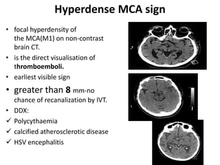
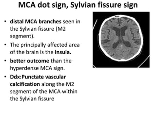
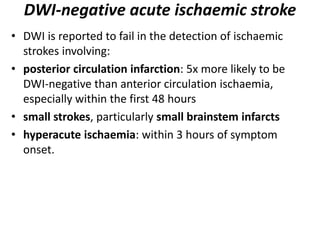
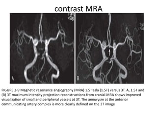
Seminar Presentation Imaging Of
CEREBROVASCULAR ACCIDENT AND
ISCHEMIC STROKE
By Dr. Irko worku (Radiology resident )
DEC 21 , 2023
1
OUTLINES
• Introduction
• Types of Ischemia
• Pathophysiology
• Principles of imaging
• Goals of imaging
• Cerebral venous infarction
• Acute cerebellar infarcts
• Vasculopathies
INTRODUCTION
• A stroke is a clinical diagnosis that refers to a sudden
onset focal neurological deficit of presumed vascular
origin.
• Stroke is generally divided into two broad categories :
• ischaemic stroke
• haemorrhagic stroke
• Stroke- clinical determination
• Infarction- pathologic term - based on imaging,
pathology, and/or persistent neurologic symptoms, with
other causes excluded.
• silent CNS infarction- If there is imaging or pathologic
evidence of an infarct but no attributable clinical
symptoms.
Clinical presentation
• An ischaemic stroke typically presents with rapid
onset neurological deficit, which is determined by the
area of the brain that is involved. The symptoms often
evolve over hours and may worsen or improve,
depending on the fate of the ischaemic penumbra.
• The vascular territory affected will determine the exact
symptoms and clinical behaviour of the lesion:
Con’t
• brain tissue is sensitive to ischemia, because
of the absence of neuronal energy stores.
• In the complete absence of blood flow, the
available energy can maintain neuronal
viability for approximately 2–3 minutes.
• However, in acute stroke, ischemia is more
often incomplete-collateral blood supply from
uninjured arterial and leptomeningeal
territories.
PATHOPHYSIOLOGY
The vascular territory
• anterior circulation infarct
– anterior cerebral artery
infarct
– middle cerebral artery
infarct
– lacunar infarct
– striatocapsular infarct
• posterior circulation infarct
– posterior cerebral artery
infarct
– cerebellar infarct
– brainstem infarct
Ageing ischaemic strokes
 radiopedia
• early hyperacute: 0 to 6 hours
• late hyperacute: 6 to 24 hours
• acute: 24 hours to 1 week
• subacute: 1 to 3 weeks
• chronic: more than 3 weeks
Goals of Imaging
Stroke Imaging modalities
• In many institutions with active stroke services which
provide reperfusion therapies, a so-called code stroke
aimed at expediting diagnosis and treatment of patients
multimodality imaging.
• Stroke protocol (CT)
 non-contrast CT (brain)
 CT perfusion (brain)
 CT angiography (aortic arch to the vertex of the skull)
• Brain MRI
• For vascular evaluation
 MRA (TOF, phase contrast MRI, contrast enhanced
angiography)
 Carotid Ultrasound/Transcranial Doppler ultrasound
Non contrast CT imaging
• Non-contrast CT of the brain remains the mainstay of
imaging in the setting of an acute stroke.
• limited sensitivity in the acute setting.
• Detection depends on :
 the territory,
 the experience of the interpreting radiologist and
 time of the scan from the onset of symptoms.
 Whether tissue is supplied by end arteries
 Pattern of collateral supply
Con’t
• The goals of CT in the acute setting are:
exclude intracranial haemorrhage, which would
preclude thrombolysis
look for any "early" features of ischaemia
exclude other intracranial pathologies that may
mimic a stroke, such as a tumour
Immediate CT finding in ischemic stroke
• hyperdense segment of a
vessel.
• hyperdense MCA sign
• MCA dot sign)
• Basilar artery hyperdense sign
• DDX- calcified cerebral
embolus.
Hyperdense MCA sign
• focal hyperdensity of
the MCA(M1) on non-contrast
brain CT.
• is the direct visualisation of
thromboemboli.
• earliest visible sign
• greater than 8 mm-no
chance of recanalization by IVT.
• DDX:
 Polycythaemia
 calcified atherosclerotic disease
 HSV encephalitis
MCA dot sign, Sylvian fissure sign
• distal MCA branches seen in
the Sylvian fissure (M2
segment).
• The principally affected area
of the brain is the insula.
• better outcome than the
hyperdense MCA sign.
• Ddx:Punctate vascular
calcification along the M2
segment of the MCA within
the Sylvian fissure
CON’T
Calcified cerebral embolus
• small in size, 2-3 mm.
 calcific AS (most common)
 mitral annular calcification
 calcified Major vessels
• If multiple ( salted pretzel sign)
• round or ovoid shape,
• higher attenuation (~160 HU)
• Ddx: haemorrhage, vessel wall
calcification, infection
(e.g. neurocysticercosis), caverno
mas,
Early hyperacute
• loss of grey-white matter differentiation, and
hypoattenuation of deep nuclei.
 Use stroke window (8w/c32)
• cortical hypodensity with associated parenchymal
swelling with resultant gyral effacement.
• The insular ribbon sign
Insular ribon sign and obscuration of the
lentiform nucleus, which appears
hypoattenuated because of cytotoxic edema
Con’t
Summary of evolution of infarction on CT
Acute
• The hypoattenuation and swelling
• petechial haemorrhages
• significant mass effect
• secondary damage .
• Subacute
• swelling starts to subside and elevation of the
attenuation of the cortex.
• affected cortex will appear near normal.(CT
fogging phenomenon)
Chronic
• residual swelling passes
• gliosis sets in eventually appearing as a region
of low density with a negative mass effect.
Quantitation of Ischemic Involvement
• the European Cooperative Acute Stroke Study trial, involvement
of more than one-third of the MCA territory depicted at
unenhanced CT was a criterion for the exclusion of patients
from thrombolytic therapy because of a potential increase in
the risk for hemorrhage.
• The Alberta Stroke Program Early CT Score (ASPECTS) was
proposed in 2001 as a means of quantitatively assessing acute
ischemia on CT images by using a 10-point topographic scoring
system.
• the MCA territory is divided into 10 regions, each of which
accounts for one point in the total score
ASPECT SCORE
• One point for each region.
• Score ---/10
• M1 to M3 are at the level of
the basal ganglia
• M4 to M6 are at the level of
the ventricles immediately
above the basal ganglia
• score less than or equal to 7
predicts a worse functional
outcome at 3 months as well as
symptomatic haemorrhage,
with thrombolysis did not have
a good clinical outcome
pc-ASPECTS
• thalami (1 point each)
• occipital lobes (1 point
each)
• midbrain (2 points)
• pons (2 points)
• cerebellar hemispheres
(1 point each)
• Score---/10
Differentiating between acute and chronic infarction on a CT
acute
• cytotoxic oedema
• Hypoattenuating area -
more dense than CSF
• Has positive mass effect
sulcal / ventricular
effacement, midline
shift/ herniation.
chronic:
• encephalomalacia;
• Wallerian degeneration
• Hypoattenuating area
has
• Negative mass effect
like-widened sulci, ex
vacuo dilatation of
ipsilateral ventricle CSF
density.
MRI
• more time consuming and
• less available
• BUT higher sensitivity and
specificity for early ischemic
infarction.
Early hyperacute
• DWI demonstrates increased
signal and reduced ADC
values.
• thromboembolism may be
detected (e.g. on SWI, GRE ).
• Slow or stagnant flow in
vessels may also be detected
as a loss of normal flow void
and high signal on T2/FLAIR
and T1 C+ (intravascular
enhancement).
Hyperacute infarct (0–6 hours)
• Diffusion is reduced in an acute infarct by two factors:
1) Shift from extracellular to intracellular water due to Na/K
ATPase pump failure.
2) Increased viscosity of infarcted brain due to cell lysis and
increased extracellular protein.
C, Diffusion-weighted image reveals extensive ganglionic and cortical
hyperintensity indicative of hyperacute infarction. D, Apparent diffusion coefficient map
reveals diffuse
hypointensity indicative of restricted diffusion.
CON’T…
• If infarction is incomplete then cortical
contrast enhancement may be seen as early as
2 to 4 hours.
• In a minority of cases(6.8%), DWI may be
normal - DWI-negative acute ischaemic
stroke
DWI-negative acute ischaemic stroke
• DWI is reported to fail in the detection of ischaemic
strokes involving:
• posterior circulation infarction: 5x more likely to be
DWI-negative than anterior circulation ischaemia,
especially within the first 48 hours
• small strokes, particularly small brainstem infarcts
• hyperacute ischaemia: within 3 hours of symptom
onset.
Acute infarct (6 hours–72 hours)
 • The acute infarct phase is characterized by increase in vasogenic
edema and mass effect.
 On imaging, there is increased sulcal effacement and mass effect.
 The mass effect peaks at 3–4 days,
 MRI shows hyperintensity of the infarct core on T2-weighted
images, best seen on FLAIR.
• The FLAIR abnormality is usually confined to the gray matter.
• DWI continues to show restricted diffusion.
• due to increased collateral flow there may be some arterial
enhancement.
• Perfusion images most commonly show increase in size of the
infarct core with resultant decrease in size of the penumbra.
T2W FLAIR
Early subacute infarct
• blood flow to the affected brain is re-established by
leptomeningeal collaterals and ingrowth of new vessels
into the region of infarction.
• The new vessels have an incomplete blood-brain barrier,
causing a continued increase in vasogenic edema and
mass effect, which peaks at 3–4 days.
• MR imaging shows marked hyperintensity on T2-
weighted images involving both gray and white matter
(vasogenic edema) (in contrast to the acute stage which
usually involves just the gray matter, cytotoxic edema).
• The ADC map becomes less dark or even resolves if there
is extensive edema;
 the DWI images typically remain bright due to underlying
T2 shine-through.
Late subacute infarct
• resolution of vasogenic edema and reduction in mass
effect.
• A key imaging finding is gyriform enhancement, which
may occasionally be confused for a neoplasm.
 Unlike a tumor, however, a subacute infarction will not
have mass effect.
• The enhancement of a subacute infarct -“2-2-2” rule,
which states that enhancement begins at 2 days, peaks
at 2 weeks, and disappears by 2 months.
• DWI may remain bright due to T2 shine-through,
although the ADC map will either return to normal or
show increased diffusivity.
Chronic infarct
• In the chronic stage of infarction, cellular debris and dead
brain tissue are removed by macrophages and replaced by
cystic encephalomalacia and gliosis.
Wallerian degeneration
Evolution of infarction
Perfusion Imaging
• Characterize microscopic flow at the capillary
level.
• The central volume principle: CBF=CBV/MTT
• The CBF of the normal brain ranges between 45
and 110 mL/min/100 g of tissue.
• Cerebral oligemia (about 20 to 40 mL/min/100 g)
is defined as under perfused asymptomatic
region of brain that will recover spontaneously.
Cont’d…
• Intravenous contrast is then administered and
various parameters of cerebral perfusion
calculated.
cerebral blood volume (CBV)
cerebral blood flow (CBF)
mean transit time (MTT)
time-to-maximum (Tmax) or time to peak (TTP)
• In patients where volume of brain at risk is
greater than the already infarcted brain by more
than 20%, treatment may result in improved
outcome.
cerebral perfusion
MRI DIFFUSION PERFUSION STUDY
• On the left we first have a
diffusion image indicating
the area with irreversible
changes (dead tissue).
• In the middle there is a
large area with
hypoperfusion.
• On the right the diffusion-
perfusion mismatch is
indicated in blue.
This is the tissue at risk.
This is the brain tissue
that maybe can be saved
with therapy. Diffusion in yellow. Perfusion in red.
Mismatch in blue is penumbra.
Con’t
Vascular evaluation
• Carotid Ultrasound/Transcranial Doppler
• Magnetic Resonance Angiography(There are
three different techniques used to generate
MRA: time-of-flight (TOF), phase contrast (PC),
and contrast enhanced angiography)
• Computed Tomographic Angiography
• Conventional Catheter Angiography
Carotid Ultrasound
Transcranial Doppler ultrasound
• is a noninvasive means used to evaluate the basal
cerebral arteries through the infratemporal fossa.
• It evaluates the flow velocity spectrum of the
cerebral vessels and can provide information
regarding
 the direction of flow,
 the patency of vessels,
 focal narrowing
 It can determine adequacy of middle cerebral
artery flow
contrast MRA
FIGURE 3-9 Magnetic resonance angiography (MRA) 1.5 Tesla (1.5T) versus 3T. A, 1.5T and
(B) 3T maximum intensity projection reconstructions from cranial MRA shows improved
visualization of small and peripheral vessels at 3T. The aneurysm at the anterior
communicating artery complex is more clearly defined on the 3T image
Contrast enhanced CTA
abrupt occlusion Of the internal carotid artery
Conventional Catheter Angiography Indications
1. if the MRA, CTA, or/and carotid ultrasound are
equivocal;
2. if MRA is contraindicated (e.g., in patients with
pacemakers);
3. if cardiac output is too low to produce a diagnostic
CTA;
4. to evaluate complex aneurysms or vascular
malformations responsible for an intracranial
hemorrhage; and
5. for the evaluation of vasculitis.
Cerebral venous infarction
• most commonly secondary to cerebral venous thrombosis and
frequently manifests with haemorrhage.
• Thrombosis of a cortical vein or a deep venous sinus is one of
the more common causes.
• of stroke in younger patients. Risk factors for venous thrombosis
include pregnancy, oral contraceptives, thrombophilia,
malignancy, and infection.
• On non contrast CT is increased density within the thrombosed
sinus or cortical vein (the cord sign).
• On contrast-enhanced CT, the empty delta sign signifies a filling
defect in the superior sagittal sinus.
• MR venogram will show lack of flow in the thrombosed vein or
dural venous sinus.
Cont’d…
• Venous thrombosis leads to venous hypertension, which may
cause infarction and parenchymal hemorrhage. There are
three characteristic patterns of venous infarction, dependent
on the location of the thrombosed vein:
• Superior sagittal sinus thrombosis infarction of the
parasagittal high convexity cortex.
• Deep venous system thrombosis infarction of the
bilateral thalami.
• Transverse sinus thrombosis infarction of the
posterior temporal lobe.
• Transverse sinus is usualy assymetric-larger
may be mistaken for thrombosis.
• newborns -normal polycythemia ->increased
vascular density, the relative hypodensity of
the brain, frequent occurrence of minimal
perinatal paratentorial hemorrhage can mimic
the appearance of sinus thrombosis.
Acute Cerebellar Infarcts
• <5% , Male predominance and a mean age of 65 years.
• The abrupt onset of posteriorly located headaches,
severe vertigo, dysarthria,nausea and vomiting,
nystagmus, ipsilateral dysmetria, and unsteadiness of
gait.
• delayed alteration of consciousness seen in 90% of
patients with mass effect due to cerebellar swelling.
• This can occur rapidly (within a few hours) or up to 10
days after the ictus.
• These infarcts are often difficult to identify on CT
because beam-hardening artifact or partial volume
averaging in the posterior fossa.
• visualize the fourth ventricle and quadrigeminal plate
cistern because subtle asymmetry.
VASCULOPATHIES
• Traditional term “vasculitis”
• endothelial damage and thrombosis produced by
circulating antigen-antibody complexes, mural
edema, and/or spasm.
• Heterogeneous group of diseases with
immunologic basis and similarity of the
appearances of many of these diseases.
• Prolonged insults may result in fibrosis and fixed
narrowing regardless of the initial insult.
• Catheter angiography remains the imaging “gold
standard”
• 3T MRA
Con’t…
• The vasculopathies can be said to affect
 extracranial and extradural arteries; (e.g FMD)
arteries at the skull base at or near the circle of
Willis; (e.g Moya moya,TB meningitis)
 secondary and tertiary branches of the carotid
and/or basilar arteries (e.g., sylvian and convexity
branches of the MCA); e.g wegner
granulomatosis, polyartritis nodosa
small perforating arteries (e.g., lenticulostriate
arteries).e.g collagen vascular disease, Sjogren
syndrome,migraine
Fibromuscular dysplasia
Reference
THANK YOU

Imaging ischemic infarction.pptx

  • 1.
    Seminar Presentation ImagingOf CEREBROVASCULAR ACCIDENT AND ISCHEMIC STROKE By Dr. Irko worku (Radiology resident ) DEC 21 , 2023 1
  • 2.
    OUTLINES • Introduction • Typesof Ischemia • Pathophysiology • Principles of imaging • Goals of imaging • Cerebral venous infarction • Acute cerebellar infarcts • Vasculopathies
  • 3.
    INTRODUCTION • A strokeis a clinical diagnosis that refers to a sudden onset focal neurological deficit of presumed vascular origin. • Stroke is generally divided into two broad categories : • ischaemic stroke • haemorrhagic stroke • Stroke- clinical determination • Infarction- pathologic term - based on imaging, pathology, and/or persistent neurologic symptoms, with other causes excluded. • silent CNS infarction- If there is imaging or pathologic evidence of an infarct but no attributable clinical symptoms.
  • 4.
    Clinical presentation • Anischaemic stroke typically presents with rapid onset neurological deficit, which is determined by the area of the brain that is involved. The symptoms often evolve over hours and may worsen or improve, depending on the fate of the ischaemic penumbra. • The vascular territory affected will determine the exact symptoms and clinical behaviour of the lesion:
  • 5.
    Con’t • brain tissueis sensitive to ischemia, because of the absence of neuronal energy stores. • In the complete absence of blood flow, the available energy can maintain neuronal viability for approximately 2–3 minutes. • However, in acute stroke, ischemia is more often incomplete-collateral blood supply from uninjured arterial and leptomeningeal territories.
  • 6.
  • 7.
    The vascular territory •anterior circulation infarct – anterior cerebral artery infarct – middle cerebral artery infarct – lacunar infarct – striatocapsular infarct • posterior circulation infarct – posterior cerebral artery infarct – cerebellar infarct – brainstem infarct
  • 8.
    Ageing ischaemic strokes radiopedia • early hyperacute: 0 to 6 hours • late hyperacute: 6 to 24 hours • acute: 24 hours to 1 week • subacute: 1 to 3 weeks • chronic: more than 3 weeks
  • 9.
  • 10.
    Stroke Imaging modalities •In many institutions with active stroke services which provide reperfusion therapies, a so-called code stroke aimed at expediting diagnosis and treatment of patients multimodality imaging. • Stroke protocol (CT)  non-contrast CT (brain)  CT perfusion (brain)  CT angiography (aortic arch to the vertex of the skull) • Brain MRI • For vascular evaluation  MRA (TOF, phase contrast MRI, contrast enhanced angiography)  Carotid Ultrasound/Transcranial Doppler ultrasound
  • 11.
    Non contrast CTimaging • Non-contrast CT of the brain remains the mainstay of imaging in the setting of an acute stroke. • limited sensitivity in the acute setting. • Detection depends on :  the territory,  the experience of the interpreting radiologist and  time of the scan from the onset of symptoms.  Whether tissue is supplied by end arteries  Pattern of collateral supply
  • 12.
    Con’t • The goalsof CT in the acute setting are: exclude intracranial haemorrhage, which would preclude thrombolysis look for any "early" features of ischaemia exclude other intracranial pathologies that may mimic a stroke, such as a tumour
  • 13.
    Immediate CT findingin ischemic stroke • hyperdense segment of a vessel. • hyperdense MCA sign • MCA dot sign) • Basilar artery hyperdense sign • DDX- calcified cerebral embolus.
  • 14.
    Hyperdense MCA sign •focal hyperdensity of the MCA(M1) on non-contrast brain CT. • is the direct visualisation of thromboemboli. • earliest visible sign • greater than 8 mm-no chance of recanalization by IVT. • DDX:  Polycythaemia  calcified atherosclerotic disease  HSV encephalitis
  • 15.
    MCA dot sign,Sylvian fissure sign • distal MCA branches seen in the Sylvian fissure (M2 segment). • The principally affected area of the brain is the insula. • better outcome than the hyperdense MCA sign. • Ddx:Punctate vascular calcification along the M2 segment of the MCA within the Sylvian fissure
  • 16.
  • 17.
    Calcified cerebral embolus •small in size, 2-3 mm.  calcific AS (most common)  mitral annular calcification  calcified Major vessels • If multiple ( salted pretzel sign) • round or ovoid shape, • higher attenuation (~160 HU) • Ddx: haemorrhage, vessel wall calcification, infection (e.g. neurocysticercosis), caverno mas,
  • 18.
    Early hyperacute • lossof grey-white matter differentiation, and hypoattenuation of deep nuclei.  Use stroke window (8w/c32) • cortical hypodensity with associated parenchymal swelling with resultant gyral effacement. • The insular ribbon sign
  • 19.
    Insular ribon signand obscuration of the lentiform nucleus, which appears hypoattenuated because of cytotoxic edema
  • 20.
  • 21.
    Summary of evolutionof infarction on CT Acute • The hypoattenuation and swelling • petechial haemorrhages • significant mass effect • secondary damage . • Subacute • swelling starts to subside and elevation of the attenuation of the cortex. • affected cortex will appear near normal.(CT fogging phenomenon)
  • 22.
    Chronic • residual swellingpasses • gliosis sets in eventually appearing as a region of low density with a negative mass effect.
  • 23.
    Quantitation of IschemicInvolvement • the European Cooperative Acute Stroke Study trial, involvement of more than one-third of the MCA territory depicted at unenhanced CT was a criterion for the exclusion of patients from thrombolytic therapy because of a potential increase in the risk for hemorrhage. • The Alberta Stroke Program Early CT Score (ASPECTS) was proposed in 2001 as a means of quantitatively assessing acute ischemia on CT images by using a 10-point topographic scoring system. • the MCA territory is divided into 10 regions, each of which accounts for one point in the total score
  • 24.
    ASPECT SCORE • Onepoint for each region. • Score ---/10 • M1 to M3 are at the level of the basal ganglia • M4 to M6 are at the level of the ventricles immediately above the basal ganglia • score less than or equal to 7 predicts a worse functional outcome at 3 months as well as symptomatic haemorrhage, with thrombolysis did not have a good clinical outcome
  • 25.
    pc-ASPECTS • thalami (1point each) • occipital lobes (1 point each) • midbrain (2 points) • pons (2 points) • cerebellar hemispheres (1 point each) • Score---/10
  • 26.
    Differentiating between acuteand chronic infarction on a CT acute • cytotoxic oedema • Hypoattenuating area - more dense than CSF • Has positive mass effect sulcal / ventricular effacement, midline shift/ herniation. chronic: • encephalomalacia; • Wallerian degeneration • Hypoattenuating area has • Negative mass effect like-widened sulci, ex vacuo dilatation of ipsilateral ventricle CSF density.
  • 27.
    MRI • more timeconsuming and • less available • BUT higher sensitivity and specificity for early ischemic infarction. Early hyperacute • DWI demonstrates increased signal and reduced ADC values. • thromboembolism may be detected (e.g. on SWI, GRE ). • Slow or stagnant flow in vessels may also be detected as a loss of normal flow void and high signal on T2/FLAIR and T1 C+ (intravascular enhancement).
  • 28.
    Hyperacute infarct (0–6hours) • Diffusion is reduced in an acute infarct by two factors: 1) Shift from extracellular to intracellular water due to Na/K ATPase pump failure. 2) Increased viscosity of infarcted brain due to cell lysis and increased extracellular protein. C, Diffusion-weighted image reveals extensive ganglionic and cortical hyperintensity indicative of hyperacute infarction. D, Apparent diffusion coefficient map reveals diffuse hypointensity indicative of restricted diffusion.
  • 29.
    CON’T… • If infarctionis incomplete then cortical contrast enhancement may be seen as early as 2 to 4 hours. • In a minority of cases(6.8%), DWI may be normal - DWI-negative acute ischaemic stroke
  • 30.
    DWI-negative acute ischaemicstroke • DWI is reported to fail in the detection of ischaemic strokes involving: • posterior circulation infarction: 5x more likely to be DWI-negative than anterior circulation ischaemia, especially within the first 48 hours • small strokes, particularly small brainstem infarcts • hyperacute ischaemia: within 3 hours of symptom onset.
  • 31.
    Acute infarct (6hours–72 hours)  • The acute infarct phase is characterized by increase in vasogenic edema and mass effect.  On imaging, there is increased sulcal effacement and mass effect.  The mass effect peaks at 3–4 days,  MRI shows hyperintensity of the infarct core on T2-weighted images, best seen on FLAIR. • The FLAIR abnormality is usually confined to the gray matter. • DWI continues to show restricted diffusion. • due to increased collateral flow there may be some arterial enhancement. • Perfusion images most commonly show increase in size of the infarct core with resultant decrease in size of the penumbra.
  • 32.
  • 33.
    Early subacute infarct •blood flow to the affected brain is re-established by leptomeningeal collaterals and ingrowth of new vessels into the region of infarction. • The new vessels have an incomplete blood-brain barrier, causing a continued increase in vasogenic edema and mass effect, which peaks at 3–4 days. • MR imaging shows marked hyperintensity on T2- weighted images involving both gray and white matter (vasogenic edema) (in contrast to the acute stage which usually involves just the gray matter, cytotoxic edema). • The ADC map becomes less dark or even resolves if there is extensive edema;  the DWI images typically remain bright due to underlying T2 shine-through.
  • 34.
    Late subacute infarct •resolution of vasogenic edema and reduction in mass effect. • A key imaging finding is gyriform enhancement, which may occasionally be confused for a neoplasm.  Unlike a tumor, however, a subacute infarction will not have mass effect. • The enhancement of a subacute infarct -“2-2-2” rule, which states that enhancement begins at 2 days, peaks at 2 weeks, and disappears by 2 months. • DWI may remain bright due to T2 shine-through, although the ADC map will either return to normal or show increased diffusivity.
  • 35.
    Chronic infarct • Inthe chronic stage of infarction, cellular debris and dead brain tissue are removed by macrophages and replaced by cystic encephalomalacia and gliosis.
  • 36.
  • 37.
  • 38.
    Perfusion Imaging • Characterizemicroscopic flow at the capillary level. • The central volume principle: CBF=CBV/MTT • The CBF of the normal brain ranges between 45 and 110 mL/min/100 g of tissue. • Cerebral oligemia (about 20 to 40 mL/min/100 g) is defined as under perfused asymptomatic region of brain that will recover spontaneously.
  • 39.
    Cont’d… • Intravenous contrastis then administered and various parameters of cerebral perfusion calculated. cerebral blood volume (CBV) cerebral blood flow (CBF) mean transit time (MTT) time-to-maximum (Tmax) or time to peak (TTP) • In patients where volume of brain at risk is greater than the already infarcted brain by more than 20%, treatment may result in improved outcome.
  • 40.
  • 41.
    MRI DIFFUSION PERFUSIONSTUDY • On the left we first have a diffusion image indicating the area with irreversible changes (dead tissue). • In the middle there is a large area with hypoperfusion. • On the right the diffusion- perfusion mismatch is indicated in blue. This is the tissue at risk. This is the brain tissue that maybe can be saved with therapy. Diffusion in yellow. Perfusion in red. Mismatch in blue is penumbra.
  • 42.
  • 43.
    Vascular evaluation • CarotidUltrasound/Transcranial Doppler • Magnetic Resonance Angiography(There are three different techniques used to generate MRA: time-of-flight (TOF), phase contrast (PC), and contrast enhanced angiography) • Computed Tomographic Angiography • Conventional Catheter Angiography
  • 44.
  • 45.
    Transcranial Doppler ultrasound •is a noninvasive means used to evaluate the basal cerebral arteries through the infratemporal fossa. • It evaluates the flow velocity spectrum of the cerebral vessels and can provide information regarding  the direction of flow,  the patency of vessels,  focal narrowing  It can determine adequacy of middle cerebral artery flow
  • 46.
    contrast MRA FIGURE 3-9Magnetic resonance angiography (MRA) 1.5 Tesla (1.5T) versus 3T. A, 1.5T and (B) 3T maximum intensity projection reconstructions from cranial MRA shows improved visualization of small and peripheral vessels at 3T. The aneurysm at the anterior communicating artery complex is more clearly defined on the 3T image
  • 47.
    Contrast enhanced CTA abruptocclusion Of the internal carotid artery
  • 48.
    Conventional Catheter AngiographyIndications 1. if the MRA, CTA, or/and carotid ultrasound are equivocal; 2. if MRA is contraindicated (e.g., in patients with pacemakers); 3. if cardiac output is too low to produce a diagnostic CTA; 4. to evaluate complex aneurysms or vascular malformations responsible for an intracranial hemorrhage; and 5. for the evaluation of vasculitis.
  • 49.
    Cerebral venous infarction •most commonly secondary to cerebral venous thrombosis and frequently manifests with haemorrhage. • Thrombosis of a cortical vein or a deep venous sinus is one of the more common causes. • of stroke in younger patients. Risk factors for venous thrombosis include pregnancy, oral contraceptives, thrombophilia, malignancy, and infection. • On non contrast CT is increased density within the thrombosed sinus or cortical vein (the cord sign). • On contrast-enhanced CT, the empty delta sign signifies a filling defect in the superior sagittal sinus. • MR venogram will show lack of flow in the thrombosed vein or dural venous sinus.
  • 50.
    Cont’d… • Venous thrombosisleads to venous hypertension, which may cause infarction and parenchymal hemorrhage. There are three characteristic patterns of venous infarction, dependent on the location of the thrombosed vein: • Superior sagittal sinus thrombosis infarction of the parasagittal high convexity cortex. • Deep venous system thrombosis infarction of the bilateral thalami. • Transverse sinus thrombosis infarction of the posterior temporal lobe.
  • 51.
    • Transverse sinusis usualy assymetric-larger may be mistaken for thrombosis. • newborns -normal polycythemia ->increased vascular density, the relative hypodensity of the brain, frequent occurrence of minimal perinatal paratentorial hemorrhage can mimic the appearance of sinus thrombosis.
  • 53.
    Acute Cerebellar Infarcts •<5% , Male predominance and a mean age of 65 years. • The abrupt onset of posteriorly located headaches, severe vertigo, dysarthria,nausea and vomiting, nystagmus, ipsilateral dysmetria, and unsteadiness of gait. • delayed alteration of consciousness seen in 90% of patients with mass effect due to cerebellar swelling. • This can occur rapidly (within a few hours) or up to 10 days after the ictus. • These infarcts are often difficult to identify on CT because beam-hardening artifact or partial volume averaging in the posterior fossa. • visualize the fourth ventricle and quadrigeminal plate cistern because subtle asymmetry.
  • 55.
    VASCULOPATHIES • Traditional term“vasculitis” • endothelial damage and thrombosis produced by circulating antigen-antibody complexes, mural edema, and/or spasm. • Heterogeneous group of diseases with immunologic basis and similarity of the appearances of many of these diseases. • Prolonged insults may result in fibrosis and fixed narrowing regardless of the initial insult. • Catheter angiography remains the imaging “gold standard” • 3T MRA
  • 56.
    Con’t… • The vasculopathiescan be said to affect  extracranial and extradural arteries; (e.g FMD) arteries at the skull base at or near the circle of Willis; (e.g Moya moya,TB meningitis)  secondary and tertiary branches of the carotid and/or basilar arteries (e.g., sylvian and convexity branches of the MCA); e.g wegner granulomatosis, polyartritis nodosa small perforating arteries (e.g., lenticulostriate arteries).e.g collagen vascular disease, Sjogren syndrome,migraine
  • 57.
  • 58.
  • 59.

Editor's Notes

  • #10 Imaging in patients with acute stroke should be targeted toward assessment of the four Ps—parenchyma, pipes, perfusion, and penumbra
  • #11 Aka code stroke CT
  • #15 Also called "Gács sign".The sensitivity of this CT sign is approximately 30%, while its specificity is high reaching 90%  High haematocrit (e.g. polycythaemia) can lead to elevated intravascular density, however, this is present throughout all visualised vessels.(third ct)
  • #16 the sensitivity of this CT sign is approximately 35%, while its specificity may be as high as 100% complete occlusion on CT angiogram.
  • #18 ‘regular’ thrombi which tend to fill up the vessel in a tubular configuration and density  50-70 HU . numerous small (<3 mm) calcific foci in the distal branches of a cerebral artery on non-contrast CT head that represent a shower of calcified cerebral emboli Cerebral calcified emboli may be the initial manifestation of significant vascular or cardiac disease 2 and prompts cardiovascular workup.  Multiple calcific emboli of same pt at different level.
  • #25 ASPECT score is a segmental assessment of the middle cerebral artery (MCA) vascular territory where 1 point is deducted from the initial score of 10 for every region involved. The lower the score the worse the outcome. 
  • #26 As is the case for the anterior circulation, the pc-ASPECTS is a 10 point scale, where points are lost for each region affected. Unlike ASPECTS, the pons and the midbrain are worth 2 points each (regardless of whether or not the changes are bilateral; any involvement of the pons, for example, deducted 2 points).
  • #37 Wallerian degeneration is the process of antegrade degeneration of the axons and their accompanying myelin sheaths following proximal axonal or neuronal cell body lesions.
  • #40 In CTP the density of the brain increases while the iodinated contrast agent passes the vascular supply, and with MRP the intensity of the brain decreases because the paramagnetic gadolinium agent causes T2 shortening (dynamic susceptibility imaging).
  • #41 The infarct core is the part of the ischaemic brain which has already infarcted or is destined to infarct regardless of therapy. It is defined as an area with prolonged MTT or Tmax, markedly decreased CBF and markedly reduced CBV. The ischaemic penumbra, which in most cases surrounds the infarct core, also has prolonged MTT or Tmax but in contrast, has only moderately reduced CBF and, importantly, near-normal or even increased CBV (due to autoregulatory vasodilatation)
  • #43 Many studies in animals and humans have investigated the threshold below which a reduction in CBF manifests neurological symptoms and those which correlate to pathologically irreversible neuronal damage 3–5. Across studies, depending on study design, neurological symptoms and ischemia have been reported to range in values from below nearly 20ml/100ml/minute to between 8–12ml/100ml/min where tissue oxygenation was no longer sufficient to support the cellular machinery
  • #45 Transcranial Doppler ultrasound is a noninvasive means used to evaluate the basal cerebral arteries through the infratemporal fossa.
  • #47 Contrast enhanced MRA (CEMRA) provides rapidly acquired (<30 seconds) high-resolution images of the extracranial and proximal intracranial vessels with typical coverage from the aortic arch to the circle of Willis
  • #48 abrupt occlusion Of the internal carotid artery
  • #57 Heterogeneous group of diseases. Vasculopathy is preferred to the traditiona term “vasculitis” because some of these diseases do not have an inflammatory component. CTA and MRA are capable of documenting proximal and occlusion and stenosis but do not provide sufficient detail for reliable detection or exclusion of vasculopathic changes in secondary or tertiary intracranial vessels
  • #59 Prototype -Fibromuscular dysplasia (FMD) of the carotid artery is a non-atherosclerotic and noninflammatory disease that can lead to stenosis and/or aneurysm of medium-sized arteries.