Downloaded 780 times

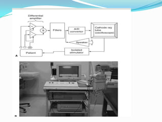


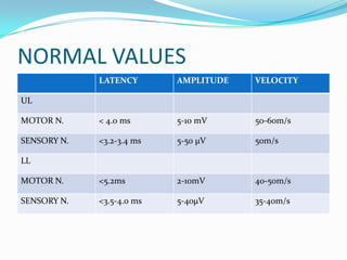


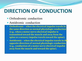
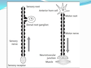


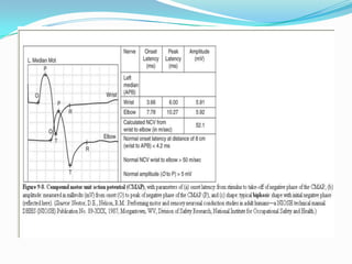


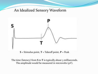


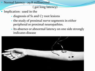


A nerve conduction study (NCS) evaluates the function of motor and sensory nerves by measuring nerve conduction velocity, latency, and amplitude. Key components of an NCS include motor nerve conduction studies, F-wave responses, sensory nerve conduction studies, H-reflexes, and repetitive stimulation tests. NCS provides information about nerve demyelination, axonal injury, and other neuropathies.


