Downloaded 1,327 times

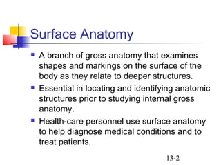
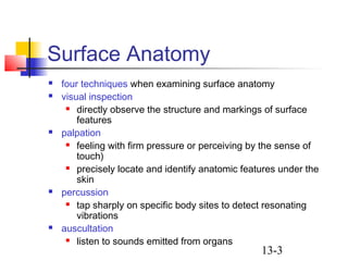
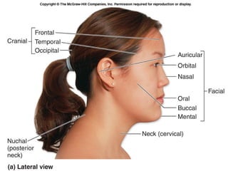
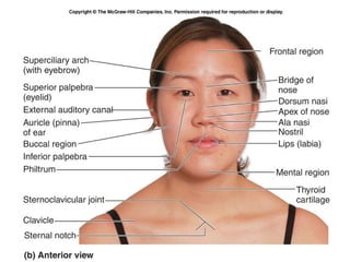


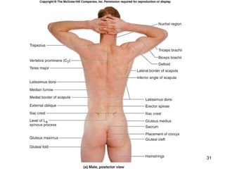
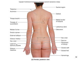
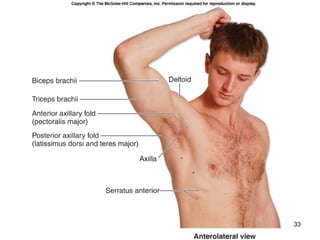
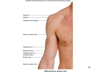
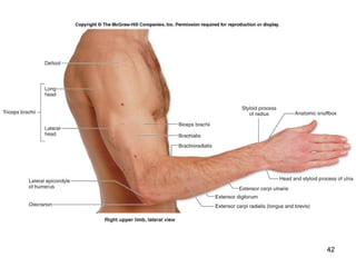
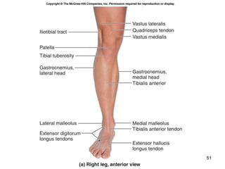
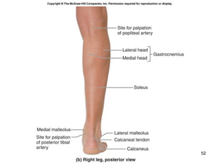
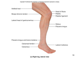
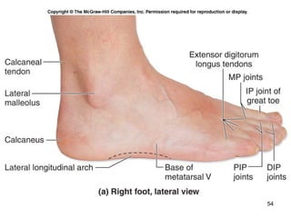

This document provides a chapter outline on surface anatomy, describing prominent anatomical landmarks that can be observed or felt on the external surfaces of the body. It covers surface features of the head and neck, thorax, back, abdomen, upper limbs, gluteal region, and thighs. Key points include descriptions of bones, muscles, blood vessels, and other structures that can be identified by visual inspection, palpation, percussion, or auscultation on the surface of various body regions. The document is intended to familiarize readers with techniques for examining surface anatomy and identifying important anatomical structures.


