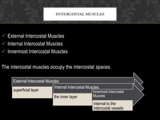
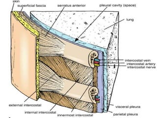
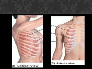
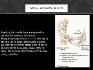
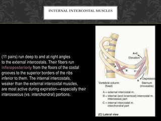
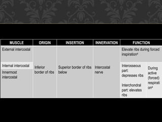
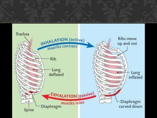
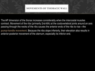
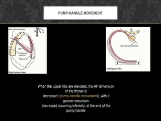
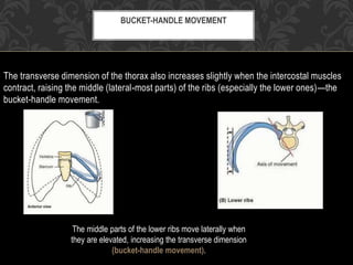
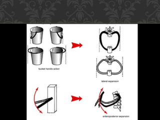
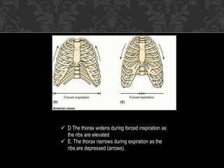
MOVEMENTS OF
THORACIC WALL
 External Intercostal Muscles
 Internal Intercostal Muscles
 Innermost Intercostal Muscles
The intercostal muscles occupy the intercostal spaces.
INTERCOSTAL MUSCLES
External Intercostal Muscles
superficial layer
Internal Intercostal Muscles
the inner layer
Innermost Intercostal
Muscles
internal to the
intercostal vessels
Anteriorly, the muscle fibers are replaced by
the external intercostal membranes.
These muscles run inferoanteriorly from the rib
above to the rib below. Each muscle attaches
superiorly to the inferior border of the rib above
and inferiorly to the superior border of the rib
below. The external intercostals are most active
during inspiration.
EXTERNAL INTERCOSTAL MUSCLES
(11 pairs) run deep to and at right angles
to the external intercostals. Their fibers run
inferoposteriorly from the floors of the costal
grooves to the superior borders of the ribs
inferior to them. The internal intercostals,
weaker than the external intercostal muscles,
are most active during expiration—especially their
interosseous (vs. interchondral) portions.
INTERNAL INTERCOSTAL MUSCLES
Similar to the internal intercostals and are essentially their deeper parts. The innermost
intercostals are separated from the internal intercostals by intercostal nerves and vessels.
These muscles occupy the lateral-most parts of the intercostal spaces. It is likely that their
actions are the same as those of the internal intercostal muscles.
INNERMOST INTERCOSTAL MUSCLES
MUSCLE ORIGIN INSERTION INNERVATION FUNCTION
External intercostal
Inferior
border of ribs
Superior border of ribs
below
Intercostal
nerve
Elevate ribs during forced
inspirationa
Internal intercostal Interosseous
part:
depresses ribs
Interchondral
part: elevates
ribs
During
active
(forced)
respirati
ona
Innermost
intercostal
Movements of the thoracic wall and the diaphragm during inspiration produce increases in
the intrathoracic volume and diameters of the thorax. Consequent pressure changes result in
air being alternately drawn into the lungs (inspiration) through the nose, mouth, larynx, and
trachea and expelled from the lungs (expiration) through the same passages. During
expiration the diaphragm, intercostal muscles, and other muscles relax, decreasing
intrathoracic volume and increasing the intrathoracic pressure. Concurrently, intra-abdominal
pressure decreases and abdominal viscera are decompressed. This allows the stretched
elastic tissue of the lungs to recoil, expelling most of the air.
MECHANISM OF RESPIRATION
The vertical dimension (height) of the central part of the thoracic
cavity increases during inspiration as contraction of the diaphragm
causes it to descend, compressing the abdominal viscera.
During expiration, the vertical dimension returns to the neutral
position as the elastic recoil of the lungs produces sub-
atmospheric pressure in the pleural cavities.
The AP dimension of the thorax increases considerably when the intercostal muscles
contract. Movement of the ribs (primarily 2nd-6th) at the costovertebral joints around an axis
passing through the necks of the ribs causes the anterior ends of the ribs to rise—the
pump-handle movement. Because the ribs slope inferiorly, their elevation also results in
anterior-posterior movement of the sternum, especially its inferior end.
MOVEMENTS OF THORACIC WALL
PUMP-HANDLE MOVEMENT
When the upper ribs are elevated, the AP dimension
of the thorax is
increased (pump-handle movement), with a
greater excursion
(increase) occurring inferiorly, at the end of the
pump handle
The transverse dimension of the thorax also increases slightly when the intercostal muscles
contract, raising the middle (lateral-most parts) of the ribs (especially the lower ones)—the
bucket-handle movement.
BUCKET-HANDLE MOVEMENT
The middle parts of the lower ribs move laterally when
they are elevated, increasing the transverse dimension
(bucket-handle movement).
The combination of rib movements (arrows) that occur
during forced inspiration increase the AP and transverse
dimensions of the thoracic cage.
The combination of all these movements moves the thoracic cage anteriorly, superiorly, and
laterally.
 D The thorax widens during forced inspiration as
the ribs are elevated
 E. The thorax narrows during expiration as the
ribs are depressed (arrows).

Movements of thoracic wall

  • 1.
  • 2.
     External IntercostalMuscles  Internal Intercostal Muscles  Innermost Intercostal Muscles The intercostal muscles occupy the intercostal spaces. INTERCOSTAL MUSCLES External Intercostal Muscles superficial layer Internal Intercostal Muscles the inner layer Innermost Intercostal Muscles internal to the intercostal vessels
  • 5.
    Anteriorly, the musclefibers are replaced by the external intercostal membranes. These muscles run inferoanteriorly from the rib above to the rib below. Each muscle attaches superiorly to the inferior border of the rib above and inferiorly to the superior border of the rib below. The external intercostals are most active during inspiration. EXTERNAL INTERCOSTAL MUSCLES
  • 6.
    (11 pairs) rundeep to and at right angles to the external intercostals. Their fibers run inferoposteriorly from the floors of the costal grooves to the superior borders of the ribs inferior to them. The internal intercostals, weaker than the external intercostal muscles, are most active during expiration—especially their interosseous (vs. interchondral) portions. INTERNAL INTERCOSTAL MUSCLES
  • 7.
    Similar to theinternal intercostals and are essentially their deeper parts. The innermost intercostals are separated from the internal intercostals by intercostal nerves and vessels. These muscles occupy the lateral-most parts of the intercostal spaces. It is likely that their actions are the same as those of the internal intercostal muscles. INNERMOST INTERCOSTAL MUSCLES
  • 8.
    MUSCLE ORIGIN INSERTIONINNERVATION FUNCTION External intercostal Inferior border of ribs Superior border of ribs below Intercostal nerve Elevate ribs during forced inspirationa Internal intercostal Interosseous part: depresses ribs Interchondral part: elevates ribs During active (forced) respirati ona Innermost intercostal
  • 9.
    Movements of thethoracic wall and the diaphragm during inspiration produce increases in the intrathoracic volume and diameters of the thorax. Consequent pressure changes result in air being alternately drawn into the lungs (inspiration) through the nose, mouth, larynx, and trachea and expelled from the lungs (expiration) through the same passages. During expiration the diaphragm, intercostal muscles, and other muscles relax, decreasing intrathoracic volume and increasing the intrathoracic pressure. Concurrently, intra-abdominal pressure decreases and abdominal viscera are decompressed. This allows the stretched elastic tissue of the lungs to recoil, expelling most of the air. MECHANISM OF RESPIRATION
  • 11.
    The vertical dimension(height) of the central part of the thoracic cavity increases during inspiration as contraction of the diaphragm causes it to descend, compressing the abdominal viscera. During expiration, the vertical dimension returns to the neutral position as the elastic recoil of the lungs produces sub- atmospheric pressure in the pleural cavities.
  • 12.
    The AP dimensionof the thorax increases considerably when the intercostal muscles contract. Movement of the ribs (primarily 2nd-6th) at the costovertebral joints around an axis passing through the necks of the ribs causes the anterior ends of the ribs to rise—the pump-handle movement. Because the ribs slope inferiorly, their elevation also results in anterior-posterior movement of the sternum, especially its inferior end. MOVEMENTS OF THORACIC WALL
  • 13.
    PUMP-HANDLE MOVEMENT When theupper ribs are elevated, the AP dimension of the thorax is increased (pump-handle movement), with a greater excursion (increase) occurring inferiorly, at the end of the pump handle
  • 14.
    The transverse dimensionof the thorax also increases slightly when the intercostal muscles contract, raising the middle (lateral-most parts) of the ribs (especially the lower ones)—the bucket-handle movement. BUCKET-HANDLE MOVEMENT The middle parts of the lower ribs move laterally when they are elevated, increasing the transverse dimension (bucket-handle movement).
  • 15.
    The combination ofrib movements (arrows) that occur during forced inspiration increase the AP and transverse dimensions of the thoracic cage. The combination of all these movements moves the thoracic cage anteriorly, superiorly, and laterally.
  • 17.
     D Thethorax widens during forced inspiration as the ribs are elevated  E. The thorax narrows during expiration as the ribs are depressed (arrows).