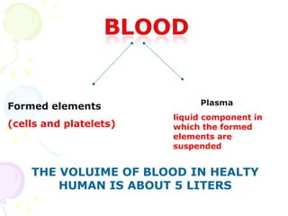
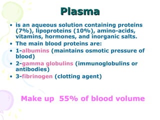
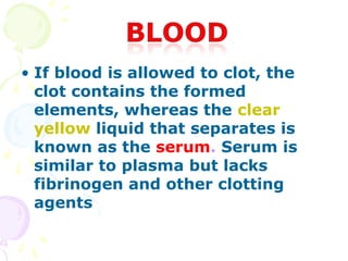
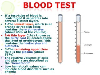
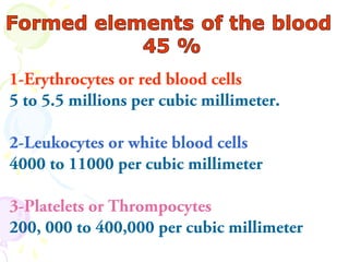
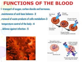
1. Plasma is the liquid component of blood in which formed elements such as cells and platelets are suspended. It makes up 55% of total blood volume.
2. When blood is centrifuged, it separates into distinct layers - the bottom layer contains erythrocytes, the thin middle layer contains leukocytes and platelets, and the remaining clear fluid is plasma.
3. The main functions of red blood cells include transport of oxygen, carbon dioxide, and hormones throughout the body. White blood cells and platelets are involved in defense against infection and clotting at the site of injury.
All Time Service Available Call Girls Marine Drive š³ 9820252231 For 18+ VIP C...
Ā
Plasma and Formed Elements in Blood
1.
2. Plasma
Formed elements
liquid component in
(cells and platelets) which the formed
elements are
suspended
THE VOLUIME OF BLOOD IN HEALTY
HUMAN IS ABOUT 5 LITERS
3. Plasma
ā¢ is an aqueous solution containing proteins
(7%), lipoproteins (10%), amino-acids,
vitamins, hormones, and inorganic salts.
ā¢ The main blood proteins are:
ā¢ 1-albumins (maintains osmotic pressure of
blood)
ā¢ 2-gamma globulins (immunoglobulins or
antibodies)
ā¢ 3-fibrinogen (clotting agent)
Make up 55% of blood volume
4. ā¢ If blood is allowed to clot, the
clot contains the formed
elements, whereas the clear
yellow liquid that separates is
known as the serum. Serum is
similar to plasma but lacks
fibrinogen and other clotting
agents
5. ā¢ If a test-tube of blood is
centrifuged it separates into
several distinct layers.
ā¢ 1-The lowest layer, which is an
orange or reddish color,
contains the erythrocytes ,
(about 45% of the volume).
ā¢ 2-A thin layer (1%) known as
the Buffy coat is located above
the layer of erythrocytes. This
contains the leukocytes and
platelets.
ā¢ 3-The remaining upper clear
fluid is the plasma (about
55%).
ā¢ The relative volumes of cells
and plasma are described as
the "hematocritā.
ā¢ Low hematocrit values can
indicate blood disorders such as
anemia
6. ā¢ A drop of blood spread
thinly on a microscope
slide and dried is
known as a blood
smear.
ā¢ various common blood
staining techniques
(Leishman, Giemsa,
Wright) are all
modifications of the
Romanovsky (1891)
staining method based
on mixtures of
methylene blue (basic
dye) and eosin (acid
dye).
7. 1-Erythrocytes or red blood cells
5 to 5.5 millions per cubic millimeter.
2-Leukocytes or white blood cells
4000 to 11000 per cubic millimeter
3-Platelets or Thrompocytes
200, 000 to 400,000 per cubic millimeter
8. 1- transport of oxygen, carbon dioxide and hormones.
.maintenance of acid-base balance- 2
.removal of waste products of cells metabolism-3
temperature control of the body- 4
. defense against infection- 5
9. ā¢ 1- SHAPE: RBCs are rounded,
biconcave, non-nucleated disc,
They form Rouleaux, in blood
stream.
12. The Adaptability of the RBC Structure to
Suit Its Function
ā¢ 1- the cell membrane is highly selective; allowing the
exchange of gases, but above all prevents the
haemoglobin from escaping into the plasma. This is
important because haemoglobin if it is free in the
plasma, it escapes in to the tissues then to the urine
2- the highly elastic cell membrane allows the cell to
change its shape and to squeeze inside narrow
capillaries, but it regains its shape in wider vessels
3- the absence of a nucleus and organoids allows
more space for more haemoglobin inside the cell
4- the biconcave surfaces of the RBCs increase its
surface area through which gaseous exchange takes
place
13. ā¢ 1- R.B.Cs may be pearā shaped (poikilocytosis)
ā¢ 2- they may be oval ( ovalocytes)
ā¢ 3- or may be biconvex (spherocytes)
ā¢ or crescent as in sickle cell anemia.
14. In hypertonic solution( with high osmotic
pressure ):
the water draw out from the RBCs which become
crenated (i.e. smaller, with notched outline).
ā¢ In hypotonic solution:
ā¢ Water is drawn inside the RBCs, and finally
rupture ,leaving remnants of their cells membrane
called āghosts ā. The rupture of RBCs and the
discharge of their Hb content called hemolysis
15. The diameter of R.B.Cs varies between 7-8Āµm
(7.5 Āµm average)
ā¢ In Macrocytic Anemias there is increase in
diameters of R.B.Cs
ā¢ In Mirocytic Anemias there is a decrease in
diameters of R.B.Cs.
ā¢ In great variation in size of the R.B.Cs is
called Anisocytosis.
16. ā¢ R.B.Cs are greenish āyellow in color,
due to the presence of Haemoglobin (Hb
%).
ā¢ A drop of blood appears red due to
overlapping of RBCs .
ā¢ The normal blood corpuscles are called
NORMOCHROMIC
17. A- HYPOCHROMIC RBC (Contains
less Hb% than usual). It has a
larger pale central area, and
poorly- stained periphery .
B- HYPERCHROMIC RBC (Contains
more Hb% than usual) It has
stained central area, and a deeply
stained periphery.
19. In normal male the average number of RBCS is
about 5 million per cubic millimeter.
In female it is about 4.5 million per cmm.
The life span of the RBCS 4 months
20. abnormalities in The
.Number of RBCs
A- REDUCE THE AMOUNT OF RBCs
1- Deficiency of iron, vitamin B12, proteins,vitamin
C, copper, calcium (i.e. deficiency anaemia).
2- haemorrhagic anaemia
3- Haemolysis or destruction of RBCs. e.g. in
increased fragility of RBCs or by bacterial toxins
(i.e. haemolytic anemia) erythrocytes have the
shape of elongated crescents (sickles) due to of
abnormal Hb.
4- Malfunction of bone marrow by various
exogenous or endogenous causes a plastic anaemia
21. B- increase the amount of
:the RBCs Polycythaemia
- Excessive production of RBCs from the
bone marrow due to hypoxia which
stimulates the bone marrow, as
a- The foetus, due to intra-uterine anoxia
e- Severe muscular exercise, due to
excessive need of oxygen by the tissues
22. ORIGIN OF RBCS ,THEIR
LIFE SPAN AND FATE
ORIGIN: BONE MARROW
LIFE SPAN: 120 DAYS.
Fate: destroyed and phagocytosed by
macrophages in spleen, bone marrow and
liver, to hemasiderin (iron-containing
pigment) and bilirubin
23. (Leukocytes (White blood cells
Leukocytes are colorless because they
contain no hemoglobin; however,
each cell has a nucleus. In blood
stream leukocytes are spherical in
shape and mobile.
IN NORMAL ADULT THE āTOTAL
LEUKOCYTIC COUNT āIS BETWEEN
4,000- 10,000 PER CUBIC MILLIMETER
24. According to type of cytoplasmic granules
and the shape of nuclei leukocytes are
classified into
ā¢ A-Granular leukocytes
ā¢ They contain specific granules and lobulated nuclei.
ā¢ 1- Neutrophils 60-75 %
ā¢ 2- Eosinphils 2-4 %
ā¢ 3- Basophils 0- 0.5 %
ā¢ B- Nongranular leukocytes
ā¢ They do not contain specific granules with non-lobulated
nuclei.
ā¢ 1- Lymphocytes 20-40 %
ā¢ 2- monocytes 3- 8 %
25. Leukocytes can leave the blood circulatory system and enter
connective tissue, where they mostly function. This ability to
cross the capillary walls is known as diapedesis.
This migration of leukocytes from capillaries into the
connective tissue is important in tissue responses to injury.
The leukocytes do not return to the blood, with the exception
of lymphocytes (recirculation).
26. Neutrophils (leukocytes or polymorphs: (
- are the most common of the leukocytes and represent
-- some 55-70% of all the circulating leukocytes.
-and are characterized by a segmented nucleus.
- In females a small "drumstick" is found (Barr's body)
on the nucleus representing a condensation of the sex-
chromatin
27. Some specific granules contain phagocytins, which are
anti-bacterial substances involved intracellular
digestion.
The non-specific granules as azurophilic contain
lysosomal , enzymes (peroxidase).
Neutrophils are phagocytic cells involved as a first line
of defense against invading microorganisms.
They are important in inflammation and at sites of
injury or wounds
Pus that develops in sites of infection is mainly
composed of dead neutrophils and dead tissue
28. CLINICAL NOTE FOR
NEUTROPHIL
ā¢ Acute inflammation
ā¢ Acute inflammation is
series of changes that
take place in
connective tissue in
response to injury or
invasion by organisms.
ā¢ In acute inflammation
the percentage of
neutrophils is
increased i.e.
neutrophilia and the
total leukocytic count
increases i.e.
leukocytosis
29. The clinical signs of
acute inflammation
ā¢ 1- Swelling :This localized Odem or increase fluid in
amorphous material of connective tissue is
responsible for the swelling at site of acute
inflammation.
ā¢ 2- Redness and hotness: histamine causes dilatation
of arterioles and capillaries . this dilatation causes
increased blood flow and is responsible for the
redness and hotness.
ā¢ 3- Pain This is caused by the pressure of the
increased amounts of the tissue fluid on the
sensitive sensory nerve endings.
4- Loss of function : this is caused by
pain
30. Eosinophils
ā¢ Eosinophils represent 1-4% of leukocytes. They typically
have bilobed nuclei and large) acidophilic granules)
ā¢ The granules are considered to be lysosomes and contain
acid phosphatase and others enzymes.
ā¢ Eosinophils are involved in selective phagocytosis.
An increase in the numbers of eosinophils (eosinophilia )is
seen in allergic reactions or helminthes parasitic
infections.
31. Basophils
ā¢ These represent only about
1% of the leukocytes.
ā¢ Their nucleus is bilobed or
S-shaped.
ā¢ They have very large,
irregular basophilic granules,
that commonly obscure the
nucleus.
ā¢ The granules (similar to
those of mast cells) contain
histamine and heparin and
show metachromatic
staining.
ā¢ In stained blood smears the
granules stain a violet color.
.
33. AGRANULOCYTES
ā¢ Lymphocytes
ā¢ Lymphocytes represent 25-30% of all the
leukocytes. Lymphocytes are involved in the
immune responses of the body .
ā¢ The most common lymphocytes are small cells
(6-8mm diameter), with very dense regular
nuclei, with relatively little cytoplasm.
ā¢ Types: T- lymphocytes and B-Lymphocytes
34. Monocytes
ā¢ Monocytes represent about 5% of the leukocytes. They
possess oval or kidney-shaped nuclei, often eccentrically
located.
ā¢ The nuclei are less intensely . The cytoplasm less is
basophilic with small azurophilic granules (lysosomes).
ā¢ Monocytes are part of the Mononuclear Phagocyte
System. They are non-terminal cells and can differentiate
into phagocytic cells such as macrophages.
ā¢ Monocytes that enter the tissues do not return to the
blood.
35. Blood platelets
ā¢ Blood platelets are derived from the cytoplasm
of megakaryocytes of the bone marrow.
ā¢ They are small with a central core of small
purple granules called granulomere surrounded
by an outer pale blue hyalomere. At the
periphery (as seen by electron microscopy) there
are peripheral or marginal microtubules .
36. (BLOOD PLATELET( THROMBOCYTES
ā¢ SHAPE: Are fragments of cells, they are rounded or
oval in shape. In blood film: they are seen singly, or
more commonly as clumps group in between RBCs
ā¢ Number:250,000-350,000per cubic mm.
ā¢ Decrease the number is called thrombocytopenia in
which the bleeding time is prolonged.
ā¢ ORIGIN: megakaryocytes in red bone marrow
37. Blood platelets
ā¢ If a blood vessel is injured, the platelets
are involved in the clotting reaction. They
aggregate at the site of injury, change
shape and help form the thrombus.
ā¢