I have listed out the LE cells structure and Microscopical examinaton of LE CELLS, Difference between tart cells and le cells, clinical symptoms and diagnostic procedure.
I have listed out the LE cells structure and Microscopical examinaton of LE CELLS, Difference between tart cells and le cells, clinical symptoms and diagnostic procedure.
It is fluid which is present in
the abdominal cavity.
The peritoneal cavity is a potential
space lined by mesothelium of the
visceral n parietal peritoneum.
An absolute eosinophil count is a blood test that measures the number of one type of white blood cells called eosinophils.
Eosinophils become active when you have certain allergic diseases, infections, and other medical conditions.
It is fluid which is present
in the pericardial cavity of
heart b/w parietal pericardium n visceral pericardium.
The pericardial cavity is a
potential space lined by
mesothelium of the visceral n parietal pericardium.
CSF:
Derived through ultrafilteration and secretion through choroid plexus, produced at the rate of 500 ml/day.
Provides physical support, collects wastes, circulates nutrients and lubricates the CNS.
Normal CSF volumes:
In Adults: 90 - 150 ml
In Neonates: 10 - 60 ml
Total CSF volume is replaced every 5-7 hours.
COLLECTION
Lumbar puncture, Cisternal puncture, Lateral cervical puncture, Shunts and cannulas
Opening pressure – 90-180 mm H2O
Approximately 15-20 cc fluid collected
LAB
REQUIRED
Opening CSF pressure
Total cell count
Differential cell count
Glucose
Total protein
OPTIONAL
Cultures, Gram stain, AFB, Fungal and bacterial
antigens, Enzymes, PCR, Cytology, Electrophoresis,
VDRL, D-Dimers
FLOW CYTOMETRY, PRINCIPLE, APPLICATION, USE IN HAEMATOLOGY, COMPONENT OF FLOW CYTOMETRY, DATA INTERPRETATION, DATA ANALYSIS, CELL SHORTING ADVANTAGES AND DISADVANTAGES, IMMUNOLOGICAL CLASSIFICATION OF ACUTE
LEUKEMIA
Flow cytometry (FCM) is a technique used to detect and measure physical and chemical characteristics of a population of cells or particles. In this process, a sample containing cells or particles is suspended in a fluid and injected into the flow cytometer instrument.
Ozempic: Preoperative Management of Patients on GLP-1 Receptor Agonists Saeid Safari
Preoperative Management of Patients on GLP-1 Receptor Agonists like Ozempic and Semiglutide
ASA GUIDELINE
NYSORA Guideline
2 Case Reports of Gastric Ultrasound
ARTIFICIAL INTELLIGENCE IN HEALTHCARE.pdfAnujkumaranit
Artificial intelligence (AI) refers to the simulation of human intelligence processes by machines, especially computer systems. It encompasses tasks such as learning, reasoning, problem-solving, perception, and language understanding. AI technologies are revolutionizing various fields, from healthcare to finance, by enabling machines to perform tasks that typically require human intelligence.
Prix Galien International 2024 Forum ProgramLevi Shapiro
June 20, 2024, Prix Galien International and Jerusalem Ethics Forum in ROME. Detailed agenda including panels:
- ADVANCES IN CARDIOLOGY: A NEW PARADIGM IS COMING
- WOMEN’S HEALTH: FERTILITY PRESERVATION
- WHAT’S NEW IN THE TREATMENT OF INFECTIOUS,
ONCOLOGICAL AND INFLAMMATORY SKIN DISEASES?
- ARTIFICIAL INTELLIGENCE AND ETHICS
- GENE THERAPY
- BEYOND BORDERS: GLOBAL INITIATIVES FOR DEMOCRATIZING LIFE SCIENCE TECHNOLOGIES AND PROMOTING ACCESS TO HEALTHCARE
- ETHICAL CHALLENGES IN LIFE SCIENCES
- Prix Galien International Awards Ceremony
Flu Vaccine Alert in Bangalore Karnatakaaddon Scans
As flu season approaches, health officials in Bangalore, Karnataka, are urging residents to get their flu vaccinations. The seasonal flu, while common, can lead to severe health complications, particularly for vulnerable populations such as young children, the elderly, and those with underlying health conditions.
Dr. Vidisha Kumari, a leading epidemiologist in Bangalore, emphasizes the importance of getting vaccinated. "The flu vaccine is our best defense against the influenza virus. It not only protects individuals but also helps prevent the spread of the virus in our communities," he says.
This year, the flu season is expected to coincide with a potential increase in other respiratory illnesses. The Karnataka Health Department has launched an awareness campaign highlighting the significance of flu vaccinations. They have set up multiple vaccination centers across Bangalore, making it convenient for residents to receive their shots.
To encourage widespread vaccination, the government is also collaborating with local schools, workplaces, and community centers to facilitate vaccination drives. Special attention is being given to ensuring that the vaccine is accessible to all, including marginalized communities who may have limited access to healthcare.
Residents are reminded that the flu vaccine is safe and effective. Common side effects are mild and may include soreness at the injection site, mild fever, or muscle aches. These side effects are generally short-lived and far less severe than the flu itself.
Healthcare providers are also stressing the importance of continuing COVID-19 precautions. Wearing masks, practicing good hand hygiene, and maintaining social distancing are still crucial, especially in crowded places.
Protect yourself and your loved ones by getting vaccinated. Together, we can help keep Bangalore healthy and safe this flu season. For more information on vaccination centers and schedules, residents can visit the Karnataka Health Department’s official website or follow their social media pages.
Stay informed, stay safe, and get your flu shot today!
These lecture slides, by Dr Sidra Arshad, offer a quick overview of physiological basis of a normal electrocardiogram.
Learning objectives:
1. Define an electrocardiogram (ECG) and electrocardiography
2. Describe how dipoles generated by the heart produce the waveforms of the ECG
3. Describe the components of a normal electrocardiogram of a typical bipolar leads (limb II)
4. Differentiate between intervals and segments
5. Enlist some common indications for obtaining an ECG
Study Resources:
1. Chapter 11, Guyton and Hall Textbook of Medical Physiology, 14th edition
2. Chapter 9, Human Physiology - From Cells to Systems, Lauralee Sherwood, 9th edition
3. Chapter 29, Ganong’s Review of Medical Physiology, 26th edition
4. Electrocardiogram, StatPearls - https://www.ncbi.nlm.nih.gov/books/NBK549803/
5. ECG in Medical Practice by ABM Abdullah, 4th edition
6. ECG Basics, http://www.nataliescasebook.com/tag/e-c-g-basics
Tom Selleck Health: A Comprehensive Look at the Iconic Actor’s Wellness Journeygreendigital
Tom Selleck, an enduring figure in Hollywood. has captivated audiences for decades with his rugged charm, iconic moustache. and memorable roles in television and film. From his breakout role as Thomas Magnum in Magnum P.I. to his current portrayal of Frank Reagan in Blue Bloods. Selleck's career has spanned over 50 years. But beyond his professional achievements. fans have often been curious about Tom Selleck Health. especially as he has aged in the public eye.
Follow us on: Pinterest
Introduction
Many have been interested in Tom Selleck health. not only because of his enduring presence on screen but also because of the challenges. and lifestyle choices he has faced and made over the years. This article delves into the various aspects of Tom Selleck health. exploring his fitness regimen, diet, mental health. and the challenges he has encountered as he ages. We'll look at how he maintains his well-being. the health issues he has faced, and his approach to ageing .
Early Life and Career
Childhood and Athletic Beginnings
Tom Selleck was born on January 29, 1945, in Detroit, Michigan, and grew up in Sherman Oaks, California. From an early age, he was involved in sports, particularly basketball. which played a significant role in his physical development. His athletic pursuits continued into college. where he attended the University of Southern California (USC) on a basketball scholarship. This early involvement in sports laid a strong foundation for his physical health and disciplined lifestyle.
Transition to Acting
Selleck's transition from an athlete to an actor came with its physical demands. His first significant role in "Magnum P.I." required him to perform various stunts and maintain a fit appearance. This role, which he played from 1980 to 1988. necessitated a rigorous fitness routine to meet the show's demands. setting the stage for his long-term commitment to health and wellness.
Fitness Regimen
Workout Routine
Tom Selleck health and fitness regimen has evolved. adapting to his changing roles and age. During his "Magnum, P.I." days. Selleck's workouts were intense and focused on building and maintaining muscle mass. His routine included weightlifting, cardiovascular exercises. and specific training for the stunts he performed on the show.
Selleck adjusted his fitness routine as he aged to suit his body's needs. Today, his workouts focus on maintaining flexibility, strength, and cardiovascular health. He incorporates low-impact exercises such as swimming, walking, and light weightlifting. This balanced approach helps him stay fit without putting undue strain on his joints and muscles.
Importance of Flexibility and Mobility
In recent years, Selleck has emphasized the importance of flexibility and mobility in his fitness regimen. Understanding the natural decline in muscle mass and joint flexibility with age. he includes stretching and yoga in his routine. These practices help prevent injuries, improve posture, and maintain mobilit
Acute scrotum is a general term referring to an emergency condition affecting the contents or the wall of the scrotum.
There are a number of conditions that present acutely, predominantly with pain and/or swelling
A careful and detailed history and examination, and in some cases, investigations allow differentiation between these diagnoses. A prompt diagnosis is essential as the patient may require urgent surgical intervention
Testicular torsion refers to twisting of the spermatic cord, causing ischaemia of the testicle.
Testicular torsion results from inadequate fixation of the testis to the tunica vaginalis producing ischemia from reduced arterial inflow and venous outflow obstruction.
The prevalence of testicular torsion in adult patients hospitalized with acute scrotal pain is approximately 25 to 50 percent
Title: Sense of Smell
Presenter: Dr. Faiza, Assistant Professor of Physiology
Qualifications:
MBBS (Best Graduate, AIMC Lahore)
FCPS Physiology
ICMT, CHPE, DHPE (STMU)
MPH (GC University, Faisalabad)
MBA (Virtual University of Pakistan)
Learning Objectives:
Describe the primary categories of smells and the concept of odor blindness.
Explain the structure and location of the olfactory membrane and mucosa, including the types and roles of cells involved in olfaction.
Describe the pathway and mechanisms of olfactory signal transmission from the olfactory receptors to the brain.
Illustrate the biochemical cascade triggered by odorant binding to olfactory receptors, including the role of G-proteins and second messengers in generating an action potential.
Identify different types of olfactory disorders such as anosmia, hyposmia, hyperosmia, and dysosmia, including their potential causes.
Key Topics:
Olfactory Genes:
3% of the human genome accounts for olfactory genes.
400 genes for odorant receptors.
Olfactory Membrane:
Located in the superior part of the nasal cavity.
Medially: Folds downward along the superior septum.
Laterally: Folds over the superior turbinate and upper surface of the middle turbinate.
Total surface area: 5-10 square centimeters.
Olfactory Mucosa:
Olfactory Cells: Bipolar nerve cells derived from the CNS (100 million), with 4-25 olfactory cilia per cell.
Sustentacular Cells: Produce mucus and maintain ionic and molecular environment.
Basal Cells: Replace worn-out olfactory cells with an average lifespan of 1-2 months.
Bowman’s Gland: Secretes mucus.
Stimulation of Olfactory Cells:
Odorant dissolves in mucus and attaches to receptors on olfactory cilia.
Involves a cascade effect through G-proteins and second messengers, leading to depolarization and action potential generation in the olfactory nerve.
Quality of a Good Odorant:
Small (3-20 Carbon atoms), volatile, water-soluble, and lipid-soluble.
Facilitated by odorant-binding proteins in mucus.
Membrane Potential and Action Potential:
Resting membrane potential: -55mV.
Action potential frequency in the olfactory nerve increases with odorant strength.
Adaptation Towards the Sense of Smell:
Rapid adaptation within the first second, with further slow adaptation.
Psychological adaptation greater than receptor adaptation, involving feedback inhibition from the central nervous system.
Primary Sensations of Smell:
Camphoraceous, Musky, Floral, Pepperminty, Ethereal, Pungent, Putrid.
Odor Detection Threshold:
Examples: Hydrogen sulfide (0.0005 ppm), Methyl-mercaptan (0.002 ppm).
Some toxic substances are odorless at lethal concentrations.
Characteristics of Smell:
Odor blindness for single substances due to lack of appropriate receptor protein.
Behavioral and emotional influences of smell.
Transmission of Olfactory Signals:
From olfactory cells to glomeruli in the olfactory bulb, involving lateral inhibition.
Primitive, less old, and new olfactory systems with different path
Pulmonary Thromboembolism - etilogy, types, medical- Surgical and nursing man...VarunMahajani
Disruption of blood supply to lung alveoli due to blockage of one or more pulmonary blood vessels is called as Pulmonary thromboembolism. In this presentation we will discuss its causes, types and its management in depth.
NVBDCP.pptx Nation vector borne disease control programSapna Thakur
NVBDCP was launched in 2003-2004 . Vector-Borne Disease: Disease that results from an infection transmitted to humans and other animals by blood-feeding arthropods, such as mosquitoes, ticks, and fleas. Examples of vector-borne diseases include Dengue fever, West Nile Virus, Lyme disease, and malaria.
Report Back from SGO 2024: What’s the Latest in Cervical Cancer?bkling
Are you curious about what’s new in cervical cancer research or unsure what the findings mean? Join Dr. Emily Ko, a gynecologic oncologist at Penn Medicine, to learn about the latest updates from the Society of Gynecologic Oncology (SGO) 2024 Annual Meeting on Women’s Cancer. Dr. Ko will discuss what the research presented at the conference means for you and answer your questions about the new developments.
Report Back from SGO 2024: What’s the Latest in Cervical Cancer?
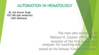
AUTOMATION IN HEMATOLOGY
1. AUTOMATION IN HEMATOLOGY
The man who started it all
Wallace H. Coulter (1913 –1998)
Inventor of the first automated
analyzer for counting and sizing cells
based on his famous ‘Coulter Principle’
Dr. Ajit Kumar Singh
PGT MD (lab medicine)
CNCI (Kolkata)
2. Necessity for Automation
Cell counts
Dx of Hemoglobinopathies
Immunophenotyping
Dx of Leukemias & Lymphomas
Coagulation Abnormalities.
4. Types of Automated Hematology
Analyzers
Semi-automated
analyzers
Measures only few
parameters
Some steps like
dilution of
blood is carried out
manually
Fully automated
analyzers
Measures multiple
parameters.
Requires only
anticoagulated blood
samples.
5. Components of a cell counter
HYDRAULICS
Aspirating unit.
Dispensers.
Diluters.
Mixing chambers.
Aperture bath.
Hemoglobinometer.
PNEUMATICS
Vacuums & Pressures for operating valves.
ELECTRICALS
Analyzers & Computing circuitary.
6. XN-550
Compact 6-part differential analyzer
Throughput of 70 samples per hour
Single sample analysis in closed mode
Fully integrated IPU including LCD color touchscreen
Only 25 µL aspiration volume in whole blood mode
More than just CBC + DIFF – added clinical values available
7. Principles of working of an automated
blood analyzer
Electrical Impedance.
Light Scatter.
Fluorescence.
Light Absorption.
Electrical Conductivity.
8. Electrical impedance
Cell counting & sizing is based on the Coulter principle - detection &
measurement of changes in electrical impedance (resistance) produced by a
blood cell as it passes through an electrical field.
Blood cells are poor conductors of electricity but are suspended in an
electrically conductive diluent.
2 chambers filled with a conductive buffered electrolyte solution separated
by a glass tube having a small aperture.
A DC current is generated between two electrolytes.
9. Electrical impedance
As a cell passes through the aperture, flow of current is impeded and a
voltage pulse is generated.
The no: of pulses indicate the no: of the blood cells.
The amplitude (height) of each pulse is proportional to the cell volume.
The requisite condition for cell counting by this method is high dilution of
sample.
10. RBC RBC Count
•MCV
•Size distribution histogram
•RDW
•Hematocrit
•MCH
•MCHC
WBC o Total Count
o 3 part differential
Lymphocyte
Mononuclear cells
Granulocyte
Platelets o Platelet count
o Platelet histograms giving
MPV
PDW
11. Optical light scatter
Each cell flows in a single line through a flow cell.
A LASER device is focused on the flow cell.
As LASER light beam strikes a cell, it is scattered in various directions.
Photodetectors capture the light.
Forward Scatter Light (FALS) ∝ to cell size.
Side Scatter Light (SS) (90°) corresponds to nuclear complexity & granularity
of cytoplasm.
Used to distinguish between granulocytes, lymphocytes & monocytes.
13. Variables measured by using OPTICAL LIGHT
SCATTER
RBC Count
The 5 part differential
Neutrophils
Eosinophils
Basophils
Lymphocytes
Monocytes
Mean Cell Volume
14. Fluorescence flow cytometry (FFC)
Fluorescence flow cytometry (FFC) is used to analyze physiological and chemical
properties of cells. It can also be used to analyze other biological particles in
urinalysis analyzers. It provides:
Information about cell size and structure
Information about a cell’s interior
In flow cytometry, we examine cells and particles while they are flowing through
a very narrow flow cell.
15. Fluorescence flow cytometry (FFC)
First a blood sample is aspirated and proportioned, then diluted to a pre-set ratio
and labelled with a proprietary fluorescence marker that binds specifically to
nucleic acids.
Next the sample is transported into the flow cell. The sample is illuminated by a
semiconductor laser beam, which can separate the cells using three different
signals:
forward-scattered light (forward scatter or FSC)
side-scattered light (side scatter or SSC)
side-fluorescence light (side fluorescence or SFL).
16. Fluorescence flow cytometry (FFC)
The intensity of the forward scatter indicates the cell volume. The side
scatter provides information about the internal cell structure and its content,
such as nucleus and granules. The side fluorescence indicates the amount of
nucleic acids present in the cell.
Cells with similar physical and chemical properties form a cluster in a graph
known as a scattergram.
17. Fluorescence flow cytometry (FFC)
The principle of fluorescence flow cytometry is used in different analysers for
haematology and urinalysis. For blood cell counts we use fluorescence flow
cytometry, e.g. for the WBC and differential, for NRBC counting and
reticulocyte measurement.
In urinalysis analysers, fluorescence technology is also used for counting
bacteria, red blood cells, white blood cells and other elements.
18. What is flow cytometry
Flow – cell in motion
Cyto – cell
Metry – measure
Measuring property of cell while in a fluid stream
The fluorescence can then be measured to determine the amount and type of
cells present in a sample. Up to thousands of particles per second can be
analysed as they pass through the liquid stream.
A beam of laser light is directed at a hydrodynamically-focused stream of fluid
that carries the cells. Several detectors are carefully placed around the stream,
at the point where the fluid passes through the light beam.
19. What is flow cytometry
One of these detectors is in line with the light beam and is used to measure
Forward Scatter or FSC. Another detector is placed perpendicular to the
stream and is used to measure Side Scatter (SSC).
Since fluorescent labels are used to detect the different cells or components,
fluorescent detectors are also in place. The suspended particles or cells,
which may range in size from 0.2 to 150μm, pass through the beam of light
and scatter the light beams.
The fluorescently labelled cell components are excited by the laser and emit
light at a longer wavelength than the light source.
20. Flow Cytometry
Measures multiple cellular & fluorescent properties of cells when they flow as
a single cell suspension through a laser beam.
Provides the following information about a cell:
• Cell size (forward scatter)
• Internal complexity or granularity (side scatter)
• Relative fluorescence intensity
21. Components of Flow Cytometry
Fluidics (The Flow System)
The sample is injected into a stream of sheath fluid within the flow chamber.
They are forced into the center of the stream forming a single file by the principle of
HYDRODYNAMIC FOCUSING.
‘Only 1 cell or particle can pass through the LASER Beam@ a given moment.’
The sample pressure is always > than the sheath pressure ensuring a high flow rate, thus allowing
more cells to enter the stream@a given moment.
• High Flow rate used for immunophenotyping analysis of cells.
• Low Flow rate used for DNA Analysis.
22. Components of Flow Cytometry
Optics
Following cell delivery, a light source like the Argon- ion LASER is required to excite the cells.
When light from a Laser Beam intersects a cell at the ‘interrogation point’, 2 events occur -
Light Scattering
Fluorescence (Emission of Light )
Light Scattered in the forward direction is detected in Forward Scatter Channel ∝ to cell size and
that
scattered@90° to axis of Laser path is detected in Side Scatter Channel ∝ to granularity of cell.
The cells tagged with fluorescence emit a momentary pulse of fluorescence.
A system of optical mirrors and filters then direct the specified wavelengths of light to the
designated photodetectors.
23. Components of Flow Cytometry
Electronics
The photodetectors - photodiodes and photomultiplier tubes convert the optical signals (photons)
to corresponding electronic signals(electrons).
The electronic signal produced is proportional to the amount of light striking a cell.
The electric current travels to the amplifier and is converted to a voltage pulse
The voltage pulse is assigned a digital value representing a channel by the Analog-to Digital
Converter (ADC) .
The channel no: is transferred to the computer which displays it to the appropriate position on the
data plot.
25. Common Applications of Flow Cytometry
1. Leukemias and lymphomas Immunophenotyping (evaluation of cell surface
markers),diagnosis,
detection of minimal residual disease, and to identify
prognostically important subgroups.
2. Paroxysmal nocturnal
hemoglobinuria
Deficiency of CD 55 and CD 59.
3. Hematopoietic stem cell
transplantation
Enumeration of CD34+ stem cells.
4. Feto -maternal hemorrhage Detection and quantitation
of foetal hemoglobin in maternal blood sample.
5. Anemias Reticulocyte count.
6. Human immunodeficiency virus
infection
For enumeration of CD4+ lymphocytes
7. Histocompatibility cross
26. Data Analysis
Data is collected and stored in the computer – can be displayed in various
formats.
Parameters – Forward Scatter, Side scatter, emitted fluorescence.
Data plots :
Single Parameter – Histogram
Two Parameters – Dot Plot
27. sodium lauryl sulphate (SLS) detection
method
Hemoglobin is a routine diagnostic parameter in each blood count. The
method recommended by the ICSH (International Committee for
Standardization in Hematology) for measuring hemoglobin concentration is
the cyan-methemoglobin method.
SLS hemoglobin detection method uses cyanide-free sodium lauryl sulphate
(SLS). The reagent lyses red blood cells and white blood cells in the sample.
The chemical reaction begins by altering the globin and then oxidising the
heme group.
Now the SLS’ hydrophilic groups can bind to the heme group and form a
stable, colored complex (SLS-HGB), which is analyzed using a photometric
method.
28. sodium lauryl sulphate (SLS) detection
method
An LED sends out monochromatic light and by moving through the mixture
light is absorbed by the SLS-HGB complexes. The absorbance is measured by a
photo sensor and is proportional to the hemoglobin concentration of the
sample.
Absorption photometric methods are usually influenced by the turbidity of the
sample itself. In blood samples, turbidity can be caused due to lipaemia or
leucocytosis. By using the SLS-HGB method these interferences can be
minimised due to the effect of the reagent.
29. PLT-F channel
The new PLT-F method is based on a Fluorocell fluorescent dye (oxazine), an
extended counting volume, and an extended counting time.
Compared with the PLT-O method, platelets are more clearly distinguished
from other blood cells using the difference in forward scattered light and the
fluorescence intensity.
The fluorescence marker specifically labels platelets and no other blood cells,
which minimises interferences and is one reason for the extremely good
correlation with the CD41/CD61 immune flow cytometry method. Another
reason is the high measurement accuracy in the low concentration range
since the PLT-F channel analyses a 5-fold larger sample volume of the
aspirated sample compared to the DC detection measurement.
30. PLT-F channel
The IPF supports quick and efficient differential diagnosis of
thrombocytopenia as it initially suggests whether its cause is in the bone
marrow or in the peripheral blood.
The membranes of the platelets are perforated by the lysing reagent but they
remain largely intact during this process. Subsequently, the fluorescence
marker specifically labels the RNA inside the platelets, avoiding interferences
with other cells or fragments of similar size.
Using the forward scattered light and the fluorescence signal, the platelets
are separated from red blood cells and white blood cells.
31. PLT-F channel
The PLT-F channel also allows the rapid and fully automated quantification of
the immature platelet fraction (IPF and IPF#). Immature platelets can be
separated from the mature platelets since they are more reactive and contain
more RNA than mature ones. This is reflected by increased fluorescence
signals, which are inversely proportional to the degree of maturity of the
platelets.
32. WBC differential channel
Analysing white blood cell differentials consists of a cytochemical reaction of
the cells with a reagent set, followed by fluorescence flow cytometric
analysis.
The WBC differential channel provides counts of 10 white blood cell
subpopulations including immature granulocytes (IG) as well as flag
information in cases of abnormalities.
The specially developed lysis reagent initially perforates the cell membranes
while leaving the cells largely intact. The fluorescence marker labels the
intracellular nucleic acids (mostly RNA) in the second step. The composition
of these two reagents effects a mild reaction with the blood cells, so that
almost all of the blood cells’ structure remains intact.
Thus, optimal separation is achieved, particularly of lymphocytes and
monocytes.
33. WBC differential channel
The prepared sample is then analysed using fluorescence flow cytometry. The
measurement signals related to side scatter (SSC) and side fluorescence (SFL)
are analysed and depicted in a scattergram.
Cells with similar cytochemical properties fall within the same area in the
scattergram and can be separated using an advanced software algorithm.