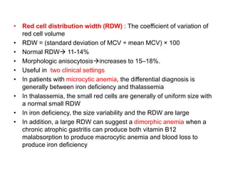
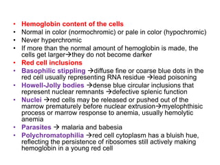
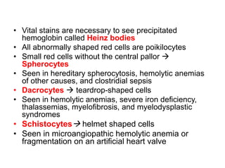
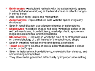
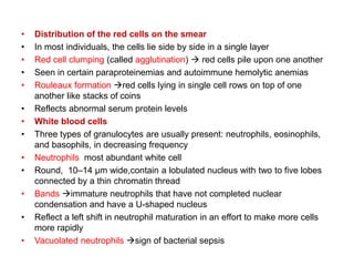
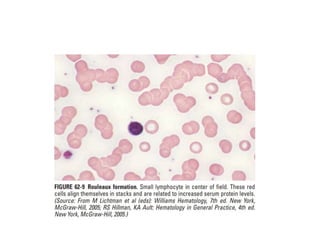
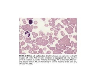
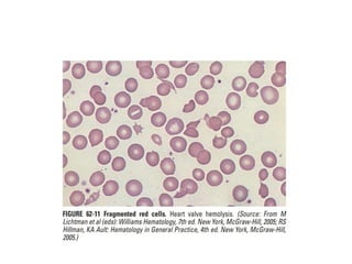
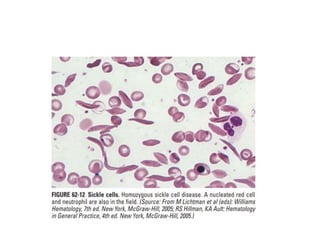
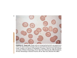
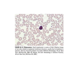
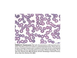
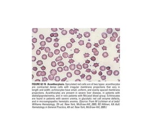
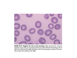
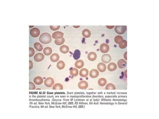
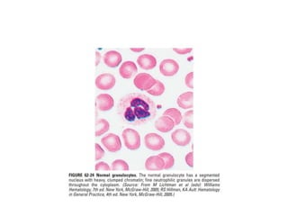
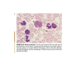
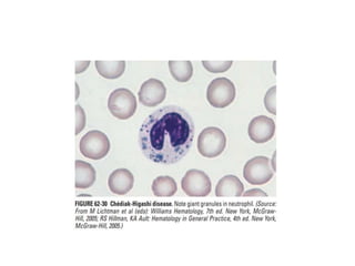
INTERPRETING PERIPHERAL
BLOOD SMEARS
Dr.G.VENKATARAMANA
MBBS DNB FAMILY MEDICINE
• The best place to examine blood cell morphology
is the feathered edge of the blood smear where
red cells lie in a single layer, side by side, just
barely touching one another but not overlapping
• Platelets Red cells White cells
• Using an oil immersion lens that magnifies the
cells 100-fold, one counts the platelets in five to
six fields, averages the number per field, and
multiplies by 20,000 to get a rough estimate of the
platelet count
• Platelets1–2 μm in diameter, blue granulated
appearance
PLATELETS
• Large platelets  sign of rapid platelet turnover (as young
platelets are often larger than old ones), rare inherited
syndromes
• If the platelet count is low, the absence of large (young)
platelets may be an indicator of marrow production problems
• Platelet clumping  associated with falsely low automated
platelet counts
• Caused by the anticoagulant into which the blood is drawn
• Neutrophil fragmentation  falsely elevated automated
platelet counts
• Absence of platelet granules  Artifact of the handling of the
blood or may indicate marrow disease or a rare congenital
anomaly, gray platelet syndrome
• Elevated platelet counts  Myeloproliferative disorder or a
reaction to systemic inflammation
RED BLOOD CELLS
• Size  by comparing the red cell to the nucleus of a
small lymphocyte.
• Both are normally about 8-μm wide
• Red cells smaller than the small lymphocyte nucleus 
Microcytic
• Red cells larger than the small lymphocyte nucleus 
Macrocytic
• Macrocytic cells also tend to be more oval than
spherical in shape and are sometimes called
macroovalocytes
• However, some patients may have both iron and
vitamin B12 deficiency, which will produce an MCV in the
normal range but wide variation in red cell size
• When the red cells vary greatly in size  anisocytosis
• When the red cells vary greatly in shapepoikilocytosis
• Red cell distribution width (RDW) : The coefficient of variation of
red cell volume
• RDW = (standard deviation of MCV Ć· mean MCV) Ɨ 100
• Normal RDW 11-14%
• Morphologic anisocytosisincreases to 15–18%.
• Useful in two clinical settings
• In patients with microcytic anemia, the differential diagnosis is
generally between iron deficiency and thalassemia
• In thalassemia, the small red cells are generally of uniform size with
a normal small RDW
• In iron deficiency, the size variability and the RDW are large
• In addition, a large RDW can suggest a dimorphic anemia when a
chronic atrophic gastritis can produce both vitamin B12
malabsorption to produce macrocytic anemia and blood loss to
produce iron deficiency
• Hemoglobin content of the cells
• Normal in color (normochromic) or pale in color (hypochromic)
• Never hyperchromic
• If more than the normal amount of hemoglobin is made, the
cells get largerthey do not become darker
• Red cell inclusions
• Basophilic stippling diffuse fine or coarse blue dots in the
red cell usually representing RNA residue lead poisoning
• Howell-Jolly bodies dense blue circular inclusions that
represent nuclear remnants defective splenic function
• Nuclei red cells may be released or pushed out of the
marrow prematurely before nuclear extrusionmyelophthisic
process or marrow response to anemia, usually hemolytic
anemia
• Parasites  malaria and babesia
• Polychromatophilia red cell cytoplasm has a bluish hue,
reflecting the persistence of ribosomes still actively making
hemoglobin in a young red cell
• Vital stains are necessary to see precipitated
hemoglobin called Heinz bodies
• All abnormally shaped red cells are poikilocytes
• Small red cells without the central pallor 
Spherocytes
• Seen in hereditary spherocytosis, hemolytic anemias
of other causes, and clostridial sepsis
• Dacrocytes  teardrop-shaped cells
• Seen in hemolytic anemias, severe iron deficiency,
thalassemias, myelofibrosis, and myelodysplastic
syndromes
• Schistocytes helmet shaped cells
• Seen in microangiopathic hemolytic anemia or
fragmentation on an artificial heart valve
• Echinocytes spiculated red cells with the spikes evenly spaced
artifact of abnormal drying of the blood smear or reflect changes
in stored blood
• Also seen in renal failure and malnutrition
• Acanthocytes spiculated red cells with the spikes irregularly
distributed
• Seen in renal disease, abetalipoproteinemia, or splenectomy
• Elliptocytes elliptical-shaped red cells inherited defect in the
red cell membrane, iron deficiency, myelodysplastic syndromes,
megaloblastic anemia, and thalassemias
• Stomatocytes  red cells in which the area of central pallor takes
on the morphology of a slit instead of the usual round shape
• Seen in Inherited red cell membrane defect ,alcoholism
• Target cells have an area of central pallor that contains a dense
center, or bull’s eye
• Seen in thalassemia, iron deficiency, cholestatic liver disease, and
some hemoglobinopathies
• They also can be generated artifactually by improper slide making
• Distribution of the red cells on the smear
• In most individuals, the cells lie side by side in a single layer
• Red cell clumping (called agglutination)  red cells pile upon one another
• Seen in certain paraproteinemias and autoimmune hemolytic anemias
• Rouleaux formation red cells lying in single cell rows on top of one
another like stacks of coins
• Reflects abnormal serum protein levels
• White blood cells
• Three types of granulocytes are usually present: neutrophils, eosinophils,
and basophils, in decreasing frequency
• Neutrophils most abundant white cell
• Round, 10–14 μm wide,contain a lobulated nucleus with two to five lobes
connected by a thin chromatin thread
• Bands immature neutrophils that have not completed nuclear
condensation and have a U-shaped nucleus
• Reflect a left shift in neutrophil maturation in an effort to make more cells
more rapidly
• Vacuolated neutrophils sign of bacterial sepsis
• Dohle bodies 1- to 2-μm blue cytoplasmic inclusions
• Reflect infections, burns, or other inflammatory states
• If the neutrophil granules are larger than normal and stain a darker
blue, ā€œtoxic granulationsā€ are said to be present suggest a
systemic inflammation
• Neutrophils with more than five nuclear lobes  megaloblastic
anemia
• Large misshapen granules  inherited Chediak-Higashi syndrome
• Eosinophils are slightly larger than neutrophils, have bilobed
nuclei, and contain large red granules
• Basophils are even more rare than eosinophils in the blood
• They have large dark blue granules and may be increased as part of
chronic myeloid leukemia.
• Lymphocytes
• Small lymphocytes small dark nucleus and scarce
cytoplasm
• Viral infections lymphocytes are larger, about the size of
neutrophils, with abundant cytoplasm and a less condensed
nuclear chromatin Reactive lymphocytes
• 1% of lymphocytes are larger and contain blue granules in a
light blue cytoplasm large granular lymphocytes
• In chronic lymphoid leukemia, the small lymphocytes are
increased in number, and many of them are ruptured in
making the blood smear, leaving a smudge of nuclear material
without a surrounding cytoplasm or cell membranesmudge
cells and are rare in the absence of chronic lymphoid
leukemia
• Monocytes  largest white blood cells, 15 to 22 μm in diameter
• Nucleusappears to be folded
• Cytoplasm gray
• Abnormal cells may appear in the blood
• Most often, the abnormal cells originate from neoplasms of bone
marrow derived cells, including lymphoid cells, myeloid cells, and
occasionally red cells
• More rarely, other types of tumors can get access to the
bloodstream, and rare epithelial malignant cells may be identified
• The chances of seeing such abnormal cells are increased by
examining blood smears made from buffy coats, the layer of cells
that is visible on top of sedimenting red cells when blood is left in the
test tube for an hour
• Smears made from finger sticks may include rare endothelial cells.
interpreting peripheral blood smears.pptx

interpreting peripheral blood smears.pptx

  • 1.
  • 2.
    • The bestplace to examine blood cell morphology is the feathered edge of the blood smear where red cells lie in a single layer, side by side, just barely touching one another but not overlapping • Platelets Red cells White cells • Using an oil immersion lens that magnifies the cells 100-fold, one counts the platelets in five to six fields, averages the number per field, and multiplies by 20,000 to get a rough estimate of the platelet count • Platelets1–2 μm in diameter, blue granulated appearance
  • 3.
    PLATELETS • Large platelets sign of rapid platelet turnover (as young platelets are often larger than old ones), rare inherited syndromes • If the platelet count is low, the absence of large (young) platelets may be an indicator of marrow production problems • Platelet clumping  associated with falsely low automated platelet counts • Caused by the anticoagulant into which the blood is drawn • Neutrophil fragmentation  falsely elevated automated platelet counts • Absence of platelet granules  Artifact of the handling of the blood or may indicate marrow disease or a rare congenital anomaly, gray platelet syndrome • Elevated platelet counts  Myeloproliferative disorder or a reaction to systemic inflammation
  • 4.
    RED BLOOD CELLS •Size  by comparing the red cell to the nucleus of a small lymphocyte. • Both are normally about 8-μm wide • Red cells smaller than the small lymphocyte nucleus  Microcytic • Red cells larger than the small lymphocyte nucleus  Macrocytic • Macrocytic cells also tend to be more oval than spherical in shape and are sometimes called macroovalocytes • However, some patients may have both iron and vitamin B12 deficiency, which will produce an MCV in the normal range but wide variation in red cell size • When the red cells vary greatly in size  anisocytosis • When the red cells vary greatly in shapepoikilocytosis
  • 5.
    • Red celldistribution width (RDW) : The coefficient of variation of red cell volume • RDW = (standard deviation of MCV Ć· mean MCV) Ɨ 100 • Normal RDW 11-14% • Morphologic anisocytosisincreases to 15–18%. • Useful in two clinical settings • In patients with microcytic anemia, the differential diagnosis is generally between iron deficiency and thalassemia • In thalassemia, the small red cells are generally of uniform size with a normal small RDW • In iron deficiency, the size variability and the RDW are large • In addition, a large RDW can suggest a dimorphic anemia when a chronic atrophic gastritis can produce both vitamin B12 malabsorption to produce macrocytic anemia and blood loss to produce iron deficiency
  • 6.
    • Hemoglobin contentof the cells • Normal in color (normochromic) or pale in color (hypochromic) • Never hyperchromic • If more than the normal amount of hemoglobin is made, the cells get largerthey do not become darker • Red cell inclusions • Basophilic stippling diffuse fine or coarse blue dots in the red cell usually representing RNA residue lead poisoning • Howell-Jolly bodies dense blue circular inclusions that represent nuclear remnants defective splenic function • Nuclei red cells may be released or pushed out of the marrow prematurely before nuclear extrusionmyelophthisic process or marrow response to anemia, usually hemolytic anemia • Parasites  malaria and babesia • Polychromatophilia red cell cytoplasm has a bluish hue, reflecting the persistence of ribosomes still actively making hemoglobin in a young red cell
  • 7.
    • Vital stainsare necessary to see precipitated hemoglobin called Heinz bodies • All abnormally shaped red cells are poikilocytes • Small red cells without the central pallor  Spherocytes • Seen in hereditary spherocytosis, hemolytic anemias of other causes, and clostridial sepsis • Dacrocytes  teardrop-shaped cells • Seen in hemolytic anemias, severe iron deficiency, thalassemias, myelofibrosis, and myelodysplastic syndromes • Schistocytes helmet shaped cells • Seen in microangiopathic hemolytic anemia or fragmentation on an artificial heart valve
  • 8.
    • Echinocytes spiculatedred cells with the spikes evenly spaced artifact of abnormal drying of the blood smear or reflect changes in stored blood • Also seen in renal failure and malnutrition • Acanthocytes spiculated red cells with the spikes irregularly distributed • Seen in renal disease, abetalipoproteinemia, or splenectomy • Elliptocytes elliptical-shaped red cells inherited defect in the red cell membrane, iron deficiency, myelodysplastic syndromes, megaloblastic anemia, and thalassemias • Stomatocytes  red cells in which the area of central pallor takes on the morphology of a slit instead of the usual round shape • Seen in Inherited red cell membrane defect ,alcoholism • Target cells have an area of central pallor that contains a dense center, or bull’s eye • Seen in thalassemia, iron deficiency, cholestatic liver disease, and some hemoglobinopathies • They also can be generated artifactually by improper slide making
  • 9.
    • Distribution ofthe red cells on the smear • In most individuals, the cells lie side by side in a single layer • Red cell clumping (called agglutination)  red cells pile upon one another • Seen in certain paraproteinemias and autoimmune hemolytic anemias • Rouleaux formation red cells lying in single cell rows on top of one another like stacks of coins • Reflects abnormal serum protein levels • White blood cells • Three types of granulocytes are usually present: neutrophils, eosinophils, and basophils, in decreasing frequency • Neutrophils most abundant white cell • Round, 10–14 μm wide,contain a lobulated nucleus with two to five lobes connected by a thin chromatin thread • Bands immature neutrophils that have not completed nuclear condensation and have a U-shaped nucleus • Reflect a left shift in neutrophil maturation in an effort to make more cells more rapidly • Vacuolated neutrophils sign of bacterial sepsis
  • 10.
    • Dohle bodies1- to 2-μm blue cytoplasmic inclusions • Reflect infections, burns, or other inflammatory states • If the neutrophil granules are larger than normal and stain a darker blue, ā€œtoxic granulationsā€ are said to be present suggest a systemic inflammation • Neutrophils with more than five nuclear lobes  megaloblastic anemia • Large misshapen granules  inherited Chediak-Higashi syndrome • Eosinophils are slightly larger than neutrophils, have bilobed nuclei, and contain large red granules • Basophils are even more rare than eosinophils in the blood • They have large dark blue granules and may be increased as part of chronic myeloid leukemia.
  • 11.
    • Lymphocytes • Smalllymphocytes small dark nucleus and scarce cytoplasm • Viral infections lymphocytes are larger, about the size of neutrophils, with abundant cytoplasm and a less condensed nuclear chromatin Reactive lymphocytes • 1% of lymphocytes are larger and contain blue granules in a light blue cytoplasm large granular lymphocytes • In chronic lymphoid leukemia, the small lymphocytes are increased in number, and many of them are ruptured in making the blood smear, leaving a smudge of nuclear material without a surrounding cytoplasm or cell membranesmudge cells and are rare in the absence of chronic lymphoid leukemia
  • 12.
    • Monocytes largest white blood cells, 15 to 22 μm in diameter • Nucleusappears to be folded • Cytoplasm gray • Abnormal cells may appear in the blood • Most often, the abnormal cells originate from neoplasms of bone marrow derived cells, including lymphoid cells, myeloid cells, and occasionally red cells • More rarely, other types of tumors can get access to the bloodstream, and rare epithelial malignant cells may be identified • The chances of seeing such abnormal cells are increased by examining blood smears made from buffy coats, the layer of cells that is visible on top of sedimenting red cells when blood is left in the test tube for an hour • Smears made from finger sticks may include rare endothelial cells.