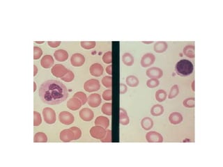
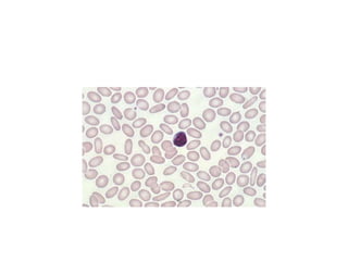
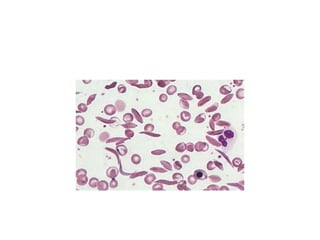
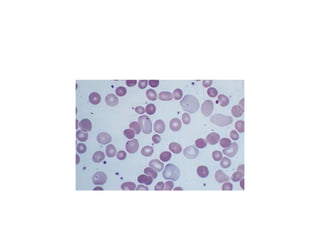
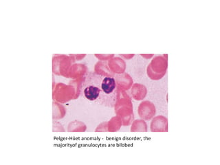
P.S
• Wright’s stain
• best place to examine blood cell morphology
is the feathered edge of the blood smear
• red cells lie in a single layer, side by side, just
barely touching one another but not
overlapping
red blood cells
• Size - by comparing the red cell to the nucleus of
a small lymphocyte
• Smaller – microcytic
• Larger – macrocytic
• vary greatly in size – anisocytosis
• vary greatly in shape – poikilocytosis
• hemoglobin content- normal in color
(normochromic ) or pale in color ( hypochromic )
Red cells shapes.
• All abnormally shaped cells – poikilocytes
• Small red cells without the central pallor are
spherocytes - hereditary spherocytosis,
hemolytic anemias of other causes, and
clostridial sepsis
• Schistocytes -helmet-shaped cells
microangiopathic hemolytic anemia or
fragmentation on an artificial heart valve
thalassemia, iron deficiency, cholestatic liver
disease, and some hemoglobinopathies.
Acanthocytes - renal disease,
abetalipoproteinemia, or splenectomy
Red cell inclusions
• Basophilic stippling —diffuse fine or coarse blue
dots - represent RNA residue— common in lead
poisoning
• Howell-Jolly bodies —dense blue circular
inclusions – represent nuclear remnants implies
defective splenic function
• Nuclei— red cells may be released or pushed out
of the marrow prematurely before nuclear
extrusion— implies a myelophthisic process or a
vigorous narrow response to anemia,usually
hemolytic anemia
• Parasites —malaria and babesia
• Polychromatophilia —cytoplasm has a bluish
hue - persistence of ribosomes still actively
making hemoglobin
• Heinz bodies - precipitated hemoglobin - Vital
stains
white blood cells
• Three types of granulocytes: neutrophils,
eosinophils, and basophils, in decreasing
frequency
• Neutrophils - lobulated nucleus with two to
five lobes
• Bands - immature neutrophils that have not
completed nuclear condensation and have a
U-shaped nucleus
• ā€œtoxic granulationsā€ - systemic inflammation
Pelger-Hüet anomaly - benign disorder, the
majorityof granulocytes are bilobed
Dƶhle bodies , can reflect infections, burns,
or other inflammatory states
Large misshapen granules may reflect the
inherited Chediak-Higashi syndrome
Normal bone marrow
Peripheral Smear & Bone Marrow

Peripheral Smear & Bone Marrow

  • 2.
    P.S • Wright’s stain •best place to examine blood cell morphology is the feathered edge of the blood smear • red cells lie in a single layer, side by side, just barely touching one another but not overlapping
  • 3.
    red blood cells •Size - by comparing the red cell to the nucleus of a small lymphocyte • Smaller – microcytic • Larger – macrocytic • vary greatly in size – anisocytosis • vary greatly in shape – poikilocytosis • hemoglobin content- normal in color (normochromic ) or pale in color ( hypochromic )
  • 8.
    Red cells shapes. •All abnormally shaped cells – poikilocytes • Small red cells without the central pallor are spherocytes - hereditary spherocytosis, hemolytic anemias of other causes, and clostridial sepsis • Schistocytes -helmet-shaped cells microangiopathic hemolytic anemia or fragmentation on an artificial heart valve
  • 12.
    thalassemia, iron deficiency,cholestatic liver disease, and some hemoglobinopathies.
  • 15.
    Acanthocytes - renaldisease, abetalipoproteinemia, or splenectomy
  • 16.
    Red cell inclusions •Basophilic stippling —diffuse fine or coarse blue dots - represent RNA residue— common in lead poisoning • Howell-Jolly bodies —dense blue circular inclusions – represent nuclear remnants implies defective splenic function • Nuclei— red cells may be released or pushed out of the marrow prematurely before nuclear extrusion— implies a myelophthisic process or a vigorous narrow response to anemia,usually hemolytic anemia
  • 17.
    • Parasites —malariaand babesia • Polychromatophilia —cytoplasm has a bluish hue - persistence of ribosomes still actively making hemoglobin • Heinz bodies - precipitated hemoglobin - Vital stains
  • 23.
    white blood cells •Three types of granulocytes: neutrophils, eosinophils, and basophils, in decreasing frequency • Neutrophils - lobulated nucleus with two to five lobes • Bands - immature neutrophils that have not completed nuclear condensation and have a U-shaped nucleus • ā€œtoxic granulationsā€ - systemic inflammation
  • 25.
    Pelger-Hüet anomaly -benign disorder, the majorityof granulocytes are bilobed
  • 26.
    Dƶhle bodies ,can reflect infections, burns, or other inflammatory states
  • 27.
    Large misshapen granulesmay reflect the inherited Chediak-Higashi syndrome
  • 29.