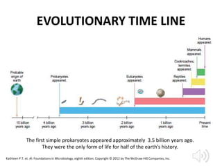
BASIC
BACTERIOLOGY
MUNAWIR
Medical Microbiology Department
Faculty of Medicine Hasanuddin University
2020
OBJECTIVES
• Distuinguish between eukariotyc and
prokariotyc cells
• Taxonomy and Classification of Bacteria
• Components of Bacteria and their functions
• Mechanism of Bacterial Infection
• Bacterial Genetic and Mechanism of antibiotic
Resistance
• Health care-associated infection
INTRODUCTION
• lens maker has evolved immensely from its early historical
beginnings.
• “animalcules”
• The scientific community acknowledged him as the “father of
protozoology and bacteriology.”
Louis Pasteur of France
Robert Koch of Germany
A V Leeuwenhoek of Dutch
• He invented pasteurization and completed some of the first
studies showing that human diseases could arise from infection
• germ theory of disease.
• Koch’s postulates, a series of proofs that verified the germ
theory and could establish whether an organism was pathogenic
and which disease it caused
• 1875, Koch used this experimental system to show that anthrax
was caused by a bacterium called Bacillus anthracis
Kathleen P.T. et. Al. Foundations in Microbiology, eighth edition. Copyright © 2012 by The McGraw-Hill
Companies, Inc.
INTRODUCTION
There are 100 million times as many bacteria
in the oceans (13 × 1028) as there are stars in
the known universe.
Bacteria were among the first life forms to appear
on Earth, and are present in most of its habitats.
Microbiology by numbers. Nature Reviews Microbiology volume9, page628 (2011)
EVOLUTIONARY TIME LINE
The first simple prokaryotes appeared approximately 3.5 billion years ago.
They were the only form of life for half of the earth’s history.
Kathleen P.T. et. Al. Foundations in Microbiology, eighth edition. Copyright © 2012 by The McGraw-Hill Companies, Inc.
Comparison of Prokaryotic and Eukaryotic Cell
Organization and Structures
. A, Prokaryotic gram-positive and gram-negative bacteria. B, Structure of the
generalized eukaryotic cell. (A, From Murray PR, Rosenthal KS, Pfaller MA: Medical
microbiology, ed 6, Philadelphia, 2009, Mosby; B, from Thibodeau GA, Patton KT:
Anatomy and physiology, ed 6, St Louis, 2007, Mosby.)
Kathleen P.T. et. Al. Foundations in Microbiology, eighth edition. Copyright © 2012 by The McGraw-Hill Companies, Inc.
COMPARISON OF PROKARYOTIC AND EUKARYOTIC CELL ORGANIZATION
Connie R. Mahon. Bacterial Cell Structure, Physiology, Metabolism, and Genetics
1
A prominent difference between prokaryotic
and eukaryotic cells is the
a. larger size of prokaryotes
b. lack of pigmentation in eukaryotes
c. presence of a nucleus in eukaryotes
d. presence of a cell wall in prokaryotes
TAXONOMY
• The formal system for organizing, classifying, and naming
living things is taxonomy
• The main taxa, or groups, in a classification scheme are
organized into several descending ranks called a hierarchy
• Classification is an orderly arrangement of organisms into
groups that indicate evolutionary relationships and history.
• Nomenclature is the process of assigning names to the
various taxonomic rankings of each microbial species
• Identification is the process of determining and recording
the traits of organisms to enable their placement in an
overall taxonomic scheme.
Kathleen P.T. et. Al. Foundations in Microbiology, eighth edition. Copyright © 2012 by The McGraw-Hill Companies, Inc.
Traditional Whittaker system of Classification
Kathleen P.T. et. Al. Foundations in Microbiology, eighth edition. Copyright © 2012 by The McGraw-Hill Companies, Inc.
The hierarchy of biological
classification's eight
major taxonomic ranks
STRAIN
A strain is a genetic variant or
subtype of a microorganism
(e.g., virus or bacterium or fungus)
a strain is a low-level taxonomic
rank used at the intraspecific level
(within a species)
A strain or variety of
bacteria is a culture
derived from a single parent
that differs in structure or
metabolism
from other cultures of that
species (also called biovars
or morphovars).
TYPE
• A type is a subspecies that can show
differences in antigenic makeup (serotype or
serovar), in susceptibility to bacterial viruses
(phage type), and in pathogenicity
(pathotype).
Kathleen P.T. et. Al. Foundations in Microbiology, eighth edition. Copyright © 2012 by The McGraw-Hill Companies, Inc.
Serotype or Serovar
serotype or serovar is a distinct variation within
a species of bacteria or virus or among immune
cells of different individuals
Phage typing
• Phage typing is a method used for detecting
single strains of bacteria
• The viruses that infect bacteria are
called bacteriophages ("phages" for short) and
some of these can only infect a single strain of
bacteria.
PATHOTYPE
PALANIAPPAN ET AL. JOURNAL OF CLINICAL MICROBIOLOGY, Apr. 2006, p. 1495–1501
2
Which is the correct order of the taxonomic
categories, going from most specific to most
general?
a. domain, kingdom, phylum, class, order, family,
genus, species
b. division, domain, kingdom, class, family, genus,
species
c. species, genus, family, order, class, phylum,
kingdom, domain
d. species, family, class, order, phylum, kingdom
3
By definition, organisms in the same_______ are
more closely related than are those in the same
________
a. order, family
b. class, phylum
c. family, genus
d. phylum, division
Prokaryotic Cell Structure
Structure of a typical bacterial cell.
Cutaway view of a rodshaped
bacterium, showing major
structural features. Note that not all
components are found in all cells.
Kathleen P.T. et. Al. Foundations in Microbiology, eighth edition. Copyright © 2012 by The McGraw-Hill Companies, Inc.
FLAGELLA
Details of the flagellar basal body and its position in the cell wall. The hook,
rings, and rod function together as a tiny device that rotates the filament 3608.
(a) Structure in gram-negative cells (b) Structure in gram-positive cells.
Kathleen P.T. et. Al. Foundations in Microbiology, eighth edition. Copyright © 2012 by The McGraw-Hill Companies, Inc.
FLAGELLA
Monotrichous flagellum on the
predatory bacterium Bdellovibrio
Lophotrichous flagella on Vibrio fi scheri, a
common marine bacterium (23,0003).
Unusual flagella on Aquaspirillum are
amphitrichous (and lophotrichous) in
arrangement and coil up into tight
loops.
An unidentified bacterium discovered inside
Paramecium cells exhibits peritrichous
flagella.
Kathleen P.T. et. Al. Foundations in Microbiology, eighth edition. Copyright © 2012 by The McGraw-Hill Companies, Inc.
FLAGELLA
(a) In general, when a polar flagellum rotates in a
counterclockwise direction, the cell swims forward in runs.
When the flagellum reverses direction and rotates clockwise,
the cell stops and tumbles.
(b) In peritrichous forms, all flagella sweep toward one end of
the cell and rotate as a single group.
(c) During tumbles, the flagella rotate in the opposite direction
and cause the cells to lose coordination.
Chemotaxis in bacteria
The operation of flagella and the mode of
locomotion in bacteria with polar and peritrichous flagella
(a) A cell moves via a random series of short runs and
tumbles when there is no attractant or repellent.
(b) The cell spends more time on runs as it gets closer to
an attractant.
Kathleen P.T. et. Al. Foundations in Microbiology, eighth edition. Copyright © 2012 by The McGraw-Hill Companies, Inc.
Fimbriae
Form and function of bacterial fimbriae.
Several cells of pathogenic Escherichia coli
covered with numerous stiff fibers called
fimbriae. Note also the dark blue masses,
which are chromosomes.
A row of E. coli cells tightly adheres by
their fimbriae to the surface of
intestinal cells.
This is how the bacterium clings and
gains access to the inside of cells
during an infection. (G 5 glycocalyx)
Kathleen P.T. et. Al. Foundations in Microbiology, eighth edition. Copyright © 2012 by The McGraw-Hill Companies, Inc.
PILI
Three bacteria in the process of
conjugating
Clearly evident are the sex pili forming mutual
conjugation bridges between a donor (upper
cell) and two recipients (two lower cells).
Fimbriae can also be seen on the donor cell.
• A pilus (also called a sex pilus ) is an elongate,
rigid tubular structure made of a special protein,
pilin .
• True pili have been found only on gram-negative
bacteria, where they are utilized primarily in a
“mating” process between cells called
conjugation, which involves a transfer of DNA
from one cell to another
• Type IV pili found in Pseudomonas bacteria carry
out a remarkable sort of twitching motility.
Kathleen P.T. et. Al. Foundations in Microbiology, eighth edition. Copyright © 2012 by The McGraw-Hill Companies, Inc.
The Bacterial Surface Coating, or Glycocalyx
• The glycocalyx develops as a coating of macromolecules to protect the cell and, in some
cases, help it adhere to its environment.
• Some bacteria are covered with a loose shield called a slime layer that evidently protects
them from dehydration and loss of nutrients.
• Bacteria produce capsules composed of repeating polysaccharide units, of protein, or of
both. A capsule is bound more tightly to the cell than a slime layer is, and it has a thicker,
gummy consistency that gives a prominently sticky (mucoid) character to the colonies of
most encapsulated bacteria
Types of glycocalyces seen through
cutaway views of cells.
a) The slime layer is a loose structure
that is easily washed off.
b) The capsule is a thick, structured
layer that is not readily removed.
Kathleen P.T. et. Al. Foundations in Microbiology, eighth edition. Copyright © 2012 by The McGraw-Hill Companies, Inc.
Capsule
Appearance of encapsulated bacteria.
(a) Close-up view of colonies of Bacillus species with and without capsules. Even at the
macroscopic level, the moist slimy character of the capsule is evident.
(b) Special staining reveals the microscopic appearance of a large, well-developed capsule
(the clear “halo” around the cells) of Klebsiella.
Kathleen P.T. et. Al. Foundations in Microbiology, eighth edition. Copyright © 2012 by The McGraw-Hill Companies, Inc.
Cell Envelopes
Comparative views of the envelopes of gram-positive and gram-negative cells
(a) A section through a gram-positive cell wall/membrane with an interpretation of the
main layers visible.
(b) A section through a gram-negative cell wall/membrane with an interpretation of its
three sandwich-style layers.
Kathleen P.T. et. Al. Foundations in Microbiology, eighth edition. Copyright © 2012 by The McGraw-Hill Companies, Inc.
A Comparison of the Detailed Structure of Gram-
positive and Gram-negative Cell Envelopes and Walls.
Kathleen P.T. et. Al. Foundations in Microbiology, eighth edition. Copyright © 2012 by The McGraw-Hill Companies, Inc.
Nontypical Cell Walls
Scanning electron micrograph of
Mycoplasma pneumoniae . Cells exhibit
extreme variation in shape due to the lack of a
cell wall.
https://bio.libretexts.org/Bookshelves/Microbiology/Book%3A_Microbiology_(Kaiser)/Unit_1%3A_Introduction_to_Microbiology_and_Prokaryotic_Cell_Anatomy/2%3A_The_Prokaryotic_Cell_-
_Bacteria/2.3%3A_The_Peptidoglycan_Cell_Wall/2.3C%3A_The_Acid-Fast_Cell_Wall
Structure of an Acid-Fast Cell Wall
Cell Membrane Structure
• A generalized version of the
fluid mosaic model of a cell
membrane indicates a
bilayer of lipids with
globular proteins embedded
to some degree in the lipid
matrix.
• This structure explains many
characteristics of
membranes, including
flexibility, solubility,
permeability, and transport.
• Appearing just beneath the cell wall is the cell, or cytoplasmic, membrane, a very
thin (5–10 nm), fl exible sheet molded completely around the cytoplasm. In general
composition, it is a lipid bilayer with proteins embedded to varying degrees
Kathleen P.T. et. Al. Foundations in Microbiology, eighth edition. Copyright © 2012 by The McGraw-Hill Companies, Inc.
Bacterial Chromosomes and Plasmids:
The Sources of Genetic Information
• DNA bacteria is not enclosed by a nuclear
membrane but instead is aggregated in a central
area of the cell called the nucleoid
• A plasmid is a small DNA molecule within a cell that
is physically separated from chromosomal DNA and
can replicate independently
Kathleen P.T. et. Al. Foundations in Microbiology, eighth edition. Copyright © 2012 by The McGraw-Hill Companies, Inc.
Ribosomes: Sites of Protein Synthesis
• A bacterial cell contains thousands of ribosomes, which are
made of RNA and protein
• a ribosome is a combination of a special type of RNA called
ribosomal RNA, or rRNA (about 60%), and protein (40%).
A model of a prokaryotic ribosome,
showing the small (30S) and large (50S)
subunits, both separate and joined.
Kathleen P.T. et. Al. Foundations in Microbiology, eighth edition. Copyright © 2012 by The McGraw-Hill Companies, Inc.
Inclusions, or Granules: Storage Bodies
Large particles (pink) of
polyhydroxybutyrate are deposited
in an insoluble, concentrated form
that provides an ample, long-term
supply of that nutrient.
A section through Aquaspirillum reveals a
chain of tiny iron magnets (magnetosomes 5
MP). These unusual bacteria use these
inclusions to orient within their habitat
Kathleen P.T. et. Al. Foundations in Microbiology, eighth edition. Copyright © 2012 by The McGraw-Hill Companies, Inc.
The Bacterial Cytoskeleton
Bacterial cytoskeleton of Bacillus.
Fluorescent stain of actin fibers
appears as fi ne helical ribbons wound
inside the cell.
• Until very recently, bacteriologists thought
bacteria lacked any real form of
cytoskeleton.
• The proteins are chemically similar to the
actin filaments universal in the cytoskeleton
of eukaryotic cells
• Presently this bacterial actin is thought to
help stabilize shape.
• It may also influence cell wall formation by
providing sites for synthesis when the wall
is being repaired or enlarged.
Kathleen P.T. et. Al. Foundations in Microbiology, eighth edition. Copyright © 2012 by The McGraw-Hill Companies, Inc.
Bacterial Endospores: An Extremely
Resistant Life Form
Development of endospores. These biological
“safety pins” are actually stages in endospore
formation of Bacillus subtilis, stained with
fluorescent proteins. The large red and blue cell
is a vegetative cell in the early stages of
sporulating. The developing spores are shown in
green and orange.
• bacterial endospore (or simply spore)
for withstanding hostile conditions
and facilitating survival
• Endospores are dormant bodies
produced by the bacteria Bacillus,
Clostridium, and Sporosarcina
• two-phase life cycle that shifts
between a vegetative cell and an
endospore
• The vegetative cell is the
metabolically active and growing
phase. When exposed to certain
environmental signals, it forms an
endospore by a process termed
sporulation.
Kathleen P.T. et. Al. Foundations in Microbiology, eighth edition. Copyright © 2012 by The McGraw-Hill Companies, Inc.
A typical sporulation cycle in Bacillus species from the active vegetative cell to release
and germination
This process takes, on average, about 10 hours. Inset is a high magnification (10,000x) cross section of a
single spore showing the dense protective layers that surround the core with its chromosome
Kathleen P.T. et. Al. Foundations in Microbiology, eighth edition. Copyright © 2012 by The McGraw-Hill Companies, Inc.
4
• Which of the following is not found in all
bacterial cells?
a. cell membrane nuclear
b. a nucleoid
c. ribosomes
d. actin cytoskeleton
5
The major locomotor structures in bacteria are
a. flagella
b. pili
c. fimbriae
d. cilia
6
Pili are tubular shafts in ______ bacteria that
serve as a means of ________
• a. gram-positive, genetic exchange
• b. gram-positive, attachment
• c. gram-negative, genetic exchange
• d. gram-negative, protection
7
Bacterial endospores function in _______
a. reproduction
b. survival
c. protein synthesis
d. storage
Common Bacterial Shapes
Kathleen P.T. et. Al. Foundations in Microbiology, eighth edition. Copyright © 2012 by The McGraw-Hill Companies, Inc.
Pleomorphism and other morphological features of
Corynebacterium
Cells are irregular in shape and size (800x). This genus typically exhibits a palisades
arrangement, with cells in parallel array (inset). Close examination will also reveal
darkly stained granules called metachromatic granules.
• Variations in cell wall
structure caused by
nutritional or slight hereditary
differences
• The cells of Corynebacterium
diphtheriae are generally
considered rod-shaped, in
culture they display club
shaped, swollen, curved, fi
lamentous, and coccoid
variations
Kathleen P.T. et. Al. Foundations in Microbiology, eighth edition. Copyright © 2012 by The McGraw-Hill Companies, Inc.
Arrangement of cocci resulting from
different planes of cell division
a) Division in one plane produces
diplococci and streptococci.
b) Division in two planes atright angles
produces tetrads and packets.
c) Division in several planes produces
irregular clusters.
Kathleen P.T. et. Al. Foundations in Microbiology, eighth edition. Copyright © 2012 by The McGraw-Hill Companies, Inc.
Unusual Forms of Medically
Significant Bacteria
• Two groups of bacteria—the
rickettsias and chlamydias—have
adapted to life inside their host
cells, where they are considered
obligate intracellular parasites
– Rickettsias are distinctive, very tiny,
gram-negative bacteria
– Bacteria of the genera Chlamydia
and Chlamydophila, termed
chlamydias, are similar to the
rickettsias in that they require host
cells for growth and metabolism
Transmission electron micrograph of the
rickettsia Coxiella burnetii
Kathleen P.T. et. Al. Foundations in Microbiology, eighth edition. Copyright © 2012 by The McGraw-Hill Companies, Inc.
Oxygen Requirements
• Aerobes: Aerobe grow in ambient air, which contains 21% oxygen and
small amount of (0.03%) of carbon dioxide
• Obligate aerobes: They have absolute requirement for oxygen in order to
grow. Pseudomonas aeruginosa, Mycobacterium tuberculosis.
• Anaerobes: Usually bacteria of this group can not grow in the presence
of oxygen, oxygen is toxic for them.
• Obligate anaerobes: These bacteria grow only under condition of high
reducing intensity and for which oxygen is toxic
• Facultative anaerobes: They are versatile organisms, capable of growth
under both aerobic and anaerobic conditions.
• Aerotolerant anaerobes: Are anaerobic bacteria that are not killed by
exposure to oxygen.
• Capnophiles: Capnophilic bacteria require increased concentration of
carbon dioxide (5% to 10%) and approximately 15% oxygen.
• Microaerophiles: Microaerophiles are those groups of bacteria that
can grow under reduced oxygen (5% to 10%) and increased carbon
dioxide (8% to 10%)
https://microbeonline.com/oxygen-requirements-for-pathogenic-bacteria/
Carbon-based Nutritional Types
• The element carbon is so key to the
structure and metabolism of all life
forms that the source of carbon
defines two basic nutritional groups:
– A heterotroph is an organism that
must obtain its carbon in an organic
form.
• they derive their nutrients from other
organisms
– An autotroph * is an organism that
uses inorganic CO 2 as its carbon source.
• they are not nutritionally dependent on
other living things.
Kathleen P.T. et. Al. Foundations in Microbiology, eighth edition. Copyright © 2012 by The McGraw-Hill Companies, Inc.
Ecological Associations among
Microorganisms
• Microbial Interactions: Microbes coexist in varied relationships in
nature.
– Symbiosis
• mutualism, a reciprocal, obligatory, and beneficial relationship between two
organisms
• commensalism, an organism receiving benefi ts from another without harming
the other organism in the relationship. One form of this is satellitism.
• Parasitism occurs between a host and an infectious agent; the host is harmed
in the interaction.
– Synergism is a mutually benefi cial but not obligatory coexistence in
which organisms cooperate to produce a reaction.
• Antagonism entails competition, inhibition, and injury directed against the
opposing organism. A special case of antagonism is antibiotic production.
– Quorum sensing is an interaction among members of a biofilm that
results in a coordinated reaction, such as secretion of inducer
molecules and enzymes .
Kathleen P.T. et. Al. Foundations in Microbiology, eighth edition. Copyright © 2012 by The McGraw-Hill Companies, Inc.
8
Which stain is used to distinguish differences
between the cell walls of medically important
bacteria?
a. simple stain
b. acridine orange stain
c. Gram stain
d. negative stain
Basic bacteriology

Basic bacteriology

  • 1.
  • 2.
    OBJECTIVES • Distuinguish betweeneukariotyc and prokariotyc cells • Taxonomy and Classification of Bacteria • Components of Bacteria and their functions • Mechanism of Bacterial Infection • Bacterial Genetic and Mechanism of antibiotic Resistance • Health care-associated infection
  • 3.
    INTRODUCTION • lens makerhas evolved immensely from its early historical beginnings. • “animalcules” • The scientific community acknowledged him as the “father of protozoology and bacteriology.” Louis Pasteur of France Robert Koch of Germany A V Leeuwenhoek of Dutch • He invented pasteurization and completed some of the first studies showing that human diseases could arise from infection • germ theory of disease. • Koch’s postulates, a series of proofs that verified the germ theory and could establish whether an organism was pathogenic and which disease it caused • 1875, Koch used this experimental system to show that anthrax was caused by a bacterium called Bacillus anthracis Kathleen P.T. et. Al. Foundations in Microbiology, eighth edition. Copyright © 2012 by The McGraw-Hill Companies, Inc.
  • 4.
    INTRODUCTION There are 100million times as many bacteria in the oceans (13 × 1028) as there are stars in the known universe. Bacteria were among the first life forms to appear on Earth, and are present in most of its habitats. Microbiology by numbers. Nature Reviews Microbiology volume9, page628 (2011)
  • 5.
    EVOLUTIONARY TIME LINE Thefirst simple prokaryotes appeared approximately 3.5 billion years ago. They were the only form of life for half of the earth’s history. Kathleen P.T. et. Al. Foundations in Microbiology, eighth edition. Copyright © 2012 by The McGraw-Hill Companies, Inc.
  • 7.
    Comparison of Prokaryoticand Eukaryotic Cell Organization and Structures . A, Prokaryotic gram-positive and gram-negative bacteria. B, Structure of the generalized eukaryotic cell. (A, From Murray PR, Rosenthal KS, Pfaller MA: Medical microbiology, ed 6, Philadelphia, 2009, Mosby; B, from Thibodeau GA, Patton KT: Anatomy and physiology, ed 6, St Louis, 2007, Mosby.) Kathleen P.T. et. Al. Foundations in Microbiology, eighth edition. Copyright © 2012 by The McGraw-Hill Companies, Inc.
  • 8.
    COMPARISON OF PROKARYOTICAND EUKARYOTIC CELL ORGANIZATION Connie R. Mahon. Bacterial Cell Structure, Physiology, Metabolism, and Genetics
  • 9.
    1 A prominent differencebetween prokaryotic and eukaryotic cells is the a. larger size of prokaryotes b. lack of pigmentation in eukaryotes c. presence of a nucleus in eukaryotes d. presence of a cell wall in prokaryotes
  • 10.
    TAXONOMY • The formalsystem for organizing, classifying, and naming living things is taxonomy • The main taxa, or groups, in a classification scheme are organized into several descending ranks called a hierarchy • Classification is an orderly arrangement of organisms into groups that indicate evolutionary relationships and history. • Nomenclature is the process of assigning names to the various taxonomic rankings of each microbial species • Identification is the process of determining and recording the traits of organisms to enable their placement in an overall taxonomic scheme. Kathleen P.T. et. Al. Foundations in Microbiology, eighth edition. Copyright © 2012 by The McGraw-Hill Companies, Inc.
  • 11.
    Traditional Whittaker systemof Classification Kathleen P.T. et. Al. Foundations in Microbiology, eighth edition. Copyright © 2012 by The McGraw-Hill Companies, Inc.
  • 12.
    The hierarchy ofbiological classification's eight major taxonomic ranks
  • 13.
    STRAIN A strain isa genetic variant or subtype of a microorganism (e.g., virus or bacterium or fungus) a strain is a low-level taxonomic rank used at the intraspecific level (within a species) A strain or variety of bacteria is a culture derived from a single parent that differs in structure or metabolism from other cultures of that species (also called biovars or morphovars).
  • 14.
    TYPE • A typeis a subspecies that can show differences in antigenic makeup (serotype or serovar), in susceptibility to bacterial viruses (phage type), and in pathogenicity (pathotype). Kathleen P.T. et. Al. Foundations in Microbiology, eighth edition. Copyright © 2012 by The McGraw-Hill Companies, Inc.
  • 15.
    Serotype or Serovar serotypeor serovar is a distinct variation within a species of bacteria or virus or among immune cells of different individuals
  • 16.
    Phage typing • Phagetyping is a method used for detecting single strains of bacteria • The viruses that infect bacteria are called bacteriophages ("phages" for short) and some of these can only infect a single strain of bacteria.
  • 17.
    PATHOTYPE PALANIAPPAN ET AL.JOURNAL OF CLINICAL MICROBIOLOGY, Apr. 2006, p. 1495–1501
  • 18.
    2 Which is thecorrect order of the taxonomic categories, going from most specific to most general? a. domain, kingdom, phylum, class, order, family, genus, species b. division, domain, kingdom, class, family, genus, species c. species, genus, family, order, class, phylum, kingdom, domain d. species, family, class, order, phylum, kingdom
  • 19.
    3 By definition, organismsin the same_______ are more closely related than are those in the same ________ a. order, family b. class, phylum c. family, genus d. phylum, division
  • 20.
    Prokaryotic Cell Structure Structureof a typical bacterial cell. Cutaway view of a rodshaped bacterium, showing major structural features. Note that not all components are found in all cells. Kathleen P.T. et. Al. Foundations in Microbiology, eighth edition. Copyright © 2012 by The McGraw-Hill Companies, Inc.
  • 21.
    FLAGELLA Details of theflagellar basal body and its position in the cell wall. The hook, rings, and rod function together as a tiny device that rotates the filament 3608. (a) Structure in gram-negative cells (b) Structure in gram-positive cells. Kathleen P.T. et. Al. Foundations in Microbiology, eighth edition. Copyright © 2012 by The McGraw-Hill Companies, Inc.
  • 22.
    FLAGELLA Monotrichous flagellum onthe predatory bacterium Bdellovibrio Lophotrichous flagella on Vibrio fi scheri, a common marine bacterium (23,0003). Unusual flagella on Aquaspirillum are amphitrichous (and lophotrichous) in arrangement and coil up into tight loops. An unidentified bacterium discovered inside Paramecium cells exhibits peritrichous flagella. Kathleen P.T. et. Al. Foundations in Microbiology, eighth edition. Copyright © 2012 by The McGraw-Hill Companies, Inc.
  • 23.
    FLAGELLA (a) In general,when a polar flagellum rotates in a counterclockwise direction, the cell swims forward in runs. When the flagellum reverses direction and rotates clockwise, the cell stops and tumbles. (b) In peritrichous forms, all flagella sweep toward one end of the cell and rotate as a single group. (c) During tumbles, the flagella rotate in the opposite direction and cause the cells to lose coordination. Chemotaxis in bacteria The operation of flagella and the mode of locomotion in bacteria with polar and peritrichous flagella (a) A cell moves via a random series of short runs and tumbles when there is no attractant or repellent. (b) The cell spends more time on runs as it gets closer to an attractant. Kathleen P.T. et. Al. Foundations in Microbiology, eighth edition. Copyright © 2012 by The McGraw-Hill Companies, Inc.
  • 24.
    Fimbriae Form and functionof bacterial fimbriae. Several cells of pathogenic Escherichia coli covered with numerous stiff fibers called fimbriae. Note also the dark blue masses, which are chromosomes. A row of E. coli cells tightly adheres by their fimbriae to the surface of intestinal cells. This is how the bacterium clings and gains access to the inside of cells during an infection. (G 5 glycocalyx) Kathleen P.T. et. Al. Foundations in Microbiology, eighth edition. Copyright © 2012 by The McGraw-Hill Companies, Inc.
  • 25.
    PILI Three bacteria inthe process of conjugating Clearly evident are the sex pili forming mutual conjugation bridges between a donor (upper cell) and two recipients (two lower cells). Fimbriae can also be seen on the donor cell. • A pilus (also called a sex pilus ) is an elongate, rigid tubular structure made of a special protein, pilin . • True pili have been found only on gram-negative bacteria, where they are utilized primarily in a “mating” process between cells called conjugation, which involves a transfer of DNA from one cell to another • Type IV pili found in Pseudomonas bacteria carry out a remarkable sort of twitching motility. Kathleen P.T. et. Al. Foundations in Microbiology, eighth edition. Copyright © 2012 by The McGraw-Hill Companies, Inc.
  • 26.
    The Bacterial SurfaceCoating, or Glycocalyx • The glycocalyx develops as a coating of macromolecules to protect the cell and, in some cases, help it adhere to its environment. • Some bacteria are covered with a loose shield called a slime layer that evidently protects them from dehydration and loss of nutrients. • Bacteria produce capsules composed of repeating polysaccharide units, of protein, or of both. A capsule is bound more tightly to the cell than a slime layer is, and it has a thicker, gummy consistency that gives a prominently sticky (mucoid) character to the colonies of most encapsulated bacteria Types of glycocalyces seen through cutaway views of cells. a) The slime layer is a loose structure that is easily washed off. b) The capsule is a thick, structured layer that is not readily removed. Kathleen P.T. et. Al. Foundations in Microbiology, eighth edition. Copyright © 2012 by The McGraw-Hill Companies, Inc.
  • 27.
    Capsule Appearance of encapsulatedbacteria. (a) Close-up view of colonies of Bacillus species with and without capsules. Even at the macroscopic level, the moist slimy character of the capsule is evident. (b) Special staining reveals the microscopic appearance of a large, well-developed capsule (the clear “halo” around the cells) of Klebsiella. Kathleen P.T. et. Al. Foundations in Microbiology, eighth edition. Copyright © 2012 by The McGraw-Hill Companies, Inc.
  • 28.
    Cell Envelopes Comparative viewsof the envelopes of gram-positive and gram-negative cells (a) A section through a gram-positive cell wall/membrane with an interpretation of the main layers visible. (b) A section through a gram-negative cell wall/membrane with an interpretation of its three sandwich-style layers. Kathleen P.T. et. Al. Foundations in Microbiology, eighth edition. Copyright © 2012 by The McGraw-Hill Companies, Inc.
  • 29.
    A Comparison ofthe Detailed Structure of Gram- positive and Gram-negative Cell Envelopes and Walls. Kathleen P.T. et. Al. Foundations in Microbiology, eighth edition. Copyright © 2012 by The McGraw-Hill Companies, Inc.
  • 30.
    Nontypical Cell Walls Scanningelectron micrograph of Mycoplasma pneumoniae . Cells exhibit extreme variation in shape due to the lack of a cell wall. https://bio.libretexts.org/Bookshelves/Microbiology/Book%3A_Microbiology_(Kaiser)/Unit_1%3A_Introduction_to_Microbiology_and_Prokaryotic_Cell_Anatomy/2%3A_The_Prokaryotic_Cell_- _Bacteria/2.3%3A_The_Peptidoglycan_Cell_Wall/2.3C%3A_The_Acid-Fast_Cell_Wall Structure of an Acid-Fast Cell Wall
  • 31.
    Cell Membrane Structure •A generalized version of the fluid mosaic model of a cell membrane indicates a bilayer of lipids with globular proteins embedded to some degree in the lipid matrix. • This structure explains many characteristics of membranes, including flexibility, solubility, permeability, and transport. • Appearing just beneath the cell wall is the cell, or cytoplasmic, membrane, a very thin (5–10 nm), fl exible sheet molded completely around the cytoplasm. In general composition, it is a lipid bilayer with proteins embedded to varying degrees Kathleen P.T. et. Al. Foundations in Microbiology, eighth edition. Copyright © 2012 by The McGraw-Hill Companies, Inc.
  • 32.
    Bacterial Chromosomes andPlasmids: The Sources of Genetic Information • DNA bacteria is not enclosed by a nuclear membrane but instead is aggregated in a central area of the cell called the nucleoid • A plasmid is a small DNA molecule within a cell that is physically separated from chromosomal DNA and can replicate independently Kathleen P.T. et. Al. Foundations in Microbiology, eighth edition. Copyright © 2012 by The McGraw-Hill Companies, Inc.
  • 33.
    Ribosomes: Sites ofProtein Synthesis • A bacterial cell contains thousands of ribosomes, which are made of RNA and protein • a ribosome is a combination of a special type of RNA called ribosomal RNA, or rRNA (about 60%), and protein (40%). A model of a prokaryotic ribosome, showing the small (30S) and large (50S) subunits, both separate and joined. Kathleen P.T. et. Al. Foundations in Microbiology, eighth edition. Copyright © 2012 by The McGraw-Hill Companies, Inc.
  • 34.
    Inclusions, or Granules:Storage Bodies Large particles (pink) of polyhydroxybutyrate are deposited in an insoluble, concentrated form that provides an ample, long-term supply of that nutrient. A section through Aquaspirillum reveals a chain of tiny iron magnets (magnetosomes 5 MP). These unusual bacteria use these inclusions to orient within their habitat Kathleen P.T. et. Al. Foundations in Microbiology, eighth edition. Copyright © 2012 by The McGraw-Hill Companies, Inc.
  • 35.
    The Bacterial Cytoskeleton Bacterialcytoskeleton of Bacillus. Fluorescent stain of actin fibers appears as fi ne helical ribbons wound inside the cell. • Until very recently, bacteriologists thought bacteria lacked any real form of cytoskeleton. • The proteins are chemically similar to the actin filaments universal in the cytoskeleton of eukaryotic cells • Presently this bacterial actin is thought to help stabilize shape. • It may also influence cell wall formation by providing sites for synthesis when the wall is being repaired or enlarged. Kathleen P.T. et. Al. Foundations in Microbiology, eighth edition. Copyright © 2012 by The McGraw-Hill Companies, Inc.
  • 36.
    Bacterial Endospores: AnExtremely Resistant Life Form Development of endospores. These biological “safety pins” are actually stages in endospore formation of Bacillus subtilis, stained with fluorescent proteins. The large red and blue cell is a vegetative cell in the early stages of sporulating. The developing spores are shown in green and orange. • bacterial endospore (or simply spore) for withstanding hostile conditions and facilitating survival • Endospores are dormant bodies produced by the bacteria Bacillus, Clostridium, and Sporosarcina • two-phase life cycle that shifts between a vegetative cell and an endospore • The vegetative cell is the metabolically active and growing phase. When exposed to certain environmental signals, it forms an endospore by a process termed sporulation. Kathleen P.T. et. Al. Foundations in Microbiology, eighth edition. Copyright © 2012 by The McGraw-Hill Companies, Inc.
  • 37.
    A typical sporulationcycle in Bacillus species from the active vegetative cell to release and germination This process takes, on average, about 10 hours. Inset is a high magnification (10,000x) cross section of a single spore showing the dense protective layers that surround the core with its chromosome Kathleen P.T. et. Al. Foundations in Microbiology, eighth edition. Copyright © 2012 by The McGraw-Hill Companies, Inc.
  • 38.
    4 • Which ofthe following is not found in all bacterial cells? a. cell membrane nuclear b. a nucleoid c. ribosomes d. actin cytoskeleton
  • 39.
    5 The major locomotorstructures in bacteria are a. flagella b. pili c. fimbriae d. cilia
  • 40.
    6 Pili are tubularshafts in ______ bacteria that serve as a means of ________ • a. gram-positive, genetic exchange • b. gram-positive, attachment • c. gram-negative, genetic exchange • d. gram-negative, protection
  • 41.
    7 Bacterial endospores functionin _______ a. reproduction b. survival c. protein synthesis d. storage
  • 42.
    Common Bacterial Shapes KathleenP.T. et. Al. Foundations in Microbiology, eighth edition. Copyright © 2012 by The McGraw-Hill Companies, Inc.
  • 43.
    Pleomorphism and othermorphological features of Corynebacterium Cells are irregular in shape and size (800x). This genus typically exhibits a palisades arrangement, with cells in parallel array (inset). Close examination will also reveal darkly stained granules called metachromatic granules. • Variations in cell wall structure caused by nutritional or slight hereditary differences • The cells of Corynebacterium diphtheriae are generally considered rod-shaped, in culture they display club shaped, swollen, curved, fi lamentous, and coccoid variations Kathleen P.T. et. Al. Foundations in Microbiology, eighth edition. Copyright © 2012 by The McGraw-Hill Companies, Inc.
  • 44.
    Arrangement of cocciresulting from different planes of cell division a) Division in one plane produces diplococci and streptococci. b) Division in two planes atright angles produces tetrads and packets. c) Division in several planes produces irregular clusters. Kathleen P.T. et. Al. Foundations in Microbiology, eighth edition. Copyright © 2012 by The McGraw-Hill Companies, Inc.
  • 45.
    Unusual Forms ofMedically Significant Bacteria • Two groups of bacteria—the rickettsias and chlamydias—have adapted to life inside their host cells, where they are considered obligate intracellular parasites – Rickettsias are distinctive, very tiny, gram-negative bacteria – Bacteria of the genera Chlamydia and Chlamydophila, termed chlamydias, are similar to the rickettsias in that they require host cells for growth and metabolism Transmission electron micrograph of the rickettsia Coxiella burnetii Kathleen P.T. et. Al. Foundations in Microbiology, eighth edition. Copyright © 2012 by The McGraw-Hill Companies, Inc.
  • 46.
    Oxygen Requirements • Aerobes:Aerobe grow in ambient air, which contains 21% oxygen and small amount of (0.03%) of carbon dioxide • Obligate aerobes: They have absolute requirement for oxygen in order to grow. Pseudomonas aeruginosa, Mycobacterium tuberculosis. • Anaerobes: Usually bacteria of this group can not grow in the presence of oxygen, oxygen is toxic for them. • Obligate anaerobes: These bacteria grow only under condition of high reducing intensity and for which oxygen is toxic • Facultative anaerobes: They are versatile organisms, capable of growth under both aerobic and anaerobic conditions. • Aerotolerant anaerobes: Are anaerobic bacteria that are not killed by exposure to oxygen. • Capnophiles: Capnophilic bacteria require increased concentration of carbon dioxide (5% to 10%) and approximately 15% oxygen. • Microaerophiles: Microaerophiles are those groups of bacteria that can grow under reduced oxygen (5% to 10%) and increased carbon dioxide (8% to 10%) https://microbeonline.com/oxygen-requirements-for-pathogenic-bacteria/
  • 47.
    Carbon-based Nutritional Types •The element carbon is so key to the structure and metabolism of all life forms that the source of carbon defines two basic nutritional groups: – A heterotroph is an organism that must obtain its carbon in an organic form. • they derive their nutrients from other organisms – An autotroph * is an organism that uses inorganic CO 2 as its carbon source. • they are not nutritionally dependent on other living things. Kathleen P.T. et. Al. Foundations in Microbiology, eighth edition. Copyright © 2012 by The McGraw-Hill Companies, Inc.
  • 48.
    Ecological Associations among Microorganisms •Microbial Interactions: Microbes coexist in varied relationships in nature. – Symbiosis • mutualism, a reciprocal, obligatory, and beneficial relationship between two organisms • commensalism, an organism receiving benefi ts from another without harming the other organism in the relationship. One form of this is satellitism. • Parasitism occurs between a host and an infectious agent; the host is harmed in the interaction. – Synergism is a mutually benefi cial but not obligatory coexistence in which organisms cooperate to produce a reaction. • Antagonism entails competition, inhibition, and injury directed against the opposing organism. A special case of antagonism is antibiotic production. – Quorum sensing is an interaction among members of a biofilm that results in a coordinated reaction, such as secretion of inducer molecules and enzymes . Kathleen P.T. et. Al. Foundations in Microbiology, eighth edition. Copyright © 2012 by The McGraw-Hill Companies, Inc.
  • 49.
    8 Which stain isused to distinguish differences between the cell walls of medically important bacteria? a. simple stain b. acridine orange stain c. Gram stain d. negative stain