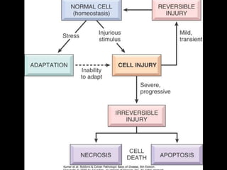
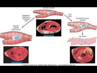
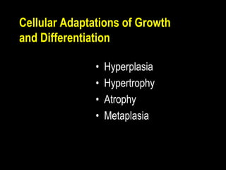
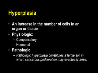
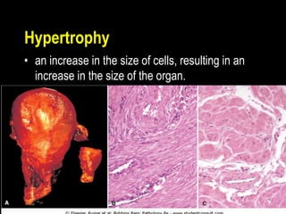
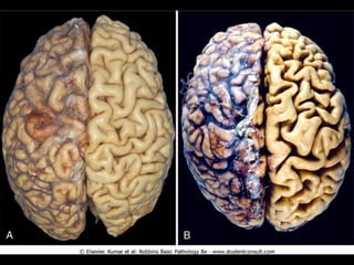
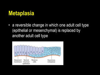
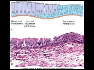
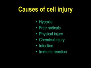
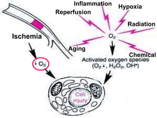
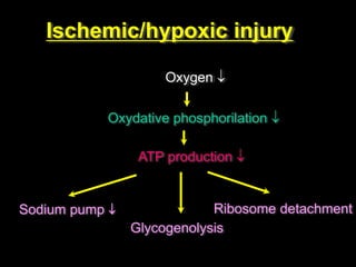
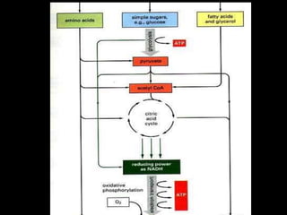
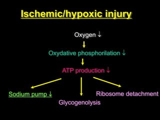
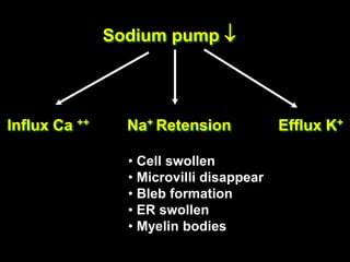
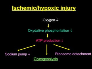
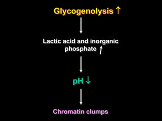
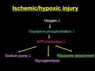
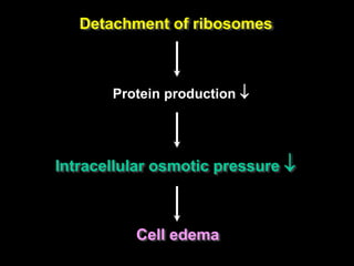
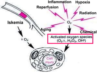
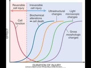
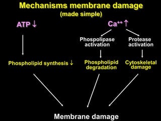
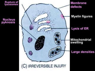
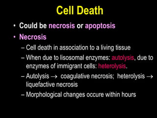
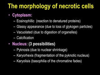
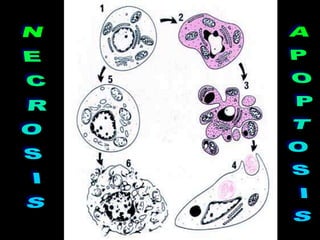
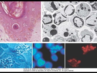
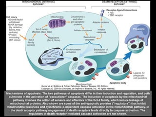
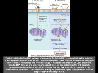
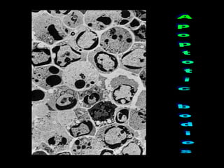
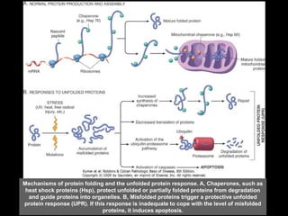
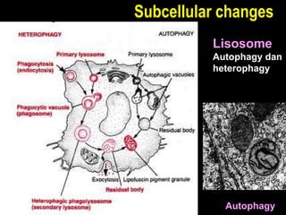
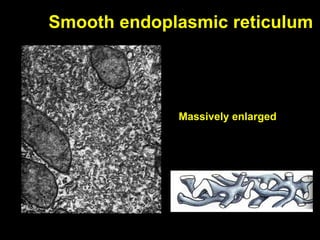
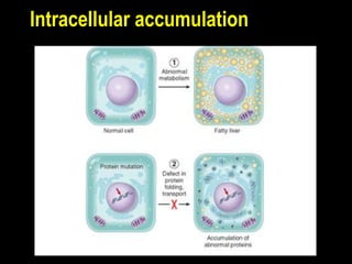
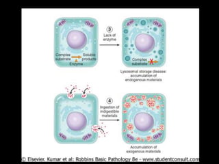
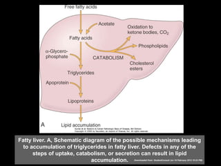
Cell Adaptation, cell Injury and
Cell Death
Mahmud Ghaznawie
Dept Pathology
Medical Faculty
Hasanuddin University
• Cellular adaptation to stress
• Hypertrophy
• Hyperplasia
• Atrophy
• Metaplasia
• Cell Injury and cell death
• Causes of cell injury
• Morphology of cell and tissue injury &
death
• Mechanisms of cell injury and death
• Necrosis and Apoptosis
• Intracellular accumulation
Learning Objectives
Plasma Membrane
Nucleus
Golgi Apparatus Mitochondria
Lisosome & peroxisome
The smooth endoplasmic reticulum
The rough endoplasmic reticulum
Cytoskeleton
MicrotubulesActin filaments
Intermediate
filaments
Cellular Adaptations of Growth
and Differentiation
• Hyperplasia
• Hypertrophy
• Atrophy
• Metaplasia
Hyperplasia
• An increase in the number of cells in an
organ or tissue
• Physiologic:
– Compensatory
– Hormonal
• Pathologic
– Pathologic hyperplasia constitutes a fertile soil in
which cancerous proliferation may eventually arise.
Hypertrophy
• an increase in the size of cells, resulting in an
increase in the size of the organ.
Atrophy
• a decrease in the size of an organ that
has reached its normal size
– Decreased workload (disuse atrophy)
– Loss of innervation (denervation atrophy)
– Diminished blood supply
– Inadequate nutrition
– Loss hormonal stimulation
– Senile atrophy
– Pressure atrophy
Metaplasia
• a reversible change in which one adult cell type
(epithelial or mesenchymal) is replaced by
another adult cell type
Causes of cell injury
• Hypoxia
• Free radicals
• Physical injury
• Chemical injury
• Infection
• Immune reaction
Inflammation Hypoxia
Chemical
Reperfusion
Radiation
Aging
Ischemia
Ischemic/hypoxic injury
Oxygen 
Oxydative phosphorilation 
ATP production 
Sodium pump 
Glycogenolysis
Ribosome detachment
Ischemic/hypoxic injury
Oxygen 
Oxydative phosphorilation 
ATP production 
Sodium pump 
Glycogenolysis
Ribosome detachment
Sodium pump 
Influx Ca ++ Na+ Retension Efflux K+
• Cell swollen
• Microvilli disappear
• Bleb formation
• ER swollen
• Myelin bodies
Ischemic/hypoxic injury
Oxygen 
Oxydative phosphorilation 
ATP production 
Sodium pump 
Glycogenolysis
Ribosome detachment
Glycogenolysis 
Lactic acid and inorganic
phosphate
pH 
Chromatin clumps
Ischemic/hypoxic injury
Oxygen 
Oxydative phosphorilation 
ATP production 
Sodium pump 
Glycogenolysis
Ribosome detachment
Detachment of ribosomes
Protein production 
Intracellular osmotic pressure 
Cell edema
Iskemia
Inflammation Hypoxia
Chemical
Reperfusion
Radiation
Aging
Injury due to Free Radicals
• Free Radicals: atoms or molecules possesing
unpaired electron in an outer orbit
• Characteristics of free radicals:
– react with any organic / inorganic substance
– the results will form a new free radicals  new
reaction chain
– the reaction will cease by itself or by enzymatic
reaction
• Three important free radicals:
– Superoxide anion radical (O2
÷)
– Hydrogen peroxide (H2O2)
– Hydroxyl ions(OH•)
• Effects of free radicals on cell membrane:
– Membrane lipid peroxidation (especially by OH•)
– Protein damage: cross-linking of amino acids,
increase protease activation
– DNA damage: single helix formation followed by cell
death of even malignant transformation (cancer)
De-activation of free radicals
• Spontaneous, because of its instability
• Endogenous/exogenous antioxidant
– Vitamine E, C and A
– Binding to storage & transport proteins (lactoferrin,
ceruloplasmine, dan trasferrin)
• Enzymatic
– Superoxide dismutase (SOD)
– Catalase
– Glutathione peroxidase
S.O.D,
Catalase,
and Gluthation peroxidase
are free radical-scavenging
enzymes
Chemical injury
• Water soluble
– Act directly (by combining with some critical molecular
component or cellular organelle)
– E.g: HgCl, cyanide, antibiotics, and chemotherapy
– Mercury binds to the sulfhydryl groups of the cell
membrane  increased membrane permeability and
inhibit ATPase-dependent transport
– Cyanide poisons mitochondrial cytochrome oxidase and
block oxidative phosphorylation
Chemical injury (cont)
• Lipid soluble
– Indirect effects (converted to reactive toxic
metabolites, which then act on target cells)
– E.g: CCl4
Myelin figures
ER swelling
Ribosomes
detachment
Mitochondrial
swelling
Small
densities
Blebs
Cell swelling
Chromatin
clumps
Autophagy
A
B
C
Reversible
Irreversible
Normal
ATP 
Phospholipid synthesis 
Ca++ 
Phospolipase
activation
Phospholipid
degradation
Cytoskeletal
damage
Membrane damage
Mechanisms membrane damage
(made simple)
Protease
activation
Membrane
defects
Myelin figures
Lysis of ER
Mitochondrial
swelling
Large densities
Nucleus
pyknosis
Rupture of
lysosomes
Cell Death
• Could be necrosis or apoptosis
• Necrosis
– Cell death in association to a living tissue
– When due to lisosomal enzymes: autolysis, due to
enzymes of immigrant cells: heterolysis.
– Autolysis  coagulative necrosis; heterolysis 
liquefactive necrosis
– Morphological changes occure within hours
The morphology of necrotic cells
• Cytoplasm:
– Eosinophillic (reaction to denatured proteins)
– Glassy appearance (due to loss of glykogen particles)
– Vacuolated (due to digestion of organelles)
– Calcification
• Nucleus: (3 possibilities)
– Pyknosis (due to nuclear shrinkage)
– Karyorhexis (fragmentation of the pyknotic nucleus)
– Karyolisis (basophilia of the chromatine fades)
Normal Necrosis
The cytoplasm
is more eosinophillic
Nuclei partially lysis
H & E staining
to show edema of the
myocardial fibres
LDH enzyme staining
to area unstained areas
Morphology of necrosis
Coagulative necrosis:
 The cell outlines are maintained
 Characteristic to hypoxic necrosis exept
on the brain.
 Occur because the
lysosomal enzymes we
also damaged
Liquefactive necrosis:
 Due to autolysis or heterolysis
 Characteristic to bacterial
infection (pus) and hypoxic
necrosis to the brain
 Gangrenous necrosis:
infected coagulative necrosis
(may then turns to liquefactive
necrosis)
Caseous necrosis
 Special form of coagulative necrosis,
spesific to tbc
 Macroscopically looks like “cheese”
 Microscopic:
amorphous mass,
granular, surrounded by
inflammatory cells
Enzymic fat
necrosis
 Destruction of fat due to pancreatic lipase
 Fatty acid formed will bind to calcium
 Microscopic: necrotic area, calcium
deposition (blue), and inflammation of the
surrounding tissue
Fibrinoid necrosis
Apoptosis
• Could be physiological or pathological
– “Programmed cell death” in embryogenesis, involusion of
hormon dependent organs, cell death in cancer, etc)
• Morphology:
– Shrinkage
– Chromatin condensation
– Formation of blebs and apoptotic bodies
– Phagocytosis of apoptotic bodies
Mechanisms of apoptosis. The two pathways of apoptosis differ in their induction and regulation, and both
culminate in the activation of "executioner" caspases. The induction of apoptosis by the mitochondrial
pathway involves the action of sensors and effectors of the Bcl-2 family, which induce leakage of
mitochondrial proteins. Also shown are some of the anti-apoptotic proteins ("regulators") that inhibit
mitochondrial leakiness and cytochrome c-dependent caspase activation in the mitochondrial pathway. In
the death receptor pathway engagement of death receptors leads directly to caspase activation. The
regulators of death receptor-mediated caspase activation are not shown.
The intrinsic (mitochondrial) pathway of apoptosis. A, Cell viability is maintained by the induction
of anti-apoptotic proteins such as Bcl-2 by survival signals. These proteins maintain the integrity of
mitochondrial membranes and prevent leakage of mitochondrial proteins. B, Loss of survival
signals, DNA damage, and other insults activate sensors that antagonize the anti-apoptotic
proteins and activate the pro-apoptotic proteins Bax and Bak, which form channels in the
mitochondrial membrane. The subsequent leakage of cytochrome c (and other proteins) leads to
caspase activation and apoptosis.
Mechanisms of protein folding and the unfolded protein response. A, Chaperones, such as
heat shock proteins (Hsp), protect unfolded or partially folded proteins from degradation
and guide proteins into organelles. B, Misfolded proteins trigger a protective unfolded
protein response (UPR). If this response is inadequate to cope with the level of misfolded
proteins, it induces apoptosis.
Subcellular changes
Autophagy
Lisosome
Autophagy dan
heterophagy
Smooth endoplasmic reticulum
Massively enlarged
Mitochondria
Enlarged
Intracellular accumulation
Fatty liver. A, Schematic diagram of the possible mechanisms leading
to accumulation of triglycerides in fatty liver. Defects in any of the
steps of uptake, catabolism, or secretion can result in lipid
accumulation. Downloaded from: StudentConsult (on 19 February 2012 10:23 PM)
Fatty change of the liver. In most cells the well-preserved nucleus is
squeezed into the displaced rim of cytoplasm about the fat vacuole.
Downloaded from: StudentConsult (on 19 February 2012 10:23 PM)
© 2005 Elsevier
Cholesterolosis. Cholesterol-laden macrophages (foam cells,
arrow) in a focus of gallbladder cholesterolosis.
Downloaded from: StudentConsult (on 19 February 2012 10:23 PM)
© 2005 Elsevier
Protein reabsorption droplets in the renal tubular epithelium.
Downloaded from: StudentConsult (on 19 February 2012 10:23 PM)
© 2005 Elsevier
Lipofuscin granules in a cardiac myocyte shown by light
microscopy Downloaded from: StudentConsult (on 19 February 2012 10:23 PM)
© 2005 Elsevier
Lipofuscin granules in a cardiac myocyte shown by electron
microscopy (note the perinuclear, intralysosomal location).
Downloaded from: StudentConsult (on 19 February 2012 10:23 PM)
© 2005 Elsevier
Hemosiderin granules in liver cells. H+E stain
showing golden-brown, finely granular pigment.Downloaded from: StudentConsult (on 19 February 2012 10:23 PM)
© 2005 Elsevier
Hemosiderin granules in liver cells. Prussian blue stain, specific for iron (seen
as blue granules). Downloaded from: StudentConsult (on 19 February 2012 10:23 PM)
© 2005 Elsevier
Dystrophic calcification of the aortic valve. View looking down onto the
unopened aortic valve in a heart with calcific aortic stenosis. It is markedly
narrowed (stenosis). The semilunar cusps are thickened and fibrotic, and
behind each cusp are irregular masses of piled-up dystrophic calcification.
© 2005 Elsevier
Conclusion
• Cell injury in the basis of any
pathologic processes
• It could be reversible or irreversible
(ended with cell death)
• The morphological changes are so
characteristic
• The mechanism of cell injury should
be beared in mind in your further
study of BMD and medicine
– Exam Questions on cell injury
– http://peir2.path.uab.edu/bmp/article_6.shtml
Mahmud Ghaznawie

Cell adaptation and injury

  • 1.
    Cell Adaptation, cellInjury and Cell Death Mahmud Ghaznawie Dept Pathology Medical Faculty Hasanuddin University
  • 2.
    • Cellular adaptationto stress • Hypertrophy • Hyperplasia • Atrophy • Metaplasia • Cell Injury and cell death • Causes of cell injury • Morphology of cell and tissue injury & death • Mechanisms of cell injury and death • Necrosis and Apoptosis • Intracellular accumulation Learning Objectives
  • 4.
  • 5.
  • 6.
    The smooth endoplasmicreticulum The rough endoplasmic reticulum
  • 7.
  • 8.
  • 12.
    Cellular Adaptations ofGrowth and Differentiation • Hyperplasia • Hypertrophy • Atrophy • Metaplasia
  • 13.
    Hyperplasia • An increasein the number of cells in an organ or tissue • Physiologic: – Compensatory – Hormonal • Pathologic – Pathologic hyperplasia constitutes a fertile soil in which cancerous proliferation may eventually arise.
  • 14.
    Hypertrophy • an increasein the size of cells, resulting in an increase in the size of the organ.
  • 15.
    Atrophy • a decreasein the size of an organ that has reached its normal size – Decreased workload (disuse atrophy) – Loss of innervation (denervation atrophy) – Diminished blood supply – Inadequate nutrition – Loss hormonal stimulation – Senile atrophy – Pressure atrophy
  • 17.
    Metaplasia • a reversiblechange in which one adult cell type (epithelial or mesenchymal) is replaced by another adult cell type
  • 20.
    Causes of cellinjury • Hypoxia • Free radicals • Physical injury • Chemical injury • Infection • Immune reaction
  • 21.
  • 22.
    Ischemic/hypoxic injury Oxygen  Oxydativephosphorilation  ATP production  Sodium pump  Glycogenolysis Ribosome detachment
  • 24.
    Ischemic/hypoxic injury Oxygen  Oxydativephosphorilation  ATP production  Sodium pump  Glycogenolysis Ribosome detachment
  • 25.
    Sodium pump  InfluxCa ++ Na+ Retension Efflux K+ • Cell swollen • Microvilli disappear • Bleb formation • ER swollen • Myelin bodies
  • 26.
    Ischemic/hypoxic injury Oxygen  Oxydativephosphorilation  ATP production  Sodium pump  Glycogenolysis Ribosome detachment
  • 27.
    Glycogenolysis  Lactic acidand inorganic phosphate pH  Chromatin clumps
  • 28.
    Ischemic/hypoxic injury Oxygen  Oxydativephosphorilation  ATP production  Sodium pump  Glycogenolysis Ribosome detachment
  • 29.
    Detachment of ribosomes Proteinproduction  Intracellular osmotic pressure  Cell edema
  • 30.
  • 31.
    Injury due toFree Radicals • Free Radicals: atoms or molecules possesing unpaired electron in an outer orbit • Characteristics of free radicals: – react with any organic / inorganic substance – the results will form a new free radicals  new reaction chain – the reaction will cease by itself or by enzymatic reaction
  • 32.
    • Three importantfree radicals: – Superoxide anion radical (O2 ÷) – Hydrogen peroxide (H2O2) – Hydroxyl ions(OH•) • Effects of free radicals on cell membrane: – Membrane lipid peroxidation (especially by OH•) – Protein damage: cross-linking of amino acids, increase protease activation – DNA damage: single helix formation followed by cell death of even malignant transformation (cancer)
  • 33.
    De-activation of freeradicals • Spontaneous, because of its instability • Endogenous/exogenous antioxidant – Vitamine E, C and A – Binding to storage & transport proteins (lactoferrin, ceruloplasmine, dan trasferrin) • Enzymatic – Superoxide dismutase (SOD) – Catalase – Glutathione peroxidase
  • 34.
    S.O.D, Catalase, and Gluthation peroxidase arefree radical-scavenging enzymes
  • 35.
    Chemical injury • Watersoluble – Act directly (by combining with some critical molecular component or cellular organelle) – E.g: HgCl, cyanide, antibiotics, and chemotherapy – Mercury binds to the sulfhydryl groups of the cell membrane  increased membrane permeability and inhibit ATPase-dependent transport – Cyanide poisons mitochondrial cytochrome oxidase and block oxidative phosphorylation
  • 36.
    Chemical injury (cont) •Lipid soluble – Indirect effects (converted to reactive toxic metabolites, which then act on target cells) – E.g: CCl4
  • 37.
  • 38.
  • 41.
    ATP  Phospholipid synthesis Ca++  Phospolipase activation Phospholipid degradation Cytoskeletal damage Membrane damage Mechanisms membrane damage (made simple) Protease activation
  • 42.
    Membrane defects Myelin figures Lysis ofER Mitochondrial swelling Large densities Nucleus pyknosis Rupture of lysosomes
  • 43.
    Cell Death • Couldbe necrosis or apoptosis • Necrosis – Cell death in association to a living tissue – When due to lisosomal enzymes: autolysis, due to enzymes of immigrant cells: heterolysis. – Autolysis  coagulative necrosis; heterolysis  liquefactive necrosis – Morphological changes occure within hours
  • 44.
    The morphology ofnecrotic cells • Cytoplasm: – Eosinophillic (reaction to denatured proteins) – Glassy appearance (due to loss of glykogen particles) – Vacuolated (due to digestion of organelles) – Calcification • Nucleus: (3 possibilities) – Pyknosis (due to nuclear shrinkage) – Karyorhexis (fragmentation of the pyknotic nucleus) – Karyolisis (basophilia of the chromatine fades)
  • 45.
    Normal Necrosis The cytoplasm ismore eosinophillic Nuclei partially lysis
  • 46.
    H & Estaining to show edema of the myocardial fibres LDH enzyme staining to area unstained areas
  • 47.
    Morphology of necrosis Coagulativenecrosis:  The cell outlines are maintained  Characteristic to hypoxic necrosis exept on the brain.  Occur because the lysosomal enzymes we also damaged
  • 49.
    Liquefactive necrosis:  Dueto autolysis or heterolysis  Characteristic to bacterial infection (pus) and hypoxic necrosis to the brain  Gangrenous necrosis: infected coagulative necrosis (may then turns to liquefactive necrosis)
  • 51.
    Caseous necrosis  Specialform of coagulative necrosis, spesific to tbc  Macroscopically looks like “cheese”  Microscopic: amorphous mass, granular, surrounded by inflammatory cells
  • 52.
    Enzymic fat necrosis  Destructionof fat due to pancreatic lipase  Fatty acid formed will bind to calcium  Microscopic: necrotic area, calcium deposition (blue), and inflammation of the surrounding tissue
  • 54.
  • 55.
    Apoptosis • Could bephysiological or pathological – “Programmed cell death” in embryogenesis, involusion of hormon dependent organs, cell death in cancer, etc) • Morphology: – Shrinkage – Chromatin condensation – Formation of blebs and apoptotic bodies – Phagocytosis of apoptotic bodies
  • 58.
    Mechanisms of apoptosis.The two pathways of apoptosis differ in their induction and regulation, and both culminate in the activation of "executioner" caspases. The induction of apoptosis by the mitochondrial pathway involves the action of sensors and effectors of the Bcl-2 family, which induce leakage of mitochondrial proteins. Also shown are some of the anti-apoptotic proteins ("regulators") that inhibit mitochondrial leakiness and cytochrome c-dependent caspase activation in the mitochondrial pathway. In the death receptor pathway engagement of death receptors leads directly to caspase activation. The regulators of death receptor-mediated caspase activation are not shown.
  • 59.
    The intrinsic (mitochondrial)pathway of apoptosis. A, Cell viability is maintained by the induction of anti-apoptotic proteins such as Bcl-2 by survival signals. These proteins maintain the integrity of mitochondrial membranes and prevent leakage of mitochondrial proteins. B, Loss of survival signals, DNA damage, and other insults activate sensors that antagonize the anti-apoptotic proteins and activate the pro-apoptotic proteins Bax and Bak, which form channels in the mitochondrial membrane. The subsequent leakage of cytochrome c (and other proteins) leads to caspase activation and apoptosis.
  • 61.
    Mechanisms of proteinfolding and the unfolded protein response. A, Chaperones, such as heat shock proteins (Hsp), protect unfolded or partially folded proteins from degradation and guide proteins into organelles. B, Misfolded proteins trigger a protective unfolded protein response (UPR). If this response is inadequate to cope with the level of misfolded proteins, it induces apoptosis.
  • 62.
  • 63.
  • 64.
  • 65.
  • 67.
    Fatty liver. A,Schematic diagram of the possible mechanisms leading to accumulation of triglycerides in fatty liver. Defects in any of the steps of uptake, catabolism, or secretion can result in lipid accumulation. Downloaded from: StudentConsult (on 19 February 2012 10:23 PM)
  • 68.
    Fatty change ofthe liver. In most cells the well-preserved nucleus is squeezed into the displaced rim of cytoplasm about the fat vacuole. Downloaded from: StudentConsult (on 19 February 2012 10:23 PM) © 2005 Elsevier
  • 69.
    Cholesterolosis. Cholesterol-laden macrophages(foam cells, arrow) in a focus of gallbladder cholesterolosis. Downloaded from: StudentConsult (on 19 February 2012 10:23 PM) © 2005 Elsevier
  • 70.
    Protein reabsorption dropletsin the renal tubular epithelium. Downloaded from: StudentConsult (on 19 February 2012 10:23 PM) © 2005 Elsevier
  • 71.
    Lipofuscin granules ina cardiac myocyte shown by light microscopy Downloaded from: StudentConsult (on 19 February 2012 10:23 PM) © 2005 Elsevier
  • 72.
    Lipofuscin granules ina cardiac myocyte shown by electron microscopy (note the perinuclear, intralysosomal location). Downloaded from: StudentConsult (on 19 February 2012 10:23 PM) © 2005 Elsevier
  • 73.
    Hemosiderin granules inliver cells. H+E stain showing golden-brown, finely granular pigment.Downloaded from: StudentConsult (on 19 February 2012 10:23 PM) © 2005 Elsevier
  • 74.
    Hemosiderin granules inliver cells. Prussian blue stain, specific for iron (seen as blue granules). Downloaded from: StudentConsult (on 19 February 2012 10:23 PM) © 2005 Elsevier
  • 75.
    Dystrophic calcification ofthe aortic valve. View looking down onto the unopened aortic valve in a heart with calcific aortic stenosis. It is markedly narrowed (stenosis). The semilunar cusps are thickened and fibrotic, and behind each cusp are irregular masses of piled-up dystrophic calcification. © 2005 Elsevier
  • 76.
    Conclusion • Cell injuryin the basis of any pathologic processes • It could be reversible or irreversible (ended with cell death) • The morphological changes are so characteristic • The mechanism of cell injury should be beared in mind in your further study of BMD and medicine
  • 77.
    – Exam Questionson cell injury – http://peir2.path.uab.edu/bmp/article_6.shtml
  • 78.