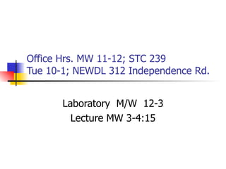
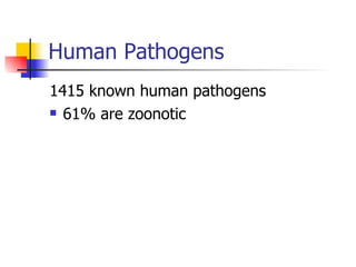
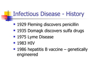
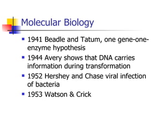
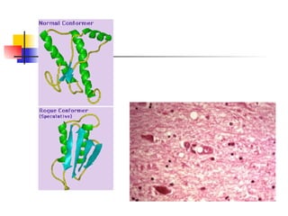
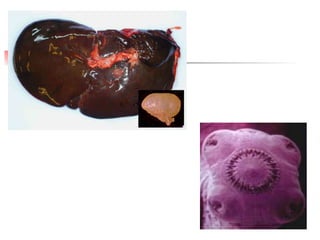
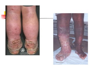
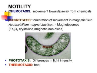
MICROBIOLOGY FALL 2011
Office Hrs. MW 11-12; STC 239 Tue 10-1; NEWDL 312 Independence Rd. Laboratory  M/W  12-3 Lecture MW 3-4:15
Syllabus
 
Dasek et al. 2000
Human Pathogens 1415 known human pathogens 61% are zoonotic
Wild Animals Involved in  MPXV Transmission (USA, 2003) Gambian Giant Rat (Crycetomys sp.) Prairie Dog (Cynomis sp.)
Ghana TX WI IL * IA 15/4 09/04 Entry and Distribution Route of  Crycetomys sp . and  Cynomys sp.  Involved in MPXV Outbreak (USA, 2003) Source: MMWR  52 (23), CDC, 2003. 15/4
Index case in Marshfield,  26 May 2003: Disseminated lesions.
  Distribution of MPXV Cases in the USA   WI IL IN OH KS MO Number of cases = 87 WI = 38 IN = 24 IL = 19 OH =  4 KS =  1 MO =  1 Source: MMWR  52 (23), CDC, 2003.
Microbiology Has been defined as the study of organisms and agents too small to be seen clearly by the naked eye – that is, the study of microorganisms
Scope and Relevance Microorganisms are exceptionally diverse, are found almost everywhere, and affect human society in countless ways.  Modern microbiology is a large discipline with many different specialties: medicine, agricultural & food sciences, ecology, genetics, and biochemistry.
Microbiology
Microbiology
Microbiology Spontaneous Generation Molecular Biology/Genetics Fermentation Infectious Disease
 
Infectious Disease - History 1798  Jenner, smallpox vaccine 1835-1844 Bassi, silkworm disease 1847-1850 Semmelweis 1849 Snow, cholera 1861 Pasteur disproves spontaneous generation 1867 Lister antiseptic surgery
 
 
Infectious Disease - History 1876-1877 Koch anthrax is caused by  Bacillus anthracis 1881 Pasteur develops anthrax vaccine 1884 Koch’s postulates published Autoclave developed Gram stain developed 1885 Pasteur develops rabies vaccine
Infectious Disease - History 1887 Petri dish developed 1890 Von Behring prepares antitoxins for diphtheria and tetanus 1899 Ross shows that mosquitoes carry malaria 1910 Ehrlich – magic bullet 1923 – Bergey’s Manual 1 st  edition
Infectious Disease - History 1929 Fleming discovers penicillin 1935 Domagk discovers sulfa drugs 1975 Lyme Disease 1983 HIV 1986 hepatitis B vaccine – genetically engineered
DISEASE Occurrence of Disease
Signs & Symptoms of Disease Fever Fatigue White Blood Cell Count Pain Muscle Aches Blood Pressure
Molecular Biology 1941 Beadle and Tatum, one gene-one-enzyme hypothesis 1944 Avery shows that DNA carries information during transformation 1952 Hershey and Chase viral infection of bacteria 1953 Watson & Crick
Molecular Biology - History 1961 Jacob & Monod propose the operon model of gene regulation 1961-1966 Nirenberg et al. elucidate the genetic code 1970 Arber & Smith – restriction endonucleases 2000 Human genome sequenced
Fermentation - History 1857 Pasteur shows that lactic fermentation is due to a microorganism 1897 Buchner prepares yeast extract that ferments
Spontaneous Generation 1799 Spallanzani attacks spontaneous generation 1861Pasteur disproves spontaneous generation
Kingdoms Animal Plant Fungi Protista Prokaryotic
Classification Kingdom Phylum Class  Order Family Genus species
 
Infectious Agents / Microrganisms Bacteria Fungi Prions Protozoans Helminths
Toxins – Exo and Endotoxins


Prokaryotes Greek – before a nucleus
 
Prokaryotes / Eukaryotes There are a number of differences between bacterial cells and plants, animals, fungi and protozoans.
Prokaryotes / Eukaryotes Size of cell 1-10 u m diameter 10-100 u m diameter Nucleus No nuclear membrane True nucleus Membrane bound organelles absent present Flagella 2 protein building blocks complex
Prokaryotes / Eukaryotes Glycocalyx Slime layer Absent Cell Wall Usually present Complex When present, simple Plasma membrane No CHO and lack sterols Sterols and CHO present Cytoplasm No cytoskeleton cytoskeleton
Prokaryotes / Eukaryotes Ribosomes Small size 70s Large 80s; small 70s  Chromosome Circular, lacks histones Linear, with histones Cell division Binary fission mitosis Sexual reproduction No meiosis meiosis
Bacterial Classification Cellular Characteristics Morphology – cell shape, cell size, arrangement of cells, arrangement of flagella, capsule, endospores


Cellular Characteristics Staining Reactions – Gram stain, acid-fast stain


Cellular Characteristics Growth and nutritional characteristics – appearance in liquid culture
Cellular Characteristics Growth and nutritional characteristics–colonial morphology
 
Cellular Characteristics Growth and nutritional characteristics  pigmentation
 
Cellular Characteristics Growth and nutritional characteristics energy sources, C, N sources,
Cellular Characteristics Growth and nutritional characteristics fermentation products
Cellular Characteristics Growth and nutritional characteristics modes of metabolism
 
Cellular Characteristics Biochemical Characteristics – cell wall constituents, pigment biochemicals, storage inclusions, antigens, RNA molecules
Cellular Characteristics Physiological and Ecological Characteristics – temperature range and optimum
Cellular Characteristics Physiological and Ecological Characteristics oxygen relationships
Cellular Characteristics Physiological and Ecological Characteristics – pH tolerance range
Cellular Characteristics Physiological and Ecological Characteristics –salt requirement and tolerance
 
Cellular Characteristics Genetic Characteristics- DNA G + C DNA hybridization
Fimbriae & Pili G- bacteria have short, fine, hairlike appendages that are thinner than flagella and not involved in motility
Fimbriae & Pili slender tubes composed of helically arranged protein subunits and are about 3 to 10 nm in diameter and up to several  u m long
FIMBRIA (s) FIMBRIAE (pl) Composition varies, contain protein Tendency to stick to each other and surfaces Bacterial attachment in aqueous environments Role in colonization    infection
 
PILUS (s) PILI (pl) Hollow, non-helical (9-10nm dia) Filamentous appendages Thinner than flagella, more numerous Example F-pilus ( SEX PILUS ) entry of genetic material during conjugation GRAM -VE BACTERIA ONLY


E.M Pili on  E. coli – N. gonorrhea
Pili & Fimbriae Some types of fimbriae attach bacteria to solid surfaces such as rocks in streams and host tissues Pili – about 1 to 10 per cell, differ from fimbriae: are larger (9 to 10 nm in diameter), they are genetically determined by sex factors or conjugative plasmids and are required for bacterial mating
Pili & Fimbriae Some bacterial viruses attach specifically to receptors on sex pili at the start of their reproductive cycle
Flagella & Motility Most motile bacteria move by use of flagella, threadlike, locomotor appendages extending outward from the plasma membrane and cell wall. Slender, rigid structures, about 20 nm across and up to 15 to 20  u m long
Flagella - Arrangements Monotrichous Amphitrichous Lophotrichous Peritrichous
Flagellar Ultrastructure Filament Hook Basal body
 
 
ARRANGEMENT OF FLAGELLA POLAR  - at one or both ends MONTRICHOUS  - single e.g.,  Vibrio  sp.  LOPHOTRICHOUS  - small tufts at same site  e.g.,  Pseudomonas  sp.
AMPHITRICHOUS  - at both poles, e.g.,  Spirillum  sp. LATERAL PERITRICHOUS  - surrounding entire cell, e.g.,  Proteus  sp.
MOTILITY CHEMOTAXIS:   movement towards/away from chemicals MAGNOTAXIS:   orientation of movement in magnetic field  Aquaspirillium magnetotacticum  - Magnetosomes  (Fe 3 O 4  crystalline magnetic iron oxide) PHOTOTAXIS:   Differences in light intensity THERMOTAXIS:  heat
Rotation of flagellar motor: reversible Clockwise (CW) or Counterclockwise (CCW) Smooth swimming/running motion Motor rotates CCW direction Flagella sweep around cell (in common axis)
Tumbling motion Motor reverse (CW direction) Flagella disperse
 
Flagellar Synthesis Complex process – involving at least 20 – 30 genes Flagellin subunits are transported through the filament’s hollow internal core. When they reach the tip, the subunits spontaneously aggregate so that the filament grows at its tip rather than at the base Self - assembly
The Mechanism of Flagellar Movement The filament is in the shape of a rigid helix, and the bacterium moves when this helix rotates
The Mechanism of Flagellar Movement Act like  propellers  on a boat
The Mechanism of Flagellar Movement E. coli  rotates 270 r.p.s.,  Vibrio alginolyticus  averages 1,100 r.p.s.
Distance and speed: 20-90  m/sec Equivalent to: 6ft human running 5 body lengths/second
AXIAL FILAMENTS Modified flagellum Long thin microfibril, inserted into a hook, entire structure enclosed in periplasmic space ENDOFLAGELLUM
 
CELL SURFACE 3 Basic Layers - GLYCOCALYX CELL WALL CELL MEMBRANE collectively termed  CELL ENVELOPE
Glycocalyx Is a network of polysaccharides extending from the surface of bacteria and other cells Aids in bacterial attachment to surfaces of solid objects in aquatic environments or to tissue surfaces in plant and animal hosts
GLYCOCALYX External mucilaginous layer Surrounds cell Shows organisation SLIME LAYER  - abundant, easily washed off    (poorly organised)  CAPSULE  - abundant, not easily washed off
Capsule Well organized and not easily washed off Composed of polysaccharides, but may be constructed of other materials i.e.  Bacillus anthracis  has a capsule of poly-D-glutamic acid Visible with the light microscope Resist phagocytosis


Capsules Contain a great deal of water – protect cell from desiccation Exclude viruses Exclude most hydrophobic toxic substances
FUNCTIONS Provide protection (drying) Block attachment of bacteriophages Antipathogenic (inhibit engulfment of pathogenic bacteria by WBC’s)  Contributes to  VIRULENCE  or  INFECTIVE ABILITY Promote attachment to surfaces Streptococcus mutans : adheres to teeth ( GLUCAN ),  DENTAL CARIES
Complement Activation Some capsules prevent formation of C3 convertase on the bacterial surface
Host response – Antibody  Subvert this type of protective host response by having capsules that resemble host polysaccharides.
CELL WALL Important in bacterial characteristics Determines shape Provides support/rigidity
STRUCTURE Composed of  PEPTIDOGLYCAN  (MUREIN) Insoluble, porous Cross-linked polymer (glycan), provides strength and rigidity N -acetyl Muramic Acid (NAM) N -acetyl Glucosamine (NAG)  1-4 glycosodic bonds
Differences in structure - basic principles of  GRAM STAIN REACTION Christian Gram 1884 Differential stain : ability of eubacterial cells to retain dye (crystal violet) after discolouration with 95% ethanol Cells retain stain:  Gram +ve  (thick cell walls)  PURPLE Cells lose stain:  Gram -ve  (thin cell walls)  RED


Peptidoglycan Structure Peptidoglycan or murein is an enormous polymer composed of many identical subunits.
Peptidoglycan Structure Most G- cell wall peptidoglycan lacks the peptide bridge.
Gram Negative Cell Walls More complex than the G+ cell walls. Peptidoglycan 5-10% of the wall weight. Braun’s lipoprotein – a small lipoprotein covalently joined to the underlying peptidoglycan and embedded in the outer membrane by its hydrophobic end. Lipopolysaccharides (LPSs)
 
Lipopolysaccharide Structure Known as  ENDOTOXIN Complex molecule: Inner most LIPID (Lipid A), achors LPS to outer membrane Polysaccharide portion (external to Lipid A) known as O-antigen O-polysaccharide long repeating sequence of sugars
LPSs Contain both lipid and carbohydrate Consist of three parts: lipid, the core polysaccharide and the O side chain
LPSs The lipid A region contains two glucosamine sugar derivatives, each with three fatty acids and phosphate or pyrophosphate attached.
LPSs Lipid A is buried in the outer membrane and the remainder of the LPS projects from the surface.
LPS The core polysaccharide is joined to lipid A. In  Salmonella  it is constructed of 10 sugars, many of them unusual in structure.
LPS The O side chain or O antigen is a short polysaccharide chain extending outward from the core. It has several peculiar sugars and varies in composition between bacterial strains.
LPS G- bacteria can rapidly change the nature of their O side chains to avoid detection. Contributes to the negative charge of the bacterial surface LPS helps stabilize membrane structure Lipid A is toxic – endotoxin Serves as a protective barrier
LPS – protective barrier Prevents or slows entry of bile salts, antibiotics, and other toxic substances.
Porin Proteins Cluster together and span the outer membrane to form a narrow channel through which molecules smaller than about 600 to 700 daltons can pass. .
Porin Proteins Larger molecules such as vitamin B 12  must be transported across the outer membrane by specific carriers. The outer membrane also prevents the loss of constituents like periplasmic enzymes.
COMPARISON OF GRM+VE /GRM-VE CELL WALLS
 
 
Periplasmic Space Contains enzymes involved in peptidoglycan synthesis and the modification of toxic compounds that could harm the cell.
Periplasmic Space Gram – contains many proteins that participate in nutrient acquisition.  Ex. hydrolytic enzymes attacking nucleic acids and phosphorylated molecules, and binding proteins involved in transport of materials into the cell. Denitrifying and chemolithoautotrophic bacteria – electron transport proteins in periplasm


CELL/CYTOPLASMIC MEMBRANE 4-5nm thick composed 1 o  phospholipids 30-40%  and protein 60-70% Phospholipid bilayer: Polar heads  (outwards into aqueous phase - membrane surface) Fatty acyl tails  (inwards - semi/liquid phase at interior)
FLUID MOSAIC MEMBRANE Davison & Danelli
ALSO CONTAINS: PROTEINS INTEGRAL  - removed by destruction i.e., Detergents PERIPHERAL  - loosely attached, easily removed i.e., Osmotic shock
 
FUNCTION Transport - control nutrients Oxidative phosphorylation (Respiration) Secretion - discharge of metabolic products Anchoring DNA (during cell division) Metabolism - enzyme sites
INTERNAL CONTENTS Cell Material divided into PROTOPLASM Granular appearance Site of biochemical activity Water 70-80% acts as solvent for nutrients, sugars, Aa’s & salts
CHROMATIN AREA no distinct membrane enclosed nucleus no mitotic apparatus BACTERIAL CHROMOSOME Typically single circular strand of DNA ( CHROMATIN   BODY ) Exception  Streptomyces  &  Borrelia  sp (Linear)  Rhodobacter sphaeroides  (2 separate chromosomes) all genes are linked Aggregated in one area ( NUCLEOID )
Bacterial Chromosome
PLASMIDS Additional to chromosome 1 or more, small circular macromolecules of DNA Capable of self-replication
Types: Fertility (F-plasmid):  genes for mating in conjugation Resistance (R-plasmids):  antibiotics, metals
Virulence factor:  enterotoxin, fimbriae, antibiotic production Colicinogenic (col-plasmids):  gene for protein ( COLICINS ) toxic to closely related bacteria (eliminates competitors) Transformation (Ti-plasmids):  plant microbiology (formation of crown gall tumors)  Agrobacterium   Metabolic:  utilization of camphor, toluene
OTHER FEATURES RIBOSOMES Located in Protoplasm RNA/PROTEIN bodies Composed of 2 sub units (70S)  Svedberg Units Sites of Protein Synthesis
MESOSOMES: Extensive invaginations (infoldings) of cyto membrane Continuous with membrane Function NOT KNOWN Corynebacterium parvum
INCLUSIONS/VACUOLES compensate for poor availability of nutrients present in Protoplast VOLUTIN GRANULES/METACHROMATIC  ( coloured ) composed of  POLYPHOSPHATE energy rich storage structures
VOLUTIN i.e., POLY-  -HYDROXYBUTYRATE serve as carbon and energy source METACHROMATIC i.e., Aquatic bacteria - colored crystals  ( Blue  or  Red  dyes) e.g.,  Corynebacterium
Cyanobacterium:  Microcystis  (12,600x) Gas vacuoles - blue Storage granules - red


Introduction structure

  • 1.
  • 2.
    Office Hrs. MW11-12; STC 239 Tue 10-1; NEWDL 312 Independence Rd. Laboratory M/W 12-3 Lecture MW 3-4:15
  • 3.
  • 4.
  • 5.
  • 6.
    Human Pathogens 1415known human pathogens 61% are zoonotic
  • 7.
    Wild Animals Involvedin MPXV Transmission (USA, 2003) Gambian Giant Rat (Crycetomys sp.) Prairie Dog (Cynomis sp.)
  • 8.
    Ghana TX WIIL * IA 15/4 09/04 Entry and Distribution Route of Crycetomys sp . and Cynomys sp. Involved in MPXV Outbreak (USA, 2003) Source: MMWR 52 (23), CDC, 2003. 15/4
  • 9.
    Index case inMarshfield, 26 May 2003: Disseminated lesions.
  • 10.
    Distributionof MPXV Cases in the USA WI IL IN OH KS MO Number of cases = 87 WI = 38 IN = 24 IL = 19 OH = 4 KS = 1 MO = 1 Source: MMWR 52 (23), CDC, 2003.
  • 11.
    Microbiology Has beendefined as the study of organisms and agents too small to be seen clearly by the naked eye – that is, the study of microorganisms
  • 12.
    Scope and RelevanceMicroorganisms are exceptionally diverse, are found almost everywhere, and affect human society in countless ways. Modern microbiology is a large discipline with many different specialties: medicine, agricultural & food sciences, ecology, genetics, and biochemistry.
  • 13.
  • 14.
  • 15.
    Microbiology Spontaneous GenerationMolecular Biology/Genetics Fermentation Infectious Disease
  • 16.
  • 17.
    Infectious Disease -History 1798 Jenner, smallpox vaccine 1835-1844 Bassi, silkworm disease 1847-1850 Semmelweis 1849 Snow, cholera 1861 Pasteur disproves spontaneous generation 1867 Lister antiseptic surgery
  • 18.
  • 19.
  • 20.
    Infectious Disease -History 1876-1877 Koch anthrax is caused by Bacillus anthracis 1881 Pasteur develops anthrax vaccine 1884 Koch’s postulates published Autoclave developed Gram stain developed 1885 Pasteur develops rabies vaccine
  • 21.
    Infectious Disease -History 1887 Petri dish developed 1890 Von Behring prepares antitoxins for diphtheria and tetanus 1899 Ross shows that mosquitoes carry malaria 1910 Ehrlich – magic bullet 1923 – Bergey’s Manual 1 st edition
  • 22.
    Infectious Disease -History 1929 Fleming discovers penicillin 1935 Domagk discovers sulfa drugs 1975 Lyme Disease 1983 HIV 1986 hepatitis B vaccine – genetically engineered
  • 23.
  • 24.
    Signs & Symptomsof Disease Fever Fatigue White Blood Cell Count Pain Muscle Aches Blood Pressure
  • 25.
    Molecular Biology 1941Beadle and Tatum, one gene-one-enzyme hypothesis 1944 Avery shows that DNA carries information during transformation 1952 Hershey and Chase viral infection of bacteria 1953 Watson & Crick
  • 26.
    Molecular Biology -History 1961 Jacob & Monod propose the operon model of gene regulation 1961-1966 Nirenberg et al. elucidate the genetic code 1970 Arber & Smith – restriction endonucleases 2000 Human genome sequenced
  • 27.
    Fermentation - History1857 Pasteur shows that lactic fermentation is due to a microorganism 1897 Buchner prepares yeast extract that ferments
  • 28.
    Spontaneous Generation 1799Spallanzani attacks spontaneous generation 1861Pasteur disproves spontaneous generation
  • 29.
    Kingdoms Animal PlantFungi Protista Prokaryotic
  • 30.
    Classification Kingdom PhylumClass Order Family Genus species
  • 31.
  • 32.
    Infectious Agents /Microrganisms Bacteria Fungi Prions Protozoans Helminths
  • 33.
    Toxins – Exoand Endotoxins
  • 34.
  • 35.
  • 36.
  • 37.
  • 38.
  • 39.
  • 40.
  • 41.
  • 42.
  • 43.
  • 44.
  • 45.
  • 46.
  • 47.
  • 48.
  • 49.
    Prokaryotes Greek –before a nucleus
  • 50.
  • 51.
    Prokaryotes / EukaryotesThere are a number of differences between bacterial cells and plants, animals, fungi and protozoans.
  • 52.
    Prokaryotes / EukaryotesSize of cell 1-10 u m diameter 10-100 u m diameter Nucleus No nuclear membrane True nucleus Membrane bound organelles absent present Flagella 2 protein building blocks complex
  • 53.
    Prokaryotes / EukaryotesGlycocalyx Slime layer Absent Cell Wall Usually present Complex When present, simple Plasma membrane No CHO and lack sterols Sterols and CHO present Cytoplasm No cytoskeleton cytoskeleton
  • 54.
    Prokaryotes / EukaryotesRibosomes Small size 70s Large 80s; small 70s Chromosome Circular, lacks histones Linear, with histones Cell division Binary fission mitosis Sexual reproduction No meiosis meiosis
  • 55.
    Bacterial Classification CellularCharacteristics Morphology – cell shape, cell size, arrangement of cells, arrangement of flagella, capsule, endospores
  • 56.
  • 57.
  • 58.
  • 59.
    Cellular Characteristics StainingReactions – Gram stain, acid-fast stain
  • 60.
  • 61.
  • 62.
  • 63.
  • 64.
  • 65.
  • 66.
  • 67.
  • 68.
  • 69.
  • 70.
  • 71.
  • 72.
  • 73.
  • 74.
    Cellular Characteristics Growthand nutritional characteristics – appearance in liquid culture
  • 75.
    Cellular Characteristics Growthand nutritional characteristics–colonial morphology
  • 76.
  • 77.
    Cellular Characteristics Growthand nutritional characteristics pigmentation
  • 78.
  • 79.
    Cellular Characteristics Growthand nutritional characteristics energy sources, C, N sources,
  • 80.
    Cellular Characteristics Growthand nutritional characteristics fermentation products
  • 81.
    Cellular Characteristics Growthand nutritional characteristics modes of metabolism
  • 82.
  • 83.
    Cellular Characteristics BiochemicalCharacteristics – cell wall constituents, pigment biochemicals, storage inclusions, antigens, RNA molecules
  • 84.
    Cellular Characteristics Physiologicaland Ecological Characteristics – temperature range and optimum
  • 85.
    Cellular Characteristics Physiologicaland Ecological Characteristics oxygen relationships
  • 86.
    Cellular Characteristics Physiologicaland Ecological Characteristics – pH tolerance range
  • 87.
    Cellular Characteristics Physiologicaland Ecological Characteristics –salt requirement and tolerance
  • 88.
  • 89.
    Cellular Characteristics GeneticCharacteristics- DNA G + C DNA hybridization
  • 90.
    Fimbriae & PiliG- bacteria have short, fine, hairlike appendages that are thinner than flagella and not involved in motility
  • 91.
    Fimbriae & Pilislender tubes composed of helically arranged protein subunits and are about 3 to 10 nm in diameter and up to several u m long
  • 92.
    FIMBRIA (s) FIMBRIAE(pl) Composition varies, contain protein Tendency to stick to each other and surfaces Bacterial attachment in aqueous environments Role in colonization  infection
  • 93.
  • 94.
    PILUS (s) PILI(pl) Hollow, non-helical (9-10nm dia) Filamentous appendages Thinner than flagella, more numerous Example F-pilus ( SEX PILUS ) entry of genetic material during conjugation GRAM -VE BACTERIA ONLY
  • 95.
  • 96.
  • 97.
  • 98.
    E.M Pili on E. coli – N. gonorrhea
  • 99.
    Pili & FimbriaeSome types of fimbriae attach bacteria to solid surfaces such as rocks in streams and host tissues Pili – about 1 to 10 per cell, differ from fimbriae: are larger (9 to 10 nm in diameter), they are genetically determined by sex factors or conjugative plasmids and are required for bacterial mating
  • 100.
    Pili & FimbriaeSome bacterial viruses attach specifically to receptors on sex pili at the start of their reproductive cycle
  • 101.
    Flagella & MotilityMost motile bacteria move by use of flagella, threadlike, locomotor appendages extending outward from the plasma membrane and cell wall. Slender, rigid structures, about 20 nm across and up to 15 to 20 u m long
  • 102.
    Flagella - ArrangementsMonotrichous Amphitrichous Lophotrichous Peritrichous
  • 103.
  • 104.
  • 105.
  • 106.
    ARRANGEMENT OF FLAGELLAPOLAR - at one or both ends MONTRICHOUS - single e.g., Vibrio sp. LOPHOTRICHOUS - small tufts at same site e.g., Pseudomonas sp.
  • 107.
    AMPHITRICHOUS -at both poles, e.g., Spirillum sp. LATERAL PERITRICHOUS - surrounding entire cell, e.g., Proteus sp.
  • 108.
    MOTILITY CHEMOTAXIS: movement towards/away from chemicals MAGNOTAXIS: orientation of movement in magnetic field Aquaspirillium magnetotacticum - Magnetosomes (Fe 3 O 4 crystalline magnetic iron oxide) PHOTOTAXIS: Differences in light intensity THERMOTAXIS: heat
  • 109.
    Rotation of flagellarmotor: reversible Clockwise (CW) or Counterclockwise (CCW) Smooth swimming/running motion Motor rotates CCW direction Flagella sweep around cell (in common axis)
  • 110.
    Tumbling motion Motorreverse (CW direction) Flagella disperse
  • 111.
  • 112.
    Flagellar Synthesis Complexprocess – involving at least 20 – 30 genes Flagellin subunits are transported through the filament’s hollow internal core. When they reach the tip, the subunits spontaneously aggregate so that the filament grows at its tip rather than at the base Self - assembly
  • 113.
    The Mechanism ofFlagellar Movement The filament is in the shape of a rigid helix, and the bacterium moves when this helix rotates
  • 114.
    The Mechanism ofFlagellar Movement Act like propellers on a boat
  • 115.
    The Mechanism ofFlagellar Movement E. coli rotates 270 r.p.s., Vibrio alginolyticus averages 1,100 r.p.s.
  • 116.
    Distance and speed:20-90  m/sec Equivalent to: 6ft human running 5 body lengths/second
  • 117.
    AXIAL FILAMENTS Modifiedflagellum Long thin microfibril, inserted into a hook, entire structure enclosed in periplasmic space ENDOFLAGELLUM
  • 118.
  • 119.
    CELL SURFACE 3Basic Layers - GLYCOCALYX CELL WALL CELL MEMBRANE collectively termed CELL ENVELOPE
  • 120.
    Glycocalyx Is anetwork of polysaccharides extending from the surface of bacteria and other cells Aids in bacterial attachment to surfaces of solid objects in aquatic environments or to tissue surfaces in plant and animal hosts
  • 121.
    GLYCOCALYX External mucilaginouslayer Surrounds cell Shows organisation SLIME LAYER - abundant, easily washed off (poorly organised) CAPSULE - abundant, not easily washed off
  • 122.
    Capsule Well organizedand not easily washed off Composed of polysaccharides, but may be constructed of other materials i.e. Bacillus anthracis has a capsule of poly-D-glutamic acid Visible with the light microscope Resist phagocytosis
  • 123.
  • 124.
  • 125.
  • 126.
    Capsules Contain agreat deal of water – protect cell from desiccation Exclude viruses Exclude most hydrophobic toxic substances
  • 127.
    FUNCTIONS Provide protection(drying) Block attachment of bacteriophages Antipathogenic (inhibit engulfment of pathogenic bacteria by WBC’s) Contributes to VIRULENCE or INFECTIVE ABILITY Promote attachment to surfaces Streptococcus mutans : adheres to teeth ( GLUCAN ), DENTAL CARIES
  • 128.
    Complement Activation Somecapsules prevent formation of C3 convertase on the bacterial surface
  • 129.
    Host response –Antibody Subvert this type of protective host response by having capsules that resemble host polysaccharides.
  • 130.
    CELL WALL Importantin bacterial characteristics Determines shape Provides support/rigidity
  • 131.
    STRUCTURE Composed of PEPTIDOGLYCAN (MUREIN) Insoluble, porous Cross-linked polymer (glycan), provides strength and rigidity N -acetyl Muramic Acid (NAM) N -acetyl Glucosamine (NAG)  1-4 glycosodic bonds
  • 132.
    Differences in structure- basic principles of GRAM STAIN REACTION Christian Gram 1884 Differential stain : ability of eubacterial cells to retain dye (crystal violet) after discolouration with 95% ethanol Cells retain stain: Gram +ve (thick cell walls) PURPLE Cells lose stain: Gram -ve (thin cell walls) RED
  • 133.
  • 134.
  • 135.
  • 136.
  • 137.
    Peptidoglycan Structure Peptidoglycanor murein is an enormous polymer composed of many identical subunits.
  • 138.
    Peptidoglycan Structure MostG- cell wall peptidoglycan lacks the peptide bridge.
  • 139.
    Gram Negative CellWalls More complex than the G+ cell walls. Peptidoglycan 5-10% of the wall weight. Braun’s lipoprotein – a small lipoprotein covalently joined to the underlying peptidoglycan and embedded in the outer membrane by its hydrophobic end. Lipopolysaccharides (LPSs)
  • 140.
  • 141.
    Lipopolysaccharide Structure Knownas ENDOTOXIN Complex molecule: Inner most LIPID (Lipid A), achors LPS to outer membrane Polysaccharide portion (external to Lipid A) known as O-antigen O-polysaccharide long repeating sequence of sugars
  • 142.
    LPSs Contain bothlipid and carbohydrate Consist of three parts: lipid, the core polysaccharide and the O side chain
  • 143.
    LPSs The lipidA region contains two glucosamine sugar derivatives, each with three fatty acids and phosphate or pyrophosphate attached.
  • 144.
    LPSs Lipid Ais buried in the outer membrane and the remainder of the LPS projects from the surface.
  • 145.
    LPS The corepolysaccharide is joined to lipid A. In Salmonella it is constructed of 10 sugars, many of them unusual in structure.
  • 146.
    LPS The Oside chain or O antigen is a short polysaccharide chain extending outward from the core. It has several peculiar sugars and varies in composition between bacterial strains.
  • 147.
    LPS G- bacteriacan rapidly change the nature of their O side chains to avoid detection. Contributes to the negative charge of the bacterial surface LPS helps stabilize membrane structure Lipid A is toxic – endotoxin Serves as a protective barrier
  • 148.
    LPS – protectivebarrier Prevents or slows entry of bile salts, antibiotics, and other toxic substances.
  • 149.
    Porin Proteins Clustertogether and span the outer membrane to form a narrow channel through which molecules smaller than about 600 to 700 daltons can pass. .
  • 150.
    Porin Proteins Largermolecules such as vitamin B 12 must be transported across the outer membrane by specific carriers. The outer membrane also prevents the loss of constituents like periplasmic enzymes.
  • 151.
    COMPARISON OF GRM+VE/GRM-VE CELL WALLS
  • 152.
  • 153.
  • 154.
    Periplasmic Space Containsenzymes involved in peptidoglycan synthesis and the modification of toxic compounds that could harm the cell.
  • 155.
    Periplasmic Space Gram– contains many proteins that participate in nutrient acquisition. Ex. hydrolytic enzymes attacking nucleic acids and phosphorylated molecules, and binding proteins involved in transport of materials into the cell. Denitrifying and chemolithoautotrophic bacteria – electron transport proteins in periplasm
  • 156.
  • 157.
  • 158.
  • 159.
    CELL/CYTOPLASMIC MEMBRANE 4-5nmthick composed 1 o phospholipids 30-40% and protein 60-70% Phospholipid bilayer: Polar heads (outwards into aqueous phase - membrane surface) Fatty acyl tails (inwards - semi/liquid phase at interior)
  • 160.
    FLUID MOSAIC MEMBRANEDavison & Danelli
  • 161.
    ALSO CONTAINS: PROTEINSINTEGRAL - removed by destruction i.e., Detergents PERIPHERAL - loosely attached, easily removed i.e., Osmotic shock
  • 162.
  • 163.
    FUNCTION Transport -control nutrients Oxidative phosphorylation (Respiration) Secretion - discharge of metabolic products Anchoring DNA (during cell division) Metabolism - enzyme sites
  • 164.
    INTERNAL CONTENTS CellMaterial divided into PROTOPLASM Granular appearance Site of biochemical activity Water 70-80% acts as solvent for nutrients, sugars, Aa’s & salts
  • 165.
    CHROMATIN AREA nodistinct membrane enclosed nucleus no mitotic apparatus BACTERIAL CHROMOSOME Typically single circular strand of DNA ( CHROMATIN BODY ) Exception Streptomyces & Borrelia sp (Linear) Rhodobacter sphaeroides (2 separate chromosomes) all genes are linked Aggregated in one area ( NUCLEOID )
  • 166.
  • 167.
    PLASMIDS Additional tochromosome 1 or more, small circular macromolecules of DNA Capable of self-replication
  • 168.
    Types: Fertility (F-plasmid): genes for mating in conjugation Resistance (R-plasmids): antibiotics, metals
  • 169.
    Virulence factor: enterotoxin, fimbriae, antibiotic production Colicinogenic (col-plasmids): gene for protein ( COLICINS ) toxic to closely related bacteria (eliminates competitors) Transformation (Ti-plasmids): plant microbiology (formation of crown gall tumors) Agrobacterium Metabolic: utilization of camphor, toluene
  • 170.
    OTHER FEATURES RIBOSOMESLocated in Protoplasm RNA/PROTEIN bodies Composed of 2 sub units (70S) Svedberg Units Sites of Protein Synthesis
  • 171.
    MESOSOMES: Extensive invaginations(infoldings) of cyto membrane Continuous with membrane Function NOT KNOWN Corynebacterium parvum
  • 172.
    INCLUSIONS/VACUOLES compensate forpoor availability of nutrients present in Protoplast VOLUTIN GRANULES/METACHROMATIC ( coloured ) composed of POLYPHOSPHATE energy rich storage structures
  • 173.
    VOLUTIN i.e., POLY- -HYDROXYBUTYRATE serve as carbon and energy source METACHROMATIC i.e., Aquatic bacteria - colored crystals ( Blue or Red dyes) e.g., Corynebacterium
  • 174.
    Cyanobacterium: Microcystis (12,600x) Gas vacuoles - blue Storage granules - red
  • 175.
  • 176.