Downloaded 130 times

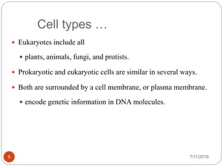
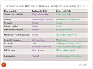
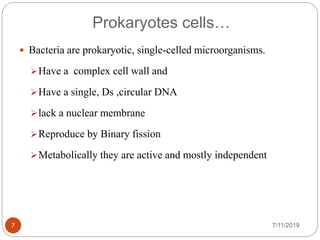
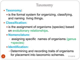
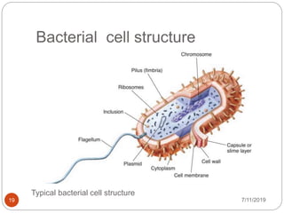
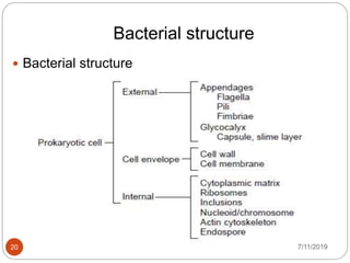


The document provides an overview of prokaryotic and eukaryotic cells, detailing their characteristics, differences, and similarities. It further discusses the taxonomy of bacteria, including classification levels, nomenclature, and methods of identification, as well as the morphology and structure of bacterial cells, including their cell wall and appendages like flagella and pili. Additionally, it explores endospores' structure, formation, and germination processes in bacteria.


