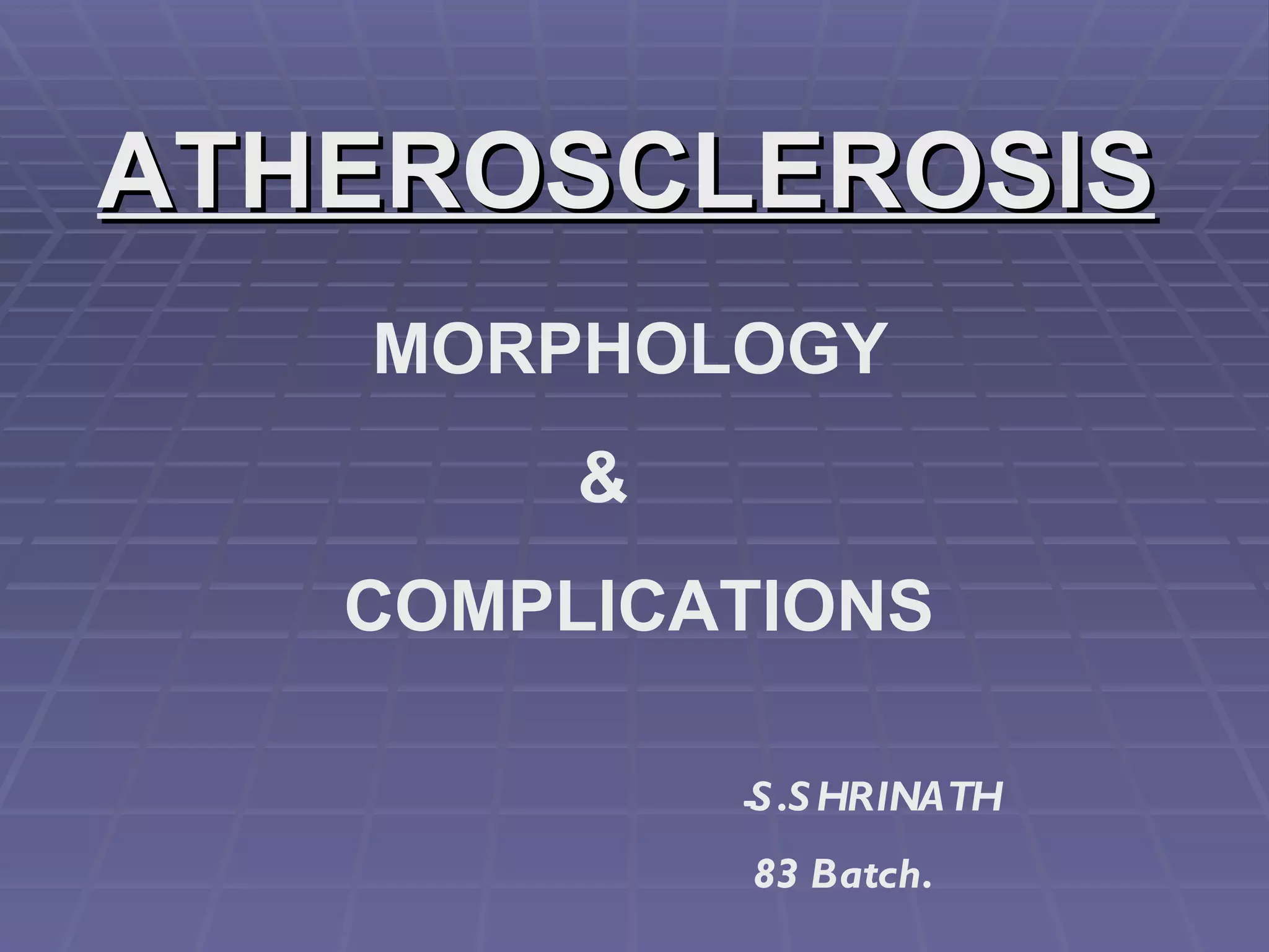
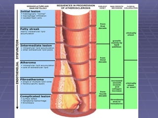
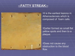
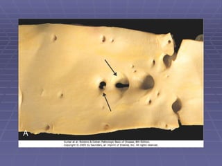
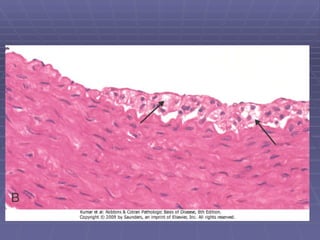
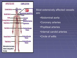
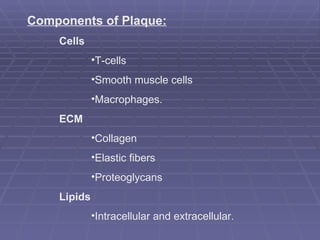
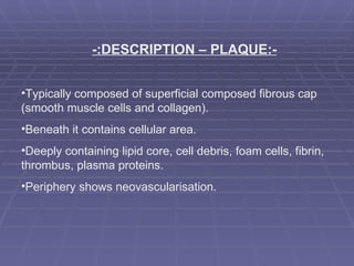
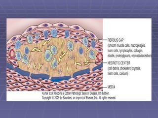
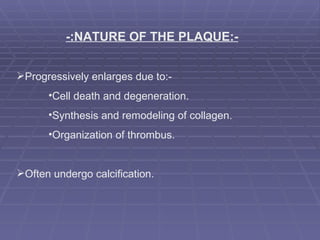
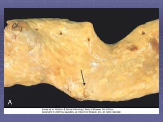
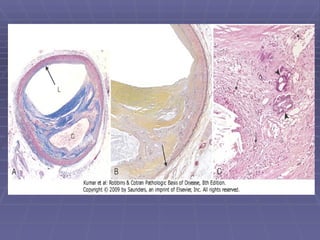
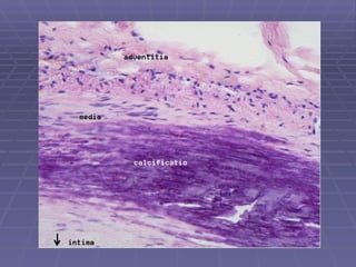
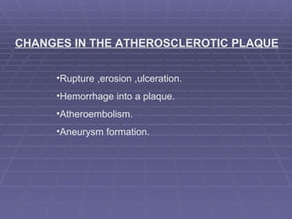
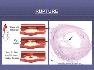
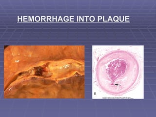
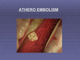
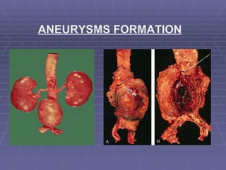
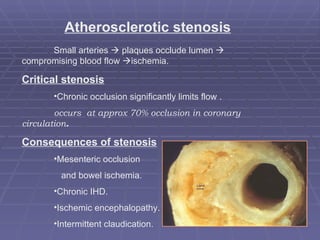
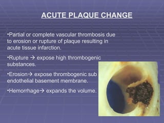
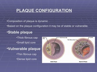
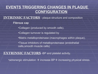
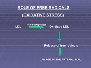
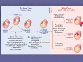
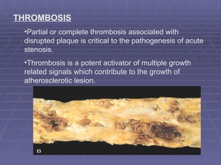
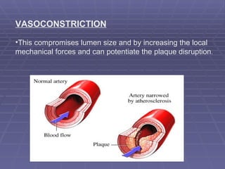
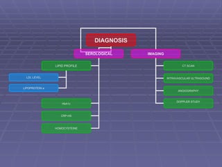
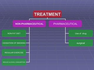
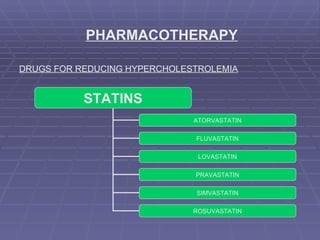
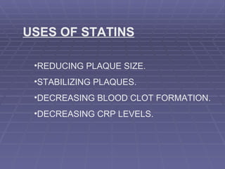
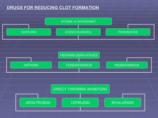
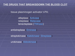
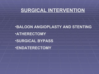
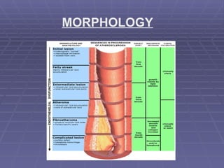
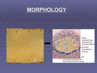
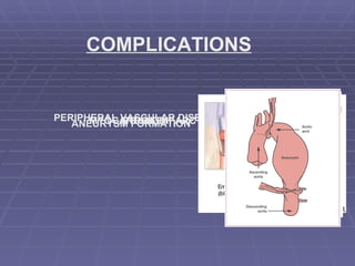
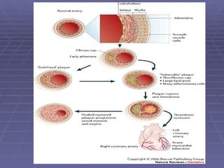
ATHEROSCLEROSIS MORPHOLOGY    &   COMPLICATIONS S.SHRINATH  83 Batch.
 
MORPHOLOGY FATTY STREAK ATHEROSCLEROTIC  PLAQUE
-:FATTY STREAK:- It is the earliest lesions in Atherosclerosis which is composed of  foam cells. Earlier formed as small flat yellow spots and then to a plaque. Does not cause any obstruction to the blood flow.
 
 
-:ATHEROSCLEROTIC PLAQUE:- Intimal thickening and lipid accumulation. Lodged in the lumen of the artery (0.3-1.5 cm in dia). Grossly they appear as white to yellow patches. Lesions are mostly eccentric but rarely circumferential. Local flow disturbances –increased susceptibility to plaque  formation.
Most extensively affected vessels are: Abdominal aorta  Coronary arteries  Popliteal arteries Internal carotid arteries Circle of willis
Components of Plaque: Cells T-cells Smooth muscle cells  Macrophages. ECM Collagen  Elastic fibers Proteoglycans Lipids Intracellular and extracellular.
-:DESCRIPTION – PLAQUE:- Typically composed of superficial composed fibrous cap  (smooth muscle cells and collagen). Beneath it contains cellular area.  Deeply containing lipid core, cell debris, foam cells, fibrin, thrombus, plasma proteins. Periphery shows neovascularisation.
 
  -:NATURE OF THE PLAQUE:- Progressively enlarges due to:-  Cell death and degeneration. Synthesis and remodeling of collagen. Organization of thrombus. Often undergo calcification.


CHANGES IN THE ATHEROSCLEROTIC PLAQUE Rupture ,erosion ,ulceration. Hemorrhage into a plaque. Atheroembolism. Aneurysm formation.
RUPTURE
HEMORRHAGE INTO PLAQUE
ATHERO EMBOLISM
ANEURYSMS FORMATION
COMPLICATIONS
-:COMPLICATIONS:- Myocardial infarction  Cerebral infarction (stroke). Aneurysm  Peripheral vascular disease (PVD).
Atherosclerotic stenosis Small arteries    plaques occlude lumen    compromising blood flow   ischemia. Critical stenosis Chronic occlusion significantly limits flow . occurs  at approx 70% occlusion in coronary  circulation . Consequences of stenosis Mesenteric occlusion  and bowel ischemia. Chronic IHD. Ischemic encephalopathy. Intermittent claudication.
ACUTE PLAQUE CHANGE Partial or complete vascular thrombosis due to erosion or rupture of plaque resulting in acute tissue infarction. Rupture    expose high thrombogenic substances. Erosion   expose thrombogenic sub endothelial basement membrane. Hemorrhage   expands the volume.
PLAQUE CONFIGURATION Composition of plaque is dynamic . Based on the plaque configuration it may be of stable or vulnerable. Stable plaque Thick fibrous cap Small lipid core Vulnerable plaque Thin fibrous cap Dense lipid core
EVENTS TRIGGERING CHANGES IN PLAQUE  CONFIGURATION INTRINSIC FACTORS  - plaque structure and composition Fibrous cap Collagen (produced by smooth cells). Collagen turnover is regulated by  Matrix metalloproteinase (macrophages within plaque), Tissue inhibitors of metalloproteinase (endothelial  cells,smooth muscle cells) EXTRINSIC FACTORS  -BP and platelet activity. adrenergic stimulation    increase BP   increasing physical stress.
ROLE OF FREE RADICALS (OXIDATIVE STRESS) LDL Oxidised LDL Release of free radicals DAMAGE TO THE ARTERIAL WALL Into macrophages
 
THROMBOSIS Partial or complete thrombosis associated with disrupted plaque is critical to the pathogenesis of acute stenosis. Thrombosis is a potent activator of multiple growth related signals which contribute to the growth of atherosclerotic lesion.
VASOCONSTRICTION This compromises lumen size and by increasing the local mechanical forces and can potentiate the plaque disruption .
CLINICAL FEATURES ON CORONARY ARTERIES: Angina Hyperhidrosis Shortness of breath Palpitations Tachycardia Weakness or dizziness Nausea ON CAROTID ARTERIES: Transient ischemic attacks  Dizziness ,Confusion ,Fainting , Coma Loss of eyesight Hemiplegia.
Claudication is the most common symptom of this condition .  Pain  Coolness, numbness Poor healing of wounds   Ulcers leading to Gangrene formation Black discoloration ON PERIPHERAL ARTERIES:
DIAGNOSIS
DIAGNOSIS SEROLOGICAL IMAGING LIPID PROFILE   HbA1c   CRP-HS HOMOCYSTEINE LDL LEVEL LIPOPROTEIN a CT SCAN INTRAVASCULAR ULTRASOUND ANGIOGRAPHY DOPPLER STUDY
TREATMENT
TREATMENT NON-PHARMACEUTICAL PHARMACEUTICAL NON-FAT DIET CESSATION OF SMOKING REGULAR EXERCISE REDUCE ALCOHOL CONSUMPTION Use of  drug surgical
PHARMACOTHERAPY DRUGS FOR REDUCING HYPERCHOLESTROLEMIA STATINS ATORVASTATIN FLUVASTATIN LOVASTATIN PRAVASTATIN SIMVASTATIN ROSUVASTATIN
USES OF STATINS REDUCING PLAQUE SIZE. STABILIZING PLAQUES. DECREASING BLOOD CLOT FORMATION. DECREASING CRP LEVELS.
DRUGS FOR REDUCING CLOT FORMATION VITAMIN –K ANTAGONIST WARFARIN ACENOCOUMAROL PHENINDIONE HEPARIN DERIVATIVES HEPARIN FONDAPARINUX INDRAPARINUX DIRECT THROMBIN INHIBITORS ARGATROBAN LEPIRUDIN BIVALURIDIN
THE DRUGS THAT BREAKSDOWN THE BLOOD CLOT tissue plasminogen activator t-PA: alteplase  ( Activase ) reteplase  ( Retavase ) tenecteplase ( TNKase ) antistreplase  ( Eminase ) streptokinase  ( Kabikinase ,  Streptase ) urokinase  ( Abbokinase )
SURGICAL INTERVENTION BALOON ANGIOPLASTY AND STENTING ATHERECTOMY SURGICAL BYPASS ENDATERECTOMY
SUMMARY
MORPHOLOGY
MORPHOLOGY
COMPLICATIONS MYOCARDIAL INFARCTION STROKE PERIPHERAL VASCULAR DISEASE ANEURYSM FORMATION
COMPLICATIONS Atherosclerotic stenosis Critical stenosis Consequences of stenosis Acute plaque change Stable plaque & vulnerable plaque Factors that bring about the Change.
 
THANK YOU

Atherosclerosis 3

  • 1.
    ATHEROSCLEROSIS MORPHOLOGY & COMPLICATIONS S.SHRINATH 83 Batch.
  • 2.
  • 3.
    MORPHOLOGY FATTY STREAKATHEROSCLEROTIC PLAQUE
  • 4.
    -:FATTY STREAK:- Itis the earliest lesions in Atherosclerosis which is composed of foam cells. Earlier formed as small flat yellow spots and then to a plaque. Does not cause any obstruction to the blood flow.
  • 5.
  • 6.
  • 7.
    -:ATHEROSCLEROTIC PLAQUE:- Intimalthickening and lipid accumulation. Lodged in the lumen of the artery (0.3-1.5 cm in dia). Grossly they appear as white to yellow patches. Lesions are mostly eccentric but rarely circumferential. Local flow disturbances –increased susceptibility to plaque formation.
  • 8.
    Most extensively affectedvessels are: Abdominal aorta Coronary arteries Popliteal arteries Internal carotid arteries Circle of willis
  • 9.
    Components of Plaque:Cells T-cells Smooth muscle cells Macrophages. ECM Collagen Elastic fibers Proteoglycans Lipids Intracellular and extracellular.
  • 10.
    -:DESCRIPTION – PLAQUE:-Typically composed of superficial composed fibrous cap (smooth muscle cells and collagen). Beneath it contains cellular area. Deeply containing lipid core, cell debris, foam cells, fibrin, thrombus, plasma proteins. Periphery shows neovascularisation.
  • 11.
  • 12.
    -:NATUREOF THE PLAQUE:- Progressively enlarges due to:- Cell death and degeneration. Synthesis and remodeling of collagen. Organization of thrombus. Often undergo calcification.
  • 13.
  • 14.
  • 15.
  • 16.
    CHANGES IN THEATHEROSCLEROTIC PLAQUE Rupture ,erosion ,ulceration. Hemorrhage into a plaque. Atheroembolism. Aneurysm formation.
  • 17.
  • 18.
  • 19.
  • 20.
  • 21.
  • 22.
    -:COMPLICATIONS:- Myocardial infarction Cerebral infarction (stroke). Aneurysm Peripheral vascular disease (PVD).
  • 23.
    Atherosclerotic stenosis Smallarteries  plaques occlude lumen  compromising blood flow  ischemia. Critical stenosis Chronic occlusion significantly limits flow . occurs at approx 70% occlusion in coronary circulation . Consequences of stenosis Mesenteric occlusion and bowel ischemia. Chronic IHD. Ischemic encephalopathy. Intermittent claudication.
  • 24.
    ACUTE PLAQUE CHANGEPartial or complete vascular thrombosis due to erosion or rupture of plaque resulting in acute tissue infarction. Rupture  expose high thrombogenic substances. Erosion  expose thrombogenic sub endothelial basement membrane. Hemorrhage  expands the volume.
  • 25.
    PLAQUE CONFIGURATION Compositionof plaque is dynamic . Based on the plaque configuration it may be of stable or vulnerable. Stable plaque Thick fibrous cap Small lipid core Vulnerable plaque Thin fibrous cap Dense lipid core
  • 26.
    EVENTS TRIGGERING CHANGESIN PLAQUE CONFIGURATION INTRINSIC FACTORS - plaque structure and composition Fibrous cap Collagen (produced by smooth cells). Collagen turnover is regulated by Matrix metalloproteinase (macrophages within plaque), Tissue inhibitors of metalloproteinase (endothelial cells,smooth muscle cells) EXTRINSIC FACTORS -BP and platelet activity. adrenergic stimulation  increase BP  increasing physical stress.
  • 27.
    ROLE OF FREERADICALS (OXIDATIVE STRESS) LDL Oxidised LDL Release of free radicals DAMAGE TO THE ARTERIAL WALL Into macrophages
  • 28.
  • 29.
    THROMBOSIS Partial orcomplete thrombosis associated with disrupted plaque is critical to the pathogenesis of acute stenosis. Thrombosis is a potent activator of multiple growth related signals which contribute to the growth of atherosclerotic lesion.
  • 30.
    VASOCONSTRICTION This compromiseslumen size and by increasing the local mechanical forces and can potentiate the plaque disruption .
  • 31.
    CLINICAL FEATURES ONCORONARY ARTERIES: Angina Hyperhidrosis Shortness of breath Palpitations Tachycardia Weakness or dizziness Nausea ON CAROTID ARTERIES: Transient ischemic attacks Dizziness ,Confusion ,Fainting , Coma Loss of eyesight Hemiplegia.
  • 32.
    Claudication is themost common symptom of this condition .  Pain Coolness, numbness Poor healing of wounds   Ulcers leading to Gangrene formation Black discoloration ON PERIPHERAL ARTERIES:
  • 33.
  • 34.
    DIAGNOSIS SEROLOGICAL IMAGINGLIPID PROFILE   HbA1c CRP-HS HOMOCYSTEINE LDL LEVEL LIPOPROTEIN a CT SCAN INTRAVASCULAR ULTRASOUND ANGIOGRAPHY DOPPLER STUDY
  • 35.
  • 36.
    TREATMENT NON-PHARMACEUTICAL PHARMACEUTICALNON-FAT DIET CESSATION OF SMOKING REGULAR EXERCISE REDUCE ALCOHOL CONSUMPTION Use of drug surgical
  • 37.
    PHARMACOTHERAPY DRUGS FORREDUCING HYPERCHOLESTROLEMIA STATINS ATORVASTATIN FLUVASTATIN LOVASTATIN PRAVASTATIN SIMVASTATIN ROSUVASTATIN
  • 38.
    USES OF STATINSREDUCING PLAQUE SIZE. STABILIZING PLAQUES. DECREASING BLOOD CLOT FORMATION. DECREASING CRP LEVELS.
  • 39.
    DRUGS FOR REDUCINGCLOT FORMATION VITAMIN –K ANTAGONIST WARFARIN ACENOCOUMAROL PHENINDIONE HEPARIN DERIVATIVES HEPARIN FONDAPARINUX INDRAPARINUX DIRECT THROMBIN INHIBITORS ARGATROBAN LEPIRUDIN BIVALURIDIN
  • 40.
    THE DRUGS THATBREAKSDOWN THE BLOOD CLOT tissue plasminogen activator t-PA: alteplase  ( Activase ) reteplase  ( Retavase ) tenecteplase ( TNKase ) antistreplase  ( Eminase ) streptokinase  ( Kabikinase ,  Streptase ) urokinase  ( Abbokinase )
  • 41.
    SURGICAL INTERVENTION BALOONANGIOPLASTY AND STENTING ATHERECTOMY SURGICAL BYPASS ENDATERECTOMY
  • 42.
  • 43.
  • 44.
  • 45.
    COMPLICATIONS MYOCARDIAL INFARCTIONSTROKE PERIPHERAL VASCULAR DISEASE ANEURYSM FORMATION
  • 46.
    COMPLICATIONS Atherosclerotic stenosisCritical stenosis Consequences of stenosis Acute plaque change Stable plaque & vulnerable plaque Factors that bring about the Change.
  • 47.
  • 48.