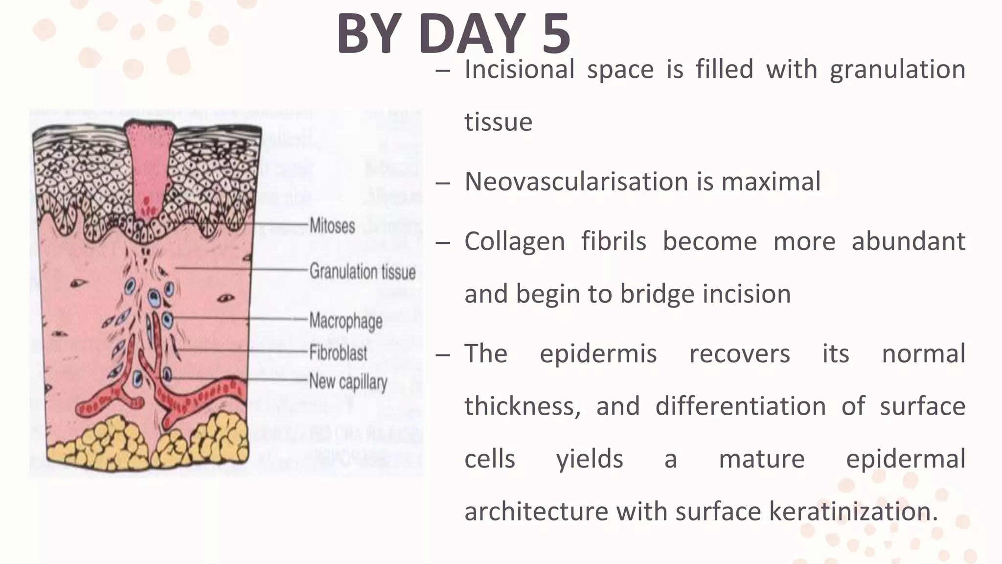
The document discusses the phases of wound healing:
1) Hemostasis and inflammation occurs within the first few days as the wound clots and immune cells clear debris.
2) Proliferation spans days 4-12 as new tissue is formed through fibroblast and endothelial cell proliferation and migration. Collagen and new blood vessels are deposited.
3) Maturation and remodeling can take months as the wound contracts and collagen is reorganized and remodeled, increasing strength of the healed tissue.
CONTENTS:
GENERAL
NORMAL FLUID CIRCULATION
EDEMA- INTRODUCTION
CAUSES
CLASSIFICATION
MAJOR TYPES
NOTE- Fonts may appear weird because the original fonts are different from the ones visible here.
The Indian Dental Academy is the Leader in continuing dental education , training dentists in all aspects of dentistry and
offering a wide range of dental certified courses in different formats.
Dentist in pune.(BDS. MDS) - Dr. Amit T. Suryawanshi. Wound healing in Dentis...All Good Things
entist in pune. (BDS. MDS) - Dr. Amit T. Suryawanshi. Seminar- Wound healing in dentistry.
Email ID- amitsuryawanshi999@gmail.com
Contact -Ph no.-9405622455
Subscribe our channel on youtube - Copy and paste this URL. https://www.youtube.com/channel/UC_gylEXTrjmEbbOTSXjuZ4Q/videos?view_as=public
Follow us on Slideshare.
The Indian Dental Academy is the Leader in continuing dental education , training dentists in all aspects of dentistry and
offering a wide range of dental certified courses in different formats.for more details please visit
www.indiandentalacademy.com
A concise and brief presentation on cleaning and shaping of root canals. Colorful and well pictured. Ideal for UG students and PG students to get a good understanding of BMP techniques.
A well pictured presentation on Endodontic Instrumentation for UG students. Best for getting a good grip on the topic as a whole. Meant to supplement not substitute standard texts.
A short slideshow covering the basics of Intrusive luxation and total avulsion, from an endodontic point of view.. Highlight are the photographs chosen with care to explain the points well. Ideal for under-graduate and Post-graduate students. Based on Grossman's Endodontic Practice, 13th Edition.
A comprehensive slideshow covering all the basics relating to dental materials and their physical properties. Based on standard text books - Phillips Science of Dental Materials (11th Edition).
A short yet comprehensive presentation on bacterial genetics, an important microbiology topic for BDS 2nd, MBBS 2nd and MD/MS /MDS 1st . Made using CP Baveja's Textbook of Microbiology. Meant as an introduction and overview with stress on some key areas.
Topics covered: Basic Principles, Synthesis of Protein, Extra Chromosomal Genetic Material, Bacterial Variation , Gene Transfer, Genetic Mechanisms of Drug Resistance, Genetic Engineering, DNA Probes, Polymerase Chain Reaction, Genetically Modified Organisms and Gene Therapy.
A Complete presentation explaining the complete morphology of Maxillary first molar, for the benefit of people like me who tried and failed to find everything in one package
Knee anatomy and clinical tests 2024.pdfvimalpl1234
This includes all relevant anatomy and clinical tests compiled from standard textbooks, Campbell,netter etc..It is comprehensive and best suited for orthopaedicians and orthopaedic residents.
New Directions in Targeted Therapeutic Approaches for Older Adults With Mantl...i3 Health
i3 Health is pleased to make the speaker slides from this activity available for use as a non-accredited self-study or teaching resource.
This slide deck presented by Dr. Kami Maddocks, Professor-Clinical in the Division of Hematology and
Associate Division Director for Ambulatory Operations
The Ohio State University Comprehensive Cancer Center, will provide insight into new directions in targeted therapeutic approaches for older adults with mantle cell lymphoma.
STATEMENT OF NEED
Mantle cell lymphoma (MCL) is a rare, aggressive B-cell non-Hodgkin lymphoma (NHL) accounting for 5% to 7% of all lymphomas. Its prognosis ranges from indolent disease that does not require treatment for years to very aggressive disease, which is associated with poor survival (Silkenstedt et al, 2021). Typically, MCL is diagnosed at advanced stage and in older patients who cannot tolerate intensive therapy (NCCN, 2022). Although recent advances have slightly increased remission rates, recurrence and relapse remain very common, leading to a median overall survival between 3 and 6 years (LLS, 2021). Though there are several effective options, progress is still needed towards establishing an accepted frontline approach for MCL (Castellino et al, 2022). Treatment selection and management of MCL are complicated by the heterogeneity of prognosis, advanced age and comorbidities of patients, and lack of an established standard approach for treatment, making it vital that clinicians be familiar with the latest research and advances in this area. In this activity chaired by Michael Wang, MD, Professor in the Department of Lymphoma & Myeloma at MD Anderson Cancer Center, expert faculty will discuss prognostic factors informing treatment, the promising results of recent trials in new therapeutic approaches, and the implications of treatment resistance in therapeutic selection for MCL.
Target Audience
Hematology/oncology fellows, attending faculty, and other health care professionals involved in the treatment of patients with mantle cell lymphoma (MCL).
Learning Objectives
1.) Identify clinical and biological prognostic factors that can guide treatment decision making for older adults with MCL
2.) Evaluate emerging data on targeted therapeutic approaches for treatment-naive and relapsed/refractory MCL and their applicability to older adults
3.) Assess mechanisms of resistance to targeted therapies for MCL and their implications for treatment selection
Title: Sense of Smell
Presenter: Dr. Faiza, Assistant Professor of Physiology
Qualifications:
MBBS (Best Graduate, AIMC Lahore)
FCPS Physiology
ICMT, CHPE, DHPE (STMU)
MPH (GC University, Faisalabad)
MBA (Virtual University of Pakistan)
Learning Objectives:
Describe the primary categories of smells and the concept of odor blindness.
Explain the structure and location of the olfactory membrane and mucosa, including the types and roles of cells involved in olfaction.
Describe the pathway and mechanisms of olfactory signal transmission from the olfactory receptors to the brain.
Illustrate the biochemical cascade triggered by odorant binding to olfactory receptors, including the role of G-proteins and second messengers in generating an action potential.
Identify different types of olfactory disorders such as anosmia, hyposmia, hyperosmia, and dysosmia, including their potential causes.
Key Topics:
Olfactory Genes:
3% of the human genome accounts for olfactory genes.
400 genes for odorant receptors.
Olfactory Membrane:
Located in the superior part of the nasal cavity.
Medially: Folds downward along the superior septum.
Laterally: Folds over the superior turbinate and upper surface of the middle turbinate.
Total surface area: 5-10 square centimeters.
Olfactory Mucosa:
Olfactory Cells: Bipolar nerve cells derived from the CNS (100 million), with 4-25 olfactory cilia per cell.
Sustentacular Cells: Produce mucus and maintain ionic and molecular environment.
Basal Cells: Replace worn-out olfactory cells with an average lifespan of 1-2 months.
Bowman’s Gland: Secretes mucus.
Stimulation of Olfactory Cells:
Odorant dissolves in mucus and attaches to receptors on olfactory cilia.
Involves a cascade effect through G-proteins and second messengers, leading to depolarization and action potential generation in the olfactory nerve.
Quality of a Good Odorant:
Small (3-20 Carbon atoms), volatile, water-soluble, and lipid-soluble.
Facilitated by odorant-binding proteins in mucus.
Membrane Potential and Action Potential:
Resting membrane potential: -55mV.
Action potential frequency in the olfactory nerve increases with odorant strength.
Adaptation Towards the Sense of Smell:
Rapid adaptation within the first second, with further slow adaptation.
Psychological adaptation greater than receptor adaptation, involving feedback inhibition from the central nervous system.
Primary Sensations of Smell:
Camphoraceous, Musky, Floral, Pepperminty, Ethereal, Pungent, Putrid.
Odor Detection Threshold:
Examples: Hydrogen sulfide (0.0005 ppm), Methyl-mercaptan (0.002 ppm).
Some toxic substances are odorless at lethal concentrations.
Characteristics of Smell:
Odor blindness for single substances due to lack of appropriate receptor protein.
Behavioral and emotional influences of smell.
Transmission of Olfactory Signals:
From olfactory cells to glomeruli in the olfactory bulb, involving lateral inhibition.
Primitive, less old, and new olfactory systems with different path
Pulmonary Thromboembolism - etilogy, types, medical- Surgical and nursing man...VarunMahajani
Disruption of blood supply to lung alveoli due to blockage of one or more pulmonary blood vessels is called as Pulmonary thromboembolism. In this presentation we will discuss its causes, types and its management in depth.
HOT NEW PRODUCT! BIG SALES FAST SHIPPING NOW FROM CHINA!! EU KU DB BK substit...GL Anaacs
Contact us if you are interested:
Email / Skype : kefaya1771@gmail.com
Threema: PXHY5PDH
New BATCH Ku !!! MUCH IN DEMAND FAST SALE EVERY BATCH HAPPY GOOD EFFECT BIG BATCH !
Contact me on Threema or skype to start big business!!
Hot-sale products:
NEW HOT EUTYLONE WHITE CRYSTAL!!
5cl-adba precursor (semi finished )
5cl-adba raw materials
ADBB precursor (semi finished )
ADBB raw materials
APVP powder
5fadb/4f-adb
Jwh018 / Jwh210
Eutylone crystal
Protonitazene (hydrochloride) CAS: 119276-01-6
Flubrotizolam CAS: 57801-95-3
Metonitazene CAS: 14680-51-4
Payment terms: Western Union,MoneyGram,Bitcoin or USDT.
Deliver Time: Usually 7-15days
Shipping method: FedEx, TNT, DHL,UPS etc.Our deliveries are 100% safe, fast, reliable and discreet.
Samples will be sent for your evaluation!If you are interested in, please contact me, let's talk details.
We specializes in exporting high quality Research chemical, medical intermediate, Pharmaceutical chemicals and so on. Products are exported to USA, Canada, France, Korea, Japan,Russia, Southeast Asia and other countries.
Lung Cancer: Artificial Intelligence, Synergetics, Complex System Analysis, S...Oleg Kshivets
RESULTS: Overall life span (LS) was 2252.1±1742.5 days and cumulative 5-year survival (5YS) reached 73.2%, 10 years – 64.8%, 20 years – 42.5%. 513 LCP lived more than 5 years (LS=3124.6±1525.6 days), 148 LCP – more than 10 years (LS=5054.4±1504.1 days).199 LCP died because of LC (LS=562.7±374.5 days). 5YS of LCP after bi/lobectomies was significantly superior in comparison with LCP after pneumonectomies (78.1% vs.63.7%, P=0.00001 by log-rank test). AT significantly improved 5YS (66.3% vs. 34.8%) (P=0.00000 by log-rank test) only for LCP with N1-2. Cox modeling displayed that 5YS of LCP significantly depended on: phase transition (PT) early-invasive LC in terms of synergetics, PT N0—N12, cell ratio factors (ratio between cancer cells- CC and blood cells subpopulations), G1-3, histology, glucose, AT, blood cell circuit, prothrombin index, heparin tolerance, recalcification time (P=0.000-0.038). Neural networks, genetic algorithm selection and bootstrap simulation revealed relationships between 5YS and PT early-invasive LC (rank=1), PT N0—N12 (rank=2), thrombocytes/CC (3), erythrocytes/CC (4), eosinophils/CC (5), healthy cells/CC (6), lymphocytes/CC (7), segmented neutrophils/CC (8), stick neutrophils/CC (9), monocytes/CC (10); leucocytes/CC (11). Correct prediction of 5YS was 100% by neural networks computing (area under ROC curve=1.0; error=0.0).
CONCLUSIONS: 5YS of LCP after radical procedures significantly depended on: 1) PT early-invasive cancer; 2) PT N0--N12; 3) cell ratio factors; 4) blood cell circuit; 5) biochemical factors; 6) hemostasis system; 7) AT; 8) LC characteristics; 9) LC cell dynamics; 10) surgery type: lobectomy/pneumonectomy; 11) anthropometric data. Optimal diagnosis and treatment strategies for LC are: 1) screening and early detection of LC; 2) availability of experienced thoracic surgeons because of complexity of radical procedures; 3) aggressive en block surgery and adequate lymph node dissection for completeness; 4) precise prediction; 5) adjuvant chemoimmunoradiotherapy for LCP with unfavorable prognosis.
Explore natural remedies for syphilis treatment in Singapore. Discover alternative therapies, herbal remedies, and lifestyle changes that may complement conventional treatments. Learn about holistic approaches to managing syphilis symptoms and supporting overall health.
Ozempic: Preoperative Management of Patients on GLP-1 Receptor Agonists Saeid Safari
Preoperative Management of Patients on GLP-1 Receptor Agonists like Ozempic and Semiglutide
ASA GUIDELINE
NYSORA Guideline
2 Case Reports of Gastric Ultrasound
These simplified slides by Dr. Sidra Arshad present an overview of the non-respiratory functions of the respiratory tract.
Learning objectives:
1. Enlist the non-respiratory functions of the respiratory tract
2. Briefly explain how these functions are carried out
3. Discuss the significance of dead space
4. Differentiate between minute ventilation and alveolar ventilation
5. Describe the cough and sneeze reflexes
Study Resources:
1. Chapter 39, Guyton and Hall Textbook of Medical Physiology, 14th edition
2. Chapter 34, Ganong’s Review of Medical Physiology, 26th edition
3. Chapter 17, Human Physiology by Lauralee Sherwood, 9th edition
4. Non-respiratory functions of the lungs https://academic.oup.com/bjaed/article/13/3/98/278874
NVBDCP.pptx Nation vector borne disease control programSapna Thakur
NVBDCP was launched in 2003-2004 . Vector-Borne Disease: Disease that results from an infection transmitted to humans and other animals by blood-feeding arthropods, such as mosquitoes, ticks, and fleas. Examples of vector-borne diseases include Dengue fever, West Nile Virus, Lyme disease, and malaria.
4. WOUND
– A wound is a break in the integrity of the skin or tissues
often which may be associated with disruption of the
structure and function.
(SRB 4th edition)
– A cut or break in the continuity of any tissue, caused by
injury or operation.
(Baillière’s 23rd Ed)
5. CLASSIFICATION OF WOUNDS
Classification based on type of wound
i. Clean incised wound
ii. Lacerated wound
iii. Bruising and contusion
iv. Hematoma
v. Puncture wound
vi. Abrasion
vii. Crush injury
viii. Injuries to bone and joint (maybe open or closed)
ix. Injuries to nerve (either clean cut or crush)
x. Injuries to arteries and veins
xi. Penetrating wounds
7. HEALING
• Healing is the body’s response to injury in an
attempt to restore normal structure and
function.
• The process of healing involves 2 distinct
processes:
A. REGENERATION
B. REPAIR
8. REGENERATION
– Regeneration: Is when healing takes place by
proliferation of parenchymal cells and usually
results in complete restoration of the original
tissues.
– The goal of all surgical procedures is regeneration
which returns the tissues to their normal
microstructure and function.
9. REPAIR
– Repair: It is a healing outcome in which tissues do
not return to their normal architecture and
function.
– Repair typically results in the formation of scar
tissue.
12. TYPES OF WOUND HEALING
– Healing by primary intention (wounds with
opposed edges)
– Healing by secondary intention (wounds with
separated edges)
13. HEALING OF WOUNDS BY
PRIMARY INTENTION
Healing of wound with following characteristics:
Clean and uninfected
Surgically incised
Without much loss of cells and tissue
Edges of wound are approximated by surgical sutures.
Wounds with opposed edges
Primary union
14. HEALING OF WOUNDS BY
PRIMARY INTENTION
– The incision causes
death of a limited number of epithelial cells and connective tissue
cells
disruption of epithelial basal membrane continuity
– The narrow incisional space immediately fills with
clotted blood containing fibrin and blood cells;
dehydration of the surface clot forms the well
known scab that covers the wound.
16. WITHIN 24 HOURS
– Neutrophils appear at margins of incision,
moving toward fibrin clot
– Epidermis at its cut edges thickens as a result of
mitotic activity of basal cells
– Within 24 to 48 hours, spurs of epithelial cells
from the both edges migrate and grow along the
cut margins of the dermis, depositing BM
components as they move. They fuse in the
midline beneath the surface scab, thus
producing a continuous but thin epithelial layer.
17. BY DAY 3
– Neutrophils replaced by macrophages
– Granulation tissue progressively invades
incision space
– Collagen fibers are now present in the
margins of the incision, but at first these
are vertically oriented.
– Epithelial cell proliferation continues,
thickening epidermal covering layer
18. BY DAY 5– Incisional space is filled with granulation
tissue
– Neovascularisation is maximal
– Collagen fibrils become more abundant
and begin to bridge incision
– The epidermis recovers its normal
thickness, and differentiation of surface
cells yields a mature epidermal
architecture with surface keratinization.
19. DURING THE SECOND WEEK
– Continued accumulation of collagen and proliferation of fibroblasts
– Leukocytic infiltrate, edema, and increased vascularity have largely
disappeared.
20. BY THE END OF
THE FIRST MONTH
– Scar comprises a cellular connective tissue devoid of
inflammatory infiltrate, covered now by intact
epidermis.
– Dermal appendages that have been destroyed in the
line of the incision are permanently lost.
– Tensile strength of the wound increases thereafter, but
it may take months for the wounded area to obtain its
maximal strength.
21. HEALING BY SECONDARY
INTENTION
– Wounds with separated edges
– Secondary union
– When there is more extensive loss of cells and tissue
– Regeneration of parenchymal cells cannot completely
reconstitute the original architecture.
– Abundant granulation tissue grows in from the margin to
complete the repair.
22. DIFFERNCES BETWEEN HEALING
BY PRIMARY AND SEONDARY
INTENTION
Secondary healing differs from primary healing in several respects:
Inflammatory reaction is more intense
Much larger amounts of granulation tissue are formed
Wound contraction occurs in large surface wounds
Substantial scar formation and thinning of the epidermis occurs
23. DIFFERNCES BETWEEN HEALING
BY PRIMARY AND SEONDARY
INTENTION
FEATURES HEALING BY PRIMARY INTENTION HEALING BY SECONDARY INTENTION
CLEANLINESS CLEAN NOT CLEAN
INFECTION NOT INFECTED INFECTED
MARGINS SURGICALLY CLEAN IRREGULAR
SUTURES USED NOT USED
HEALING SMALL GRANULATION TISSUE LARGE GRANULATION TISSUE
OUT COME LINEAR SCAR IRREGULAR WOUND
COMPLICATION NOT FREQUENT FREQUENT
26. PHASES OF WOUND HEALING
– Normal wound healing follows a predictable
pattern that can be divided into overlapping
phases:
– Hemostasis and inflammation
– Proliferation
– Maturation and remodeling
28. HEMOSTASIS AND
INFLAMMATION
– Hemostasis precedes and initiates inflammation, with
the ensuing release of chemotactic factors from the
wound site.
– Wounding > disrupts tissue integrity > division of blood
vessels and direct exposure of extracellular matrix to
platelets >platelet aggregation, degranulation, and
activation of the coagulation cascade
29. HEMOSTASIS AND
INFLAMMATION
– Platelet -granules release a number of wound-active substances,
such as platelet-derived growth factor (PDGF), transforming
growth factor beta (TGF), platelet-activating factor, fibronectin,
and serotonin.
– In addition to achieving hemostasis, the fibrin clot serves as
scaffolding for the migration into the wound of inflammatory cells
such as polymorphonuclear leukocytes (PMNs, neutrophils) and
monocytes.
31. PMN: POLYMORPHONEUCLEAR
LEUKOCYTES
– The first infiltrating cells to enter the wound site,
peaking at 24 to 48 hours
– Stimulated by:
– Increased vascular permeability
– local prostaglandin release
– The presence of chemotactic substances( co,IL-
1,TNF,TGF beta, PF 4,bacterial product)
32. PMN: POLYMORPHONEUCLEAR
LEUKOCYTES
– Primary role:
– Phagocytosis of bacteria and tissue debris
– Major source of cytokines early during inflammation, especially TNF-
alpha
– Release proteases such as collagenases, which participate in matrix
and ground substance degradation in the early phase of wound
healing.
– No role in collagen deposition or acquisition of mechanical
wound strength.
33. MACROPHAGE
– The second population of inflammatory cells that invades
the wound.
– Derived from circulating monocytes, macrophages
achieve significant numbers in the wound by 48 to 96
hours postinjury.
– Present until wound healing is complete
34. LYMPHOCYTE
– Less numerous than macrophages
– T-lymphocyte numbers peak at about 1 week postinjury
– It bridge the transition from the inflammatory to the
proliferative phase of healing.
– The role in wound healing is not fully defined
– Believed to play an active role in the modulation of the
wound environment
35. PROLIFERATION
– Is the second phase of wound healing and roughly spans
days 4 through 12
– It is during this phase that tissue continuity is re-
established.
– Fibroblasts and endothelial cells are the last cell
populations to infiltrate the healing wound.
– The strongest chemotactic factor for fibroblasts is PDGF
37. FIBROBLASTS
– Upon entering the wound environment, recruited fibroblasts first need to
proliferate
– Then become activated by the cytokines and growth factors released from
wound macrophages.
– Primary function is matrix synthesis remodeling
– Fibroblasts isolated from wounds synthesize more collagen than nonwound
fibroblasts
– They proliferate less, and they actively carry out matrix contraction.
– Cytokine-rich wound environment and lactate plays a significant role in this
phenotypic alteration and activation.
38. ENDOTHELIAL CELLS
– Endothelial cells migrate from intact venules
close to the wound.
– Proliferate extensively during this phase of
healing.
– These cells participate in the formation of
new capillaries (angiogenesis)
– Their migration, replication, and new capillary
tubule formation are under the influence of
such cytokines and growth factors as TNF-,
TGF, and VEGF
39. MATRIX SYNTHESIS
Collagen biochemistry
– Its deposition, maturation, and subsequent
remodeling are essential to the functional
integrity of the wound.
– Type I collagen is the major component of
extracellular matrix in skin.
– Type III, which is also normally present in
skin, becomes more prominent and
important during the repair process.
40. MATRIX SYNTHESIS
– Collagen synthesis is highly dependent on systemic
factors such as :
– adequate oxygen supply,
– the presence of sufficient nutrients (amino acids and carbohydrates)
– cofactors (vitamins and trace metals), and
– the local wound environment (vascular supply and lack of infection)
41. PROTEOGLYCAN SYNTHESIS
– Glycosaminoglycans comprise a large portion of the
"ground substance" that makes up granulation tissue.
– The major glycosaminoglycans present in wounds are
dermatan and chondroitin sulfate.
– It is thought that the assembly of collagen subunits
into fibrils and fibers is dependent on the lattice
provided by the sulfated proteoglycans.
– As scar collagen is deposited, the proteoglycans are
incorporated into the collagen scaffolding.
– With scar maturation and collagen remodeling, the
content of proteoglycans gradually diminishes.
42. MATURATION AND
REMODELING
– Begins during the fibroplastic phase, and is characterized
by a reorganization of previously synthesized collagen.
– The net wound collagen content is the result of a balance
between collagenolysis and collagen synthesis.
– Collagenolysis is the result of collagenase activity, a class
of matrix metalloproteinases that require activation
43. MATURATION AND
REMODELING
– Wound strength and mechanical integrity in the fresh wound are
determined by both the quantity and quality of the newly
deposited collagen.
– The deposition of matrix at the wound site follows a characteristic
pattern:
– Fibronectin and collagen type III constitute the early matrix scaffolding,
– glycosaminoglycans and proteoglycans represent the next significant matrix
components, and
– collagen type I is the final matrix.
45. MATURATION AND
REMODELING
– Fibril formation and fibril cross-linking result in decreased collagen
solubility, increased strength, and increased resistance to
enzymatic degradation of the collagen matrix
– Scar remodeling continues for many (6 to 12) months postinjury,
gradually resulting in a mature, avascular, and acellular scar.
– The mechanical strength of the scar never achieves that of the
uninjured tissue.
46. EPITHELIALIZATION
– Is a process restoring external barrier characterized
primarily by proliferation and migration of epithelial
cells adjacent to the wound.
– Begins within 1 day of injury and is seen as thickening
of the epidermis at the wound edge.
– Marginal basal cells at the edge of the wound lose
their firm attachment to the underlying dermis,
enlarge, and begin to migrate across the surface of
the provisional matrix.
47. EPITHELIALIZATION
– Re-epithelialization is complete in less than 48 hours in the case of
approximated incised wounds.
– Take substantially longer in the case of larger wounds, in which
there is a significant epidermal/dermal defect.
– If only the epithelium and superficial dermis are damaged, then
repair consists primarily of re-epithelialization with minimal or no
fibroplasia and granulation tissue formation.
49. BONE FRACTURE HEALING
– Most of the phases of healing resemble those observed in dermal
healing
– 1st stage: Hematoma formation consists of an accumulation of
blood at the fracture site, which also contains devitalized soft
tissue, dead bone, and necrotic marrow.
– 2nd stage : liquefaction and degradation of nonviable products at
the fracture site.
50. BONE FRACTURE HEALING
– 3rd stage : soft callus stage, 3-4 days after injury, soft
tissue forms a bridge between the fractured bone
segments.
– 4th stage : hard callus stage, consists of mineralization of
the soft callus and conversion to bone.
– 5th stage : Remodeling , the excessive callus is
reabsorbed and the marrow cavity is recanalized.
53. AGE
– The increased incidence of comorbidity
may contribute to impaired wound
healing
– Noncollagenous protein accumulation at
wounded sites is decreased with aging
which may impair the mechanical
properties of scarring in elderly patients.
54. HYPOXIA,ANEMIA,
HYPOPERFUSION
– Low oxygen tension has a profoundly deleterious
effect on all aspects of wound healing.
– Optimal collagen synthesis requires oxygen as a
cofactor, particularly for the hydroxylation steps.
– Increasing FiO2 of inspired air for brief periods
during and immediately after surgery results in
enhanced collagen deposition and in decreased
rates of wound infection after elective surgery.
55. STEROID AND
CHEMOTHERAPEUTIC DRUG
– Steroids inhibit the inflammatory phase of wound
healing (angiogenesis, neutrophil and macrophage
migration, and fibroblast proliferation) and the
release of lysosomal enzymes.
– Steroids used after the first 3 to 4 days postinjury do
not affect wound healing as severely as when they
are used in the immediate postoperative period.
56. STEROID AND
CHEMOTHERAPEUTIC DRUG
– Steroids also inhibit epithelialization and contraction
– Topical application of vitamin A may help
– All chemotherapeutic antimetabolite drugs adversely affect wound
healing by inhibiting early cell proliferation and wound DNA and
protein synthesis.
– Delay in the use of such drugs for about 2 weeks postinjury
appears to lessen the wound healing impairment.
57. METABOLIC DISORDERS
– Uncontrolled diabetes results in reduced inflammation,
angiogenesis, and collagen synthesis
– The large- and small-vessel disease that is the hallmark
of advanced diabetes contributes to local hypoxemia.
– Defects in granulocyte function, capillary ingrowth, and
fibroblast proliferation all have been described in
diabetes.
– Uremia also has been associated with disordered
wound healing.
58. NUTRITION
Protein and energy
– Malnutrition correlates clinically with enhanced rates of wound
complications and increased wound failure.
– During protein-calorie malnutrition
– impaired healing response and
– reduced cell-mediated immunity, and
– phagocytosis, and intracellular killing of bacteria by macrophages and
neutrophils
– Arginine appears most active in terms of enhancing wound
fibroplasia.
59. Cont..
Vitamins
– The vitamins most closely involved with wound healing are vitamin C and
vitamin A.
– Vitamin C deficiency leads to a defect in wound healing, particularly via a failure
in collagen synthesis and cross-linking.
– Vitamin A increases the inflammatory response in wound healing, probably by
increasing the lability of lysosomal membranes.
60. Cont…
Minerals
– There are over 150 known enzymes for which zinc is either an integral part or
an essential cofactor, and many of these enzymes are critical to wound healing.
– Zinc deficiency:
– decreased fibroblast proliferation,
– decreased collagen synthesis,
– impaired overall wound strength, and
– delayed epithelialization.
62. EXCESSIVE SCAR FORMATION
– Excessive healing results in a raised, thickened
scar, with both functional and cosmetic
complications.
– If it stays within margins of wound it is
hypertrophic. Keloids extend beyond the
confines of the original injury.
– Dark skinned, ages of 2-40. Wound in the
presternal or deltoid area, wounds that cross
langerhans lines.
63. KELOIDS AND HYPERTROPHIC
SCARS
– Keloids more familial
– Hypertrophic scars develop soon after
injury, keloids up to a year later.
– Hypertrophic scars may subside in time,
keloids rarely do.
– Hypertrophic scars more likely to cause
contracture over joint surface.
64. KELOIDS AND HYPERTROPHIC
SCARS
– Both from an overall increase in the quantity of collagen
synthesized.
– Recent evidence suggests that the fibroblasts within
keloids are different from those within normal dermis in
terms of their responsiveness.
– No modality of treatment is predictably effective for
these lesions.
Major factors affecting local oxygen delivery include hypoperfusion either for systemic reasons (low volume or cardiac failure) or due to local causes (arterial insufficiency, local vasoconstriction, or excessive tension on tissues). The level of vasoconstriction of the subcutaneous capillary bed is exquisitely responsive to fluid status, temperature, and hyperactive sympathetic tone as is often induced by postoperative pain. Correction of these factors can have a remarkable influence on wound outcome, particularly on decreasing wound infection rates.53–55 Mild to moderate normovolemic anemia does not appear to adversely affect wound oxygen tension and collagen synthesis, unless the hematocrit falls below 15%.55
In clean, noninfected, and well-perfused experimental wounds in human diabetic volunteers, type I diabetes mellitus was noted to decrease wound collagen accumulation in the wound, independent of the degree of glycemic control. Type II diabetic patients showed no effect on collagen accretion when compared to healthy, age-matched controls.63 Furthermore, the diabetic wound appears to be lacking in sufficient growth factor levels, which signal normal healing. It remains unclear whether decreased collagen synthesis or an increased breakdown due to an abnormally high proteolytic wound environment is responsible.
Careful preoperative correction of blood sugar levels improves the outcome of wounds in diabetic patients. Increasing the inspired oxygen tension, judicious use of antibiotics, and correction of other coexisting metabolic abnormalities all can result in improved wound healing.
The clinician must pay close attention to the nutritional status of patients
with wounds, because wound failure or wound infections may be no more
than a reflection of poor nutrition.
Clinically, it is extremely rare to encounter pure energy or protein malnutrition, and the vast majority of patients exhibit combined protein-energy malnutrition. Such patients have diminished hydroxyproline accumulation (an index
of collagen deposition) into subcutaneously implanted polytetrafluoroethylene
tubes when compared to normally nourished patients
The possible role of single amino acids in enhanced wound healing has been
studied for the last several decades. Arginine appears most active in terms of
enhancing wound fibroplasia. Indeed, arginine supplementation in both rats and
humans has been shown to increase wound collagen deposition. Conversely,
arginine deficiency in rats results in decreased wound-breaking strength and
wound-collagen accumulation.
Vitamin C deficiency leads to a defect in wound healing, particularly via a failure in collagen synthesis and cross-linking. Biochemically,
vitamin C is required for the conversion of proline and lysine to hydroxyproline and hydroxylysine. Vitamin C deficiency also has been associated with
an increased incidence of wound infection, and if wound infection does occur, it tends to be more severe. These effects are believed to be because of an
associated impairment in neutrophil function, decreased complement activity,
and decreased walling-off of bacteria secondary to insufficient collagen deposition