SlideShare a Scribd company logo
PATHOLOGY OF
BLOOD VESSELS
Angel R Reyes Jr, RMT, MD, FPSP, CHA DipHLM
1.NORMAL VESSELS
• 3 concentric layer; intima, media & adventitia.
 Intima; internal elastic lamina
 Media;external elastic lamina
 Adventitia; vasa vasorum
• Arteries;
 Elastic(large): aorta, innominate, subclavian,
 Muscular(medium-sized): renal and coronary
 Small arteries and arterioles
• Capillaries
• Veins;
 post-capillary venules, collecting venules, veins.
3
4
Arteries
• Large arteries are elastic (conducting) arteries
– pressure reservoirs
• Medium arteries are muscular (distributing)
arteries – more smooth muscle
• Contraction or relaxation of muscle changes
the size of the lumen, and so controls the
blood pressure in the vessel.
5
6
Capillaries
• Only a single layer of endothelium and a
basement membrane
• Connect arterioles and venules
• Functional part of system
• True capillaries begin at a precapillary
sphincter which controls blood flow through
the capillary
7
8
Veins
• Relatively thin; less elastic
• Larger in diameter than arteries
• Have valves to prevent backflow of blood
• Flow to heart is assisted by contraction of
skeletal muscles
9
10
11
12
Control of systemic circulation
• Nervous control – innervated by sympathetic
nervous system ONLY
• Cardiac control center (primarily in medulla
oblongata)
• Heart has both Sympathetic and
Parasympathetic innervations.
13
• Baroreceptors and chemoreceptors:
– Monitor pressure
– Monitor blood levels of O2, CO2 and H+
– Send information to cardiovascular center, which
responds
Vascular organization
• Large elastic arteries > medium-sized arteries
> small arteries >arterioles > capillaries > post-
capillary venules > collecting venules > veins
Lymphatics
• Endothelium-lined channels that drain excess
interstitial tissue fluid, eventually returning it
to blood via the thoracic ducts.
2.VASCULAR WALL CELLS AND THEIR
RESPONSE TO INJURY
• Endothelial cells;
Elaboration of Anticoagulant, Antithrombotic,
Fibrinolytic Regulators
Elaboration of Prothrombotic Molecules
Extracellular Matrix Production (Collagen,
Proteoglycans)
Modulation of Blood Flow and Vascular Reactivity
Regulation of Inflammation and Immunity
Regulation of Cell Growth
Oxidation of LDL
Cont..
• Vascular Smooth Muscle Cells
proliferate when appropriately stimulated
synthesize ECM collagen, elastin, and proteoglycans
elaborate growth factors and cytokines
vasoconstriction or vasodilation
Response of Vascular Wall Cells to
Injury
• Endothelial injury contributes to a host of pathologies
including thrombosis, atherosclerosis, and
hypertensive vascular lesions.
• Injury to the vessel wall results in a healing response,
involving intimal expansion by proliferating SMCs and
newly synthesized ECM.
• The recruitment and activation of the SMCs in this
process involves signals from cells eg ECs, and
mediators derived from coagulation and complement
cascades.
• Therefore, intimal thickening is a stereotyped
Response to Vascular Injury
3.CONGENITAL ANOMALIES
• Rarely symptomatic;
Developmental (berry) aneurysms occur in cerebral
vessels.
Arteriovenous fistulas are direct connections between
arteries and veins that bypass the intervening
capillaries.
Fibromuscular dysplasia is a focal irregular thickening
of the walls of medium and large muscular arteries.
4.ARTERIOSCLEROSIS
• Arterial wall thickening and loss of elasticity.
Arteriolosclerosis affects small arteries and arterioles with
two anatomic variants hyaline and hyperplastic.
Mönckeberg medial calcific sclerosis; calcific deposits in
muscular arteries.
Atherosclerosis.
5.ATHEROSCLEROSIS
• General descriptions;
intimal lesions called atheromas (also called
atheromatous or atherosclerotic plaques), that
protrude into vascular lumina.
plaque consists of a raised lesion with a soft, yellow,
grumous core of cholesterol (esters) covered by a firm,
white fibrous cap.
obstructs blood flow & weaken the underlying media
and rupture, causing acute catastrophic vessel
thrombosis.
causes ischemic heart disease (IHD) .
Epidemiology
• Causes more morbidity and mortality (roughly
half of all deaths) in the Western world than any
other disorder.
• The mortality rate for IHD in the United States is
among the highest in the world and is
approximately five times higher than that in
Japan.
• Japanese who immigrate to the United States and
adopt American lifestyles and dietary customs
acquire the same predisposition to
atherosclerosis as the homegrown population.
Major Constitutional Risk Factors for
IHD
• Nonmodifiable factors;
Age; between ages 40 and 60, the incidence of
myocardial infarction in men increases fivefold.
Gender; premenopausal women are relatively
protected against atherosclerosis and its
consequences compared with age-matched men.
Genetics; well-established familial predisposition to
atherosclerosis and IHD is multifactorial:such as
hypertension or diabetes, familial
hypercholesterolemia, that result in excessively high
blood lipid levels.
• Major Modifiable Factors;
 Hyperlipidemia; esp. hypercholesteremia i.e. LDL
cholesterol has an essential physiologic role
delivering cholesterol to peripheral tissues while
mobilizes cholesterol from developing and existing
atheromas.
Hypertension; increase the risk of IHD by
approximately 60% in comparison with
normotensive populations.
Cigarette Smoking; Prolonged (years) smoking of
one pack of cigarettes or more daily increases the
death rate from IHD by 200%.
Diabetes Mellitus; induces hypercholesterolemia,
the incidence of myocardial infarction is twice as
high in diabetic as in nondiabetic individuals.
• Additional Factors;
Inflammation as marked by C-reactive protein.
Hyperhomocystinemia.
Lipoprotein a.
Factors Affecting Hemostasis.
Other Factors; like lack of exercise; competitive,
stressful lifestyle ("type A" personality); and
obesity.
Pathogenesis
• Response-to-injury hypothesis;
Atherosclerosis as a chronic inflammatory response
of the arterial wall to endothelial injury.
Lesion progression occurs through interactions of
modified lipoproteins, monocyte-derived
macrophages, T lymphocytes, and the normal
cellular constituents of the arterial wall.
• Central tenets of the hypothesis;
Chronic endothelial injury, with resultant endothelial
dysfunction, causing (among other things) increased
permeability, leukocyte adhesion, and thrombosis.
Accumulation of lipoproteins (mainly LDL and its oxidized
forms) in the vessel wall.
Monocyte adhesion to the endothelium, followed by
migration into the intima and transformation into
macrophages and foam cells.
Platelet adhesionFactor release from activated platelets,
macrophages, and vascular wall cells, inducing
 SMC recruitment, either from the media or from circulating
precursors.
SMC proliferation and ECM production.
Lipid accumulation both extracellularly and within cells
(macrophages and SMCs).
Morphology
• Fatty Streaks; composed of lipid-filled foam cells but are not significantly
raised, begin as multiple minute yellow, flat spots that can coalesce into
elongated streaks,
• Atherosclerotic Plaque; a.k.a fibrous or fibrofatty plaques impinge on the
lumen of the artery and grossly appear white to yellow; thrombosis
superimposed over the surface of ulcerated plaques is red-brown in color.
• Atherosclerotic plaques have three principal components: (1) cells, including
SMCs, macrophages, and T cells; (2) ECM, including collagen, elastic fibers,
and proteoglycans; and (3) intracellular and extracellular lipid.
• Parts of the plaque(on cross-section):
 Superficial fibrous cap is composed of SMCs and relatively dense collagen.
 “Shoulder”- beneath and to the side of the cap = more cellular area containing macrophages,
T cells, and SMCs.
 Necrotic core; deep to the fibrous cap, containing cholesterol (esters), debris from dead cells,
foam cells (lipid-laden macrophages and SMCs), fibrin, variably organized thrombus, and other
plasma proteins; the cholesterol content is frequently present as crystalline aggregates that
are washed out during routine tissue processing and leave behind only empty "clefts.“
 Neovascularization at the periphery of the lesions,
m
• Plaque changes;
Rupture, ulceration, or erosion; exposes the
bloodstream to highly thrombogenic substances and
induces thrombus formation and occlude the lumen
and lead to downstream ischemia.
Hemorrhage into a plaque
Atheroembolism; plaque rupture can discharge debris
into the bloodstream, producing microemboli
composed of plaque contents.
Aneurysm formation; Atherosclerosis-induced pressure
or ischemic atrophy of the underlying media, with loss
of elastic tissue, causes weakness of the vessel wall and
development of aneurysms that may rupture
Natural History of Atherosclerosis
• Preclinical and clinical phase.
Prevention of Atherosclerotic Vascular
Disease
• Primary prevention programs; cessation of
cigarette smoking, control of hypertension,
weight loss, exercise, and lowering total and LDL
blood cholesterol levels while increasing HDL
(e.g., by diet or through statins)
• Secondary prevention programs; use of aspirin
(anti-platelet agent), statins, and beta blockers (to
limit cardiac demand), as well as surgical
interventions (e.g., coronary artery bypass
surgery, carotid endarterectomy). These can
successfully reduce recurrent myocardial or
cerebral events.
6.HYPERTENSIVE VASCULAR DISEASE
• General description;
Elevated blood pressure is called hypertension.
Remains asymptomatic until late in its course.
Contributes to the pathogenesis of coronary
heart disease and cerebrovascular accidents,
causes cardiac hypertrophy and heart failure,
aortic dissection, and renal failure.
Regulation of Blood Pressure
• BP= CO X PR
• RAA System
• Vasodilators
• Adrenal aldosterone
• ANP
Pathogenesis of Hypertension
• 90% to 95% of hypertension is idiopathic (essential
hypertension), which is compatible with long life,
unless a myocardial infarction, cerebrovascular
accident, or other complication supervenes.
• Most of the remainder of "benign hypertension" is
secondary to renal disease or, less often, to narrowing
of the renal artery, usually by an atheromatous plaque
(renovascular hypertension).
• Accelerated or malignant hypertension, the clinical
syndrome is characterized by severe hypertension
(diastolic pressure over 120mmHg), renal failure, and
retinal hemorrhages and exudates, with or without
papilledema
• Essential Hypertension;
 alterations in renal sodium homeostasis and/or vessel wall tone
or structure underlie essential hypertension.
 interplay of multiple genetic and environmental factors
affecting cardiac output and/or peripheral resistance.
• Secondary Hypertension;
 Renal: acute glomerulonephritis, chronic renal disease, polycystic
disease, renal artery stenosis, renal vasculitis, renin-producing
tumors.
 Endocrine: Adrenocortical hyperfunction, Exogenous hormones,
Pheochromocytoma, Acromegaly, Hypo/hyperthyroidism,
Pregnancy-induced.
 Cardiovascular: Coarctation of aorta , polyarteritis nodosa
increased intravascular volume , increased cardiac output ,
rigidity of the aorta.
 Neurologic: psychogenic , increased intracranial pressure , sleep
apnea, acute stress (surgery).
Vascular Pathology in Hypertension
• Accelerating atherogenesis
• Potentiate both aortic dissection and
cerebrovascular hemorrhage
• Two forms of small blood vessel disease
Hyaline Arteriolosclerosis: a homogeneous pink hyaline
thickening of the walls of arterioles with loss of underlying
structural detail and with narrowing of the lumen
 Hyperplastic Arteriolosclerosis. Related to acute or severe
elevations of blood pressure, characteristic of malignant
hypertension , associated with "onion-skin," concentric,
laminated thickening of the walls of arterioles with luminal
narrowing ,the laminations consist of SMCs and thickened,
duplicated basement membrane.
7.ANEURYSMS AND DISSECTIONS
• General description;
aneurysm is a localized abnormal dilation of a blood vessel
or the heart
 "true" aneurysm.
 false aneurysm (pseudoaneurysm): pulsating hematoma.
arterial dissection arises when blood enters the wall of the
artery, as a hematoma dissecting between its layers.
 Causes; atherosclerosis and cystic medial degeneration of
the arterial media, wall-weakening factors like include
trauma, congenital defects (e.g., berry aneurysms), infections
(mycotic aneurysms), or syphilis, and vasculitis.
 Mycotic aneurysms can originate (1) from embolization of a
septic thrombus, usually as a complication of infective
endocarditis; (2) as an extension of an adjacent suppurative
process; or (3) by circulating organisms directly infecting the
arterial wall.
Abdominal Aortic Aneurysm
• Definition
Out pouching of the aorta, most commonly in
abdominal aorta, following
atherosclerotic destruction of the aortic media,
leading to vessel wall weakness
• Morphology
 Big fusiform or sacular bulge out the side of the
abdominal aorta of varying width and length below
the renal arteries and above the bifurcation of the
aorta.
 Often contains atheromatous ulcers with thrombi
(source of atherothrombotic origin)
 May be so large that it compresses nearby vessels or
the thrombus causes occlusion syndromes
Special Variants
• Inflammatory AAA = Macrophages, Giant Cells,
Lymphocytes, Plasma cells, AAA
• Mycotic AAA = Supuritic lesions with organisms hiding
inside (salmonella)
S
• Pathogenesis
 Atherosclerosis is number 1 cause
Cystic Medial Degeneration = Collagen.
Aneurysms come from abnormal collagen
(Marfan’s) or an abnormality of collagen
remodeling (increased degradation decreased
synthesis caused by an immune reaction)
resulting in an inherently weakened aortic wall
• Clinical Course
 Rupture is often fatal. Hemorrhage occurs
into the peritoneum. ↑size = ↑risk
 Obstruction of a vessel (mesenteric, renal,
vertebral) leading to ischemic injury
 Direct Compression of the ureter or of the
vertebrae (casing vertebral erosion)
 Embolism from the thrombus that’s sitting
inside
 Tumor = AAA is a palpable mass that may
Syphilitic Aneurysm
• Cause
obliterative endarteritis characteristic of the
tertiary stage of syphilis can involve small
vessels in any part of the body.
Syphilis (stage 3) has a vascular predilection
for small vessels, especially of the aorta
Infection with subsequent Inflammation of
the vasa vasorum leads to obstruction,
inducing ischemia and obliterative
endarteritis of the aorta, causing death of
muscle and elastic tissue, called syphilitic
aortitis
• Morphology
Starts in the adventitia with vessels with
inflammation reactions in adventitia.
syphilitic aortitis.
Muscle that dies is replaced with fibrous
scar tissue in the media, contraction of
which causes the “tree‐barking”
appearance
Effects thoracic aorta with possible
dilation of aortic valve leading to
insufficiency
• Presentation
All causes by the encroachment on
mediastinal tissues
•Crush the lungs = Dyspnea Pain
from rib and vertebral erosions
Death from rupture
•Crush the esophagus = Dysphagia,
Cardiac Disease from valve
involvement Aortic Regurgitation
Aortic Dissection
• Definition
blood splays apart the laminar planes of the media
to form a blood-filled channel within the aortic wall
ruptures through the adventitia and into various
spaces, where it causes either massive hemorrhage
or cardiac tamponade (hemorrhage into the
pericardial sac).
men aged 40 to 60 years, with antecedent
hypertension (> 90% of cases ), and younger
patients with systemic or localized abnormalities of
connective tissue affecting the aorta ( Marfan’s )
• Morphology
A single intimal tear cuts into but not through the
media of the ascending aorta creating a blood
filled pocket within the aorta, between layers
Dissection can continue in both direction (towards
the heart and towards the femoral)
Usually rupture outwards, but can rupture back
inwards
– Outward = hemorrhage = fatal
– Inward = second intimal tear back into the normal lumen,
forming a new vascular channel (“double‐barrel Aorta”)
which can, over time, endothelize and become a permanent
vessel.
No distinguishing histology except for warning
signs = elastic tissue fragmentation and medial
degeneration
• Pathogenesis
HTN is primary causative agent, if you see HTN pick
Dissecting Hematoma
Medial weakening is not required for dissection, but
cystic medial necrosis from
Marfan’s or Ehler’s Danlos (weak elastic layer)
predisposes dissection
If you have a dissection, pick HTN. If HTN isn’t there,
pick Marfans
Once dissection has happened, arterial blood pressure
favors hematoma
• Clinical Course
If the dissection is Proximal subclavian/carotid
(type A = bad), if distal (type B = better)
Pain in the chest radiating to the back is a tell
tale sign
Death usually results from rupture
If dissection involves aortic root, you can get
valve problems (regurgitation, murmur
Classification of dissections
8.VASCULITIS
• General description;
Inflammation of vessel walls.
2 mechanisms: immune-mediated
inflammation and direct invasion of vascular
walls by infectious pathogens.
Noninfectious Vasculitis.
Infectious Vasculitis.
Vasculitis Associated with Other Disorders .
Non-infectious Vasculitis.
• Classification;
Large-Vessel Vasculitis
» Giant-cell (temporal) arteritis
» Takayasu arteritis
Medium-Vessel Vasculitis
» Polyarteritis nodosa
» Kawasaki disease
Small-Vessel Vasculitis
» Wegener granulomatosis
» Churg-Strauss syndrome
» Microscopic polyangiitis
• Pathogenesis;
 immune complex deposition; Antibody and
complement are typically detected in vasculitic
lesions, DNA-anti-DNA complexes(SLE) and drug
hypersensitivity.Can be 2ndary to viral infections.
antineutrophil cytoplasmic antibodies (ANCAs);
circulating antibodies that react with neutrophil
cytoplasmic antigens. Cytoplasmic localization (c-
ANCA) >>proteinase-3 (PR3) and Perinuclear
localization (p-ANCA) >>myeloperoxidase (MPO).
anti-endothelial cell antibodies; Antibodies to ECs
may predispose to certain vasculitides, for example
Kawasaki disease.
• Giant-Cell (Temporal) Arteritis
Most common form of Vasculitis, especially in Elderly
Women
affects the arteries in the head-esp. the temporal
arteries-but also the vertebral and ophthalmic arteries &
the aorta (giant-cell aortitis).
T cell-mediated immune response to an unknown vessel
wall antigen.
nodular intimal thickening with reduction of the lumen
and occasional thrombosis.
granulomatous inflammation within the inner media
centered on the internal elastic membrane.
fragmentation of the internal elastic lamina
Head pain, vision disturbances /blindness(ophthalmic
artery involved)
Responds well to steroids.
• Takayasu Arteritis
"pulseless disease“
transmural fibrous thickening of the
aorta-particularly the aortic arch and
great vessels.
severe luminal narrowing of the
major branch vessels- loss of pulse.
in women younger than 40 years of
age(Japanese population)
intimal hyperplasia and irregular
thickening of the vessel wall
adventitial mononuclear infiltrates >>
mononuclear inflammation in the media
>> granulomatous inflammation(giant cells
and patchy medial necrosis)
reduced blood pressure and weaker
pulses in the upper extremities with
coldness or numbness of the fingers;
ocular disturbances, and neurologic
deficits. Claudication of the legs(distal
aorta involved); pulmonary
hypertension(PA involved). Narrowing of
the coronary ostia >>MI, systemic
hypertension(renal arteries involved)
• Polyarteritis Nodosa
Involves renal and visceral vessels but not the
pulmonary circulation.
segmental transmural necrotizing inflammation of
small to medium-sized arteries
part of the vessel circumference involved, with a
predilection for branch points.
weakens the arterial wall >>aneurysms or even
rupture.
Impaired perfusion >>in the distribution of
affected vessels
transmural inflammation >>fibrinoid necrosis
>>fibrous /nodular thickening of the vessel wall
that can extend into the adventitia(which may
coexist)
• Kawasaki Disease
self-limited illness of infancy and childhood (80% of
patients are younger than 4 years)
coronary arteritis >>aneurysms that rupture or
thrombose, causing AMI.
leading cause of acquired heart disease in children.
delayed-type hypersensitivity -T cells to
uncharacterized vascular antigen.
pronounced inflammation affecting the entire thickness
of the vessel wall.
Clinical Course: mucocutaneous lymph node syndrome
with conjunctival & oral erythema and erosion, edema
of the hands and feet, erythema of the palms and
soles, a desquamative rash, and cervical lymph node
enlargement
• Microscopic Polyangiitis
• necrotizing vasculitis that affects capillaries , arterioles
and venules.
• hypersensitivity vasculitis or leukocytoclastic vasculitis.
• necrotizing glomerulonephritis (90% of patients) and
pulmonary capillaritis present.
• an antibody response to antigens such as drugs (e.g.,
penicillin), microorganisms (e.g., streptococci),
heterologous proteins, or tumor proteins.
• segmental fibrinoid necrosis of the media- with focal
transmural necrotizing lesions
• granulomatous inflammation is absent.
• infiltrating and fragmenting neutrophils- in postcapillary
venules.
• little or no immunoglobulin can be seen in most lesions
(so-called "pauci-immune" injury).
• Wegener Granulomatosis
Triad
– Acute necrotizing granulomas of the upper respiratory tract
– Necrotizing or granulomatous vasculitis
– Renal disease in the form of focal necrotizing, often crescentic,
glomerulonephritis.
cell-mediated hypersensitivity response, possibly to an
inhaled infectious or other environmental agent.
The upper respiratory tract lesions range from
inflammatory sinusitis with mucosal granulomas to
ulcerative lesions of the nose, palate, or pharynx,
rimmed by granulomas with geographic patterns of
central necrosis and accompanying vasculitis
renal lesions
Clinical Features
• persistent pneumonitis with bilateral nodular and
cavitary infiltrates
• chronic sinusitis (90%),
• mucosal ulcerations of the nasopharynx (75%), and
evidence of renal disease (80%).
• Other features include rashes, muscle pains,
articular involvement, mononeuritis or
polyneuritis, and fever
• Allergic granulomatosis and angiitis (Churg-
Strauss syndrome) has association with allergic
rhinitis, bronchial asthma, and peripheral
eosinophilia; p-ANCAs are present in roughly half
the patients.
• Thromboangiitis Obliterans
Buerger Disease
segmental, thrombosing acute and chronic
inflammation(tibial and radial arteries)
exclusively in heavy smokers of cigarettes, usually
beginning before age 35.
direct toxicity to endothelium by some tobacco
products, or an idiosyncratic immune response to
the same agents
sharply segmental acute and chronic vasculitis of
medium-sized and small arteries, predominantly of
the extremities.
superficial nodular phlebitis, cold sensitivity of the
Raynaud type in the hands, and instep claudication.
Vasculitis Associated with Other
Disorders
• Disorders; rheumatoid arthritis, SLE,
malignancy, or systemic illnesses such as
mixed cryoglobulinemia, antiphospholipid
antibody syndrome and Henoch-Schönlein
purpura
• Rheumatoid vasculitis
• Lupus vasculitis
Infectious Vasculitis
• Direct invasion of infectious agents;
Aspergillus and Mucor species
mycotic aneurysms
can induce thrombosis and infarction.
9.RAYNAUD PHENOMENON
• Results from an exaggerated vasoconstriction
of digital arteries and arterioles
• Two types;
Primary Raynaud phenomenon; exaggeration of
central and local vasomotor responses to cold or
emotion(common in young women)
Secondary Raynaud phenomenon; vascular
insufficiency of the extremities due to arterial disease
from other entities (SLE, scleroderma, Buerger disease
or atherosclerosis).
10.VEINS AND LYMPHATICS
• Varicose Veins
Superficial veins (usually of the legs) become
distended, more cosmetic than anything else
Obesity, jobs with legs dependent (barber, surgeon),
and pregnancy are disposing factors
Stasis of blood, rupture of valves, or thinning of walls
allows distention
May cause ulceration, but are generally asymptomatic
(do not cause emboli)
Can also be in the esophagus (esopheal varices) and in
the anus (hemorrhoids).
• Thrombophlebitis and Phlebothrombosis
Aka Deep Vein Thrombosis, DVT.
The distinction between thrombosis of the vein
(phlebothrombosis) and inflammation of the
Vein with thrombosis (thrombophlebitis) is not
relevant, they are all DVTs
Presents with pain in the calf with red, tender lesions,
with a positive Homan’s Sign(dorsiflexion induces pain)
Risk increases with Estrogen, Birth Control, Smoking,
Age, Hypercoagulability
Can result in pulmonary embolism or edema
• Superior and Inferior Vena Caval Syndromes
Something blocks these large veins (invasive neoplasm,
mural thrombosis) that causes a back up distally,
without a failure of the heart
Massive edema inferiorly for IVC (ankle, ascites, pelvis)
or superiorly for SVC (face, neck, arms)
• Lymphangitis and Lymphedema
Infection gets into the lymph nodes.
Red streaks from site of penetration, follows along
lymph tract, finished at lymph node.
Lymphadenopathy is present with PMNs and
Lymphocytes infiltrating site of infection.
Caused by Cancer or Virulent Bacteria (Staph).
11.TUMORS
• Classification of Vascular Tumors and Tumor-like
Conditions
 Benign Neoplasms, Developmental and Acquired Conditions
– Hemangioma
– Capillary hemangioma
– Cavernous hemangioma
– Pyogenic granuloma Lymphangioma
– Simple (capillary) lymphangioma
– Cavernous lymphangioma (cystic hygroma)
– Glomus tumor
– Vascular ectasias
– Nevus flammeus
– Spider telangiectasia (arterial spider)
– Hereditary hemorrhagic telangiectasis (Osler-Weber-Rendu disease)
– Reactive vascular proliferations
– Bacillary angiomatosis
 Intermediate-Grade Neoplasms
– Kaposi sarcoma
– Hemangioendotheliom
 Malignant Neoplasms
– Angiosarcoma
– Hemangiopericytoma
• Benign Tumors and Tumor-like Conditions
Hemangioma
– Capillary Hemangioma
– Cavernous Hemangioma
– Pyogenic Granuloma
Lymphangioma
– Simple (Capillary) Lymphangioma
– Cavernous Lymphangioma (Cystic Hygroma)
 Glomus Tumor (Glomangioma)
Vascular Ectasias
– Nevus Flammeus
– Spider Telangiectasia
– Hereditary Hemorrhagic Telangiectasia (Osler-Weber-Rendu
Disease)
Bacillary Angiomatosis
• Intermediate-Grade (Borderline Low-Grade
Malignant) Tumors
Kaposi Sarcoma
– Chronic KS
– Lymphadenopathic KS (also called African or endemic KS)
– Transplant-associated KS
– AIDS-associated (epidemic) KS
Hemangioendothelioma
– Epithelioid hemangioendothelioma
• Malignant Tumors
Angiosarcoma
 Hepatic angiosarcomas
 Lymphangiosarcoma
Hemangiopericytoma
12.PATHOLOGY OF VASCULAR
INTERVENTION
• Endovascular Stenting
• Vascular Replacement
• THANK YOU

More Related Content

Similar to WUP Pathology of BLOOD VESSELS in philippines .pptx

atherosclerosis.pptx
atherosclerosis.pptxatherosclerosis.pptx
atherosclerosis.pptxajaybhat49
 
2- ATHEROSCLEROSIS and its pathophysiology
2- ATHEROSCLEROSIS and its pathophysiology2- ATHEROSCLEROSIS and its pathophysiology
2- ATHEROSCLEROSIS and its pathophysiologywajidullah9551
 
Heartworm anrd arterosclerosis in dog
Heartworm anrd arterosclerosis in dogHeartworm anrd arterosclerosis in dog
Heartworm anrd arterosclerosis in dogpankaj patel
 
Diseases of vessels
Diseases of vesselsDiseases of vessels
Diseases of vesselsadolescent4u
 
atherosclerosis.pptx
atherosclerosis.pptxatherosclerosis.pptx
atherosclerosis.pptxRaufAnnaby2
 
5. Blood Vessels.pptx
5. Blood Vessels.pptx5. Blood Vessels.pptx
5. Blood Vessels.pptxmariaidrees3
 
Atherosclerosis and arteriosclerosis .pdf
Atherosclerosis and arteriosclerosis .pdfAtherosclerosis and arteriosclerosis .pdf
Atherosclerosis and arteriosclerosis .pdfNurHikmah865262
 
Cardiac pathology
Cardiac pathologyCardiac pathology
Cardiac pathologySpringer
 
The vascular biology of atherosclerosis
The vascular biology of atherosclerosisThe vascular biology of atherosclerosis
The vascular biology of atherosclerosisKunal Mahajan
 
Vulnerable plaque
Vulnerable plaqueVulnerable plaque
Vulnerable plaqueAmit Gulati
 
IHD ema_2020.pdf
IHD ema_2020.pdfIHD ema_2020.pdf
IHD ema_2020.pdfImtiyaz60
 
Acute Coronary Syndrome.ppt
Acute Coronary Syndrome.pptAcute Coronary Syndrome.ppt
Acute Coronary Syndrome.pptssuserb186f8
 
Arteriosclerosis and venous disease
Arteriosclerosis and venous diseaseArteriosclerosis and venous disease
Arteriosclerosis and venous diseasepankaj patel
 

Similar to WUP Pathology of BLOOD VESSELS in philippines .pptx (20)

atherosclerosis.pptx
atherosclerosis.pptxatherosclerosis.pptx
atherosclerosis.pptx
 
Vascular system
Vascular systemVascular system
Vascular system
 
3 angina pectoris
3 angina pectoris3 angina pectoris
3 angina pectoris
 
2- ATHEROSCLEROSIS and its pathophysiology
2- ATHEROSCLEROSIS and its pathophysiology2- ATHEROSCLEROSIS and its pathophysiology
2- ATHEROSCLEROSIS and its pathophysiology
 
Heartworm anrd arterosclerosis in dog
Heartworm anrd arterosclerosis in dogHeartworm anrd arterosclerosis in dog
Heartworm anrd arterosclerosis in dog
 
Diseases of vessels
Diseases of vesselsDiseases of vessels
Diseases of vessels
 
Atherosclerosis.pptx
Atherosclerosis.pptxAtherosclerosis.pptx
Atherosclerosis.pptx
 
atherosclerosis.pptx
atherosclerosis.pptxatherosclerosis.pptx
atherosclerosis.pptx
 
4 myocardial infraction
4 myocardial infraction4 myocardial infraction
4 myocardial infraction
 
5. Blood Vessels.pptx
5. Blood Vessels.pptx5. Blood Vessels.pptx
5. Blood Vessels.pptx
 
Atherosclerosis and arteriosclerosis .pdf
Atherosclerosis and arteriosclerosis .pdfAtherosclerosis and arteriosclerosis .pdf
Atherosclerosis and arteriosclerosis .pdf
 
Arteriosclerosis 6
Arteriosclerosis 6Arteriosclerosis 6
Arteriosclerosis 6
 
Blood vessels and cvs
Blood vessels and cvsBlood vessels and cvs
Blood vessels and cvs
 
Cardiac pathology
Cardiac pathologyCardiac pathology
Cardiac pathology
 
The vascular biology of atherosclerosis
The vascular biology of atherosclerosisThe vascular biology of atherosclerosis
The vascular biology of atherosclerosis
 
Vulnerable plaque
Vulnerable plaqueVulnerable plaque
Vulnerable plaque
 
IHD ema_2020.pdf
IHD ema_2020.pdfIHD ema_2020.pdf
IHD ema_2020.pdf
 
Acute Coronary Syndrome.ppt
Acute Coronary Syndrome.pptAcute Coronary Syndrome.ppt
Acute Coronary Syndrome.ppt
 
Arteriosclerosis and venous disease
Arteriosclerosis and venous diseaseArteriosclerosis and venous disease
Arteriosclerosis and venous disease
 
Atherosclerosis
AtherosclerosisAtherosclerosis
Atherosclerosis
 

Recently uploaded

Impact of cancers therapies on the loss in cardiac function, myocardial fffic...
Impact of cancers therapies on the loss in cardiac function, myocardial fffic...Impact of cancers therapies on the loss in cardiac function, myocardial fffic...
Impact of cancers therapies on the loss in cardiac function, myocardial fffic...Catherine Liao
 
HOT NEW PRODUCT! BIG SALES FAST SHIPPING NOW FROM CHINA!! EU KU DB BK substit...
HOT NEW PRODUCT! BIG SALES FAST SHIPPING NOW FROM CHINA!! EU KU DB BK substit...HOT NEW PRODUCT! BIG SALES FAST SHIPPING NOW FROM CHINA!! EU KU DB BK substit...
HOT NEW PRODUCT! BIG SALES FAST SHIPPING NOW FROM CHINA!! EU KU DB BK substit...GL Anaacs
 
Ophthalmology Clinical Tests for OSCE exam
Ophthalmology Clinical Tests for OSCE examOphthalmology Clinical Tests for OSCE exam
Ophthalmology Clinical Tests for OSCE examKafrELShiekh University
 
Antiplatelets in IHD, Dose Duration, DAPT vs SAPT
Antiplatelets in IHD, Dose Duration, DAPT vs SAPTAntiplatelets in IHD, Dose Duration, DAPT vs SAPT
Antiplatelets in IHD, Dose Duration, DAPT vs SAPTAkashGanganePatil1
 
Final CAPNOCYTOPHAGA INFECTION by Gauri Gawande.pptx
Final CAPNOCYTOPHAGA INFECTION by Gauri Gawande.pptxFinal CAPNOCYTOPHAGA INFECTION by Gauri Gawande.pptx
Final CAPNOCYTOPHAGA INFECTION by Gauri Gawande.pptxgauripg8
 
ANATOMY OF THE LOWER URINARY TRACT AND MALE [Autosaved] [Autosaved].pptx
ANATOMY OF THE LOWER URINARY TRACT AND MALE [Autosaved] [Autosaved].pptxANATOMY OF THE LOWER URINARY TRACT AND MALE [Autosaved] [Autosaved].pptx
ANATOMY OF THE LOWER URINARY TRACT AND MALE [Autosaved] [Autosaved].pptxBright Chipili
 
Evaluation of antidepressant activity of clitoris ternatea in animals
Evaluation of antidepressant activity of clitoris ternatea in animalsEvaluation of antidepressant activity of clitoris ternatea in animals
Evaluation of antidepressant activity of clitoris ternatea in animalsShweta
 
EATING DISORDERS (Psychiatry-7)by dr Shivam sharma.pptx
EATING DISORDERS (Psychiatry-7)by dr Shivam sharma.pptxEATING DISORDERS (Psychiatry-7)by dr Shivam sharma.pptx
EATING DISORDERS (Psychiatry-7)by dr Shivam sharma.pptxShivam Sharma
 
Report Back from SGO 2024: What’s the Latest in Cervical Cancer?
Report Back from SGO 2024: What’s the Latest in Cervical Cancer?Report Back from SGO 2024: What’s the Latest in Cervical Cancer?
Report Back from SGO 2024: What’s the Latest in Cervical Cancer?bkling
 
Relationship between vascular system disfunction, neurofluid flow and Alzheim...
Relationship between vascular system disfunction, neurofluid flow and Alzheim...Relationship between vascular system disfunction, neurofluid flow and Alzheim...
Relationship between vascular system disfunction, neurofluid flow and Alzheim...Catherine Liao
 
TEST BANK For Wong’s Essentials of Pediatric Nursing, 11th Edition by Marilyn...
TEST BANK For Wong’s Essentials of Pediatric Nursing, 11th Edition by Marilyn...TEST BANK For Wong’s Essentials of Pediatric Nursing, 11th Edition by Marilyn...
TEST BANK For Wong’s Essentials of Pediatric Nursing, 11th Edition by Marilyn...kevinkariuki227
 
TEST BANK For Williams' Essentials of Nutrition and Diet Therapy, 13th Editio...
TEST BANK For Williams' Essentials of Nutrition and Diet Therapy, 13th Editio...TEST BANK For Williams' Essentials of Nutrition and Diet Therapy, 13th Editio...
TEST BANK For Williams' Essentials of Nutrition and Diet Therapy, 13th Editio...kevinkariuki227
 
Surat @ℂall @Girls ꧁❤8527049040❤꧂@ℂall @Girls Service Vip Top Model Safe
Surat @ℂall @Girls ꧁❤8527049040❤꧂@ℂall @Girls Service Vip Top Model SafeSurat @ℂall @Girls ꧁❤8527049040❤꧂@ℂall @Girls Service Vip Top Model Safe
Surat @ℂall @Girls ꧁❤8527049040❤꧂@ℂall @Girls Service Vip Top Model SafeSavita Shen $i11
 
Ocular injury ppt Upendra pal optometrist upums saifai etawah
Ocular injury  ppt  Upendra pal  optometrist upums saifai etawahOcular injury  ppt  Upendra pal  optometrist upums saifai etawah
Ocular injury ppt Upendra pal optometrist upums saifai etawahpal078100
 
The History of Diagnostic Medical imaging
The History of Diagnostic Medical imagingThe History of Diagnostic Medical imaging
The History of Diagnostic Medical imagingYahye Mohamed
 
Temporal, Infratemporal & Pterygopalatine BY Dr.RIG.pptx
Temporal, Infratemporal & Pterygopalatine BY Dr.RIG.pptxTemporal, Infratemporal & Pterygopalatine BY Dr.RIG.pptx
Temporal, Infratemporal & Pterygopalatine BY Dr.RIG.pptxDr. Rabia Inam Gandapore
 
For Better Surat #ℂall #Girl Service ❤85270-49040❤ Surat #ℂall #Girls
For Better Surat #ℂall #Girl Service ❤85270-49040❤ Surat #ℂall #GirlsFor Better Surat #ℂall #Girl Service ❤85270-49040❤ Surat #ℂall #Girls
For Better Surat #ℂall #Girl Service ❤85270-49040❤ Surat #ℂall #GirlsSavita Shen $i11
 
Prix Galien International 2024 Forum Program
Prix Galien International 2024 Forum ProgramPrix Galien International 2024 Forum Program
Prix Galien International 2024 Forum ProgramLevi Shapiro
 
The hemodynamic and autonomic determinants of elevated blood pressure in obes...
The hemodynamic and autonomic determinants of elevated blood pressure in obes...The hemodynamic and autonomic determinants of elevated blood pressure in obes...
The hemodynamic and autonomic determinants of elevated blood pressure in obes...Catherine Liao
 
Compare home pulse pressure components collected directly from home
Compare home pulse pressure components collected directly from homeCompare home pulse pressure components collected directly from home
Compare home pulse pressure components collected directly from homeCatherine Liao
 

Recently uploaded (20)

Impact of cancers therapies on the loss in cardiac function, myocardial fffic...
Impact of cancers therapies on the loss in cardiac function, myocardial fffic...Impact of cancers therapies on the loss in cardiac function, myocardial fffic...
Impact of cancers therapies on the loss in cardiac function, myocardial fffic...
 
HOT NEW PRODUCT! BIG SALES FAST SHIPPING NOW FROM CHINA!! EU KU DB BK substit...
HOT NEW PRODUCT! BIG SALES FAST SHIPPING NOW FROM CHINA!! EU KU DB BK substit...HOT NEW PRODUCT! BIG SALES FAST SHIPPING NOW FROM CHINA!! EU KU DB BK substit...
HOT NEW PRODUCT! BIG SALES FAST SHIPPING NOW FROM CHINA!! EU KU DB BK substit...
 
Ophthalmology Clinical Tests for OSCE exam
Ophthalmology Clinical Tests for OSCE examOphthalmology Clinical Tests for OSCE exam
Ophthalmology Clinical Tests for OSCE exam
 
Antiplatelets in IHD, Dose Duration, DAPT vs SAPT
Antiplatelets in IHD, Dose Duration, DAPT vs SAPTAntiplatelets in IHD, Dose Duration, DAPT vs SAPT
Antiplatelets in IHD, Dose Duration, DAPT vs SAPT
 
Final CAPNOCYTOPHAGA INFECTION by Gauri Gawande.pptx
Final CAPNOCYTOPHAGA INFECTION by Gauri Gawande.pptxFinal CAPNOCYTOPHAGA INFECTION by Gauri Gawande.pptx
Final CAPNOCYTOPHAGA INFECTION by Gauri Gawande.pptx
 
ANATOMY OF THE LOWER URINARY TRACT AND MALE [Autosaved] [Autosaved].pptx
ANATOMY OF THE LOWER URINARY TRACT AND MALE [Autosaved] [Autosaved].pptxANATOMY OF THE LOWER URINARY TRACT AND MALE [Autosaved] [Autosaved].pptx
ANATOMY OF THE LOWER URINARY TRACT AND MALE [Autosaved] [Autosaved].pptx
 
Evaluation of antidepressant activity of clitoris ternatea in animals
Evaluation of antidepressant activity of clitoris ternatea in animalsEvaluation of antidepressant activity of clitoris ternatea in animals
Evaluation of antidepressant activity of clitoris ternatea in animals
 
EATING DISORDERS (Psychiatry-7)by dr Shivam sharma.pptx
EATING DISORDERS (Psychiatry-7)by dr Shivam sharma.pptxEATING DISORDERS (Psychiatry-7)by dr Shivam sharma.pptx
EATING DISORDERS (Psychiatry-7)by dr Shivam sharma.pptx
 
Report Back from SGO 2024: What’s the Latest in Cervical Cancer?
Report Back from SGO 2024: What’s the Latest in Cervical Cancer?Report Back from SGO 2024: What’s the Latest in Cervical Cancer?
Report Back from SGO 2024: What’s the Latest in Cervical Cancer?
 
Relationship between vascular system disfunction, neurofluid flow and Alzheim...
Relationship between vascular system disfunction, neurofluid flow and Alzheim...Relationship between vascular system disfunction, neurofluid flow and Alzheim...
Relationship between vascular system disfunction, neurofluid flow and Alzheim...
 
TEST BANK For Wong’s Essentials of Pediatric Nursing, 11th Edition by Marilyn...
TEST BANK For Wong’s Essentials of Pediatric Nursing, 11th Edition by Marilyn...TEST BANK For Wong’s Essentials of Pediatric Nursing, 11th Edition by Marilyn...
TEST BANK For Wong’s Essentials of Pediatric Nursing, 11th Edition by Marilyn...
 
TEST BANK For Williams' Essentials of Nutrition and Diet Therapy, 13th Editio...
TEST BANK For Williams' Essentials of Nutrition and Diet Therapy, 13th Editio...TEST BANK For Williams' Essentials of Nutrition and Diet Therapy, 13th Editio...
TEST BANK For Williams' Essentials of Nutrition and Diet Therapy, 13th Editio...
 
Surat @ℂall @Girls ꧁❤8527049040❤꧂@ℂall @Girls Service Vip Top Model Safe
Surat @ℂall @Girls ꧁❤8527049040❤꧂@ℂall @Girls Service Vip Top Model SafeSurat @ℂall @Girls ꧁❤8527049040❤꧂@ℂall @Girls Service Vip Top Model Safe
Surat @ℂall @Girls ꧁❤8527049040❤꧂@ℂall @Girls Service Vip Top Model Safe
 
Ocular injury ppt Upendra pal optometrist upums saifai etawah
Ocular injury  ppt  Upendra pal  optometrist upums saifai etawahOcular injury  ppt  Upendra pal  optometrist upums saifai etawah
Ocular injury ppt Upendra pal optometrist upums saifai etawah
 
The History of Diagnostic Medical imaging
The History of Diagnostic Medical imagingThe History of Diagnostic Medical imaging
The History of Diagnostic Medical imaging
 
Temporal, Infratemporal & Pterygopalatine BY Dr.RIG.pptx
Temporal, Infratemporal & Pterygopalatine BY Dr.RIG.pptxTemporal, Infratemporal & Pterygopalatine BY Dr.RIG.pptx
Temporal, Infratemporal & Pterygopalatine BY Dr.RIG.pptx
 
For Better Surat #ℂall #Girl Service ❤85270-49040❤ Surat #ℂall #Girls
For Better Surat #ℂall #Girl Service ❤85270-49040❤ Surat #ℂall #GirlsFor Better Surat #ℂall #Girl Service ❤85270-49040❤ Surat #ℂall #Girls
For Better Surat #ℂall #Girl Service ❤85270-49040❤ Surat #ℂall #Girls
 
Prix Galien International 2024 Forum Program
Prix Galien International 2024 Forum ProgramPrix Galien International 2024 Forum Program
Prix Galien International 2024 Forum Program
 
The hemodynamic and autonomic determinants of elevated blood pressure in obes...
The hemodynamic and autonomic determinants of elevated blood pressure in obes...The hemodynamic and autonomic determinants of elevated blood pressure in obes...
The hemodynamic and autonomic determinants of elevated blood pressure in obes...
 
Compare home pulse pressure components collected directly from home
Compare home pulse pressure components collected directly from homeCompare home pulse pressure components collected directly from home
Compare home pulse pressure components collected directly from home
 

WUP Pathology of BLOOD VESSELS in philippines .pptx

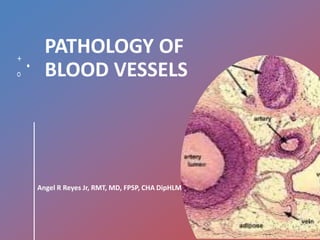
  • 1. PATHOLOGY OF BLOOD VESSELS Angel R Reyes Jr, RMT, MD, FPSP, CHA DipHLM
  • 2. 1.NORMAL VESSELS • 3 concentric layer; intima, media & adventitia.  Intima; internal elastic lamina  Media;external elastic lamina  Adventitia; vasa vasorum • Arteries;  Elastic(large): aorta, innominate, subclavian,  Muscular(medium-sized): renal and coronary  Small arteries and arterioles • Capillaries • Veins;  post-capillary venules, collecting venules, veins.
  • 3. 3
  • 4. 4 Arteries • Large arteries are elastic (conducting) arteries – pressure reservoirs • Medium arteries are muscular (distributing) arteries – more smooth muscle • Contraction or relaxation of muscle changes the size of the lumen, and so controls the blood pressure in the vessel.
  • 5. 5
  • 6. 6 Capillaries • Only a single layer of endothelium and a basement membrane • Connect arterioles and venules • Functional part of system • True capillaries begin at a precapillary sphincter which controls blood flow through the capillary
  • 7. 7
  • 8. 8 Veins • Relatively thin; less elastic • Larger in diameter than arteries • Have valves to prevent backflow of blood • Flow to heart is assisted by contraction of skeletal muscles
  • 9. 9
  • 10. 10
  • 11. 11
  • 12. 12 Control of systemic circulation • Nervous control – innervated by sympathetic nervous system ONLY • Cardiac control center (primarily in medulla oblongata) • Heart has both Sympathetic and Parasympathetic innervations.
  • 13. 13 • Baroreceptors and chemoreceptors: – Monitor pressure – Monitor blood levels of O2, CO2 and H+ – Send information to cardiovascular center, which responds
  • 14. Vascular organization • Large elastic arteries > medium-sized arteries > small arteries >arterioles > capillaries > post- capillary venules > collecting venules > veins
  • 15. Lymphatics • Endothelium-lined channels that drain excess interstitial tissue fluid, eventually returning it to blood via the thoracic ducts.
  • 16. 2.VASCULAR WALL CELLS AND THEIR RESPONSE TO INJURY • Endothelial cells; Elaboration of Anticoagulant, Antithrombotic, Fibrinolytic Regulators Elaboration of Prothrombotic Molecules Extracellular Matrix Production (Collagen, Proteoglycans) Modulation of Blood Flow and Vascular Reactivity Regulation of Inflammation and Immunity Regulation of Cell Growth Oxidation of LDL
  • 17. Cont.. • Vascular Smooth Muscle Cells proliferate when appropriately stimulated synthesize ECM collagen, elastin, and proteoglycans elaborate growth factors and cytokines vasoconstriction or vasodilation
  • 18. Response of Vascular Wall Cells to Injury • Endothelial injury contributes to a host of pathologies including thrombosis, atherosclerosis, and hypertensive vascular lesions. • Injury to the vessel wall results in a healing response, involving intimal expansion by proliferating SMCs and newly synthesized ECM. • The recruitment and activation of the SMCs in this process involves signals from cells eg ECs, and mediators derived from coagulation and complement cascades. • Therefore, intimal thickening is a stereotyped Response to Vascular Injury
  • 19.
  • 20. 3.CONGENITAL ANOMALIES • Rarely symptomatic; Developmental (berry) aneurysms occur in cerebral vessels. Arteriovenous fistulas are direct connections between arteries and veins that bypass the intervening capillaries. Fibromuscular dysplasia is a focal irregular thickening of the walls of medium and large muscular arteries.
  • 21. 4.ARTERIOSCLEROSIS • Arterial wall thickening and loss of elasticity. Arteriolosclerosis affects small arteries and arterioles with two anatomic variants hyaline and hyperplastic. Mönckeberg medial calcific sclerosis; calcific deposits in muscular arteries. Atherosclerosis.
  • 22. 5.ATHEROSCLEROSIS • General descriptions; intimal lesions called atheromas (also called atheromatous or atherosclerotic plaques), that protrude into vascular lumina. plaque consists of a raised lesion with a soft, yellow, grumous core of cholesterol (esters) covered by a firm, white fibrous cap. obstructs blood flow & weaken the underlying media and rupture, causing acute catastrophic vessel thrombosis. causes ischemic heart disease (IHD) .
  • 23. Epidemiology • Causes more morbidity and mortality (roughly half of all deaths) in the Western world than any other disorder. • The mortality rate for IHD in the United States is among the highest in the world and is approximately five times higher than that in Japan. • Japanese who immigrate to the United States and adopt American lifestyles and dietary customs acquire the same predisposition to atherosclerosis as the homegrown population.
  • 24. Major Constitutional Risk Factors for IHD • Nonmodifiable factors; Age; between ages 40 and 60, the incidence of myocardial infarction in men increases fivefold. Gender; premenopausal women are relatively protected against atherosclerosis and its consequences compared with age-matched men. Genetics; well-established familial predisposition to atherosclerosis and IHD is multifactorial:such as hypertension or diabetes, familial hypercholesterolemia, that result in excessively high blood lipid levels.
  • 25. • Major Modifiable Factors;  Hyperlipidemia; esp. hypercholesteremia i.e. LDL cholesterol has an essential physiologic role delivering cholesterol to peripheral tissues while mobilizes cholesterol from developing and existing atheromas. Hypertension; increase the risk of IHD by approximately 60% in comparison with normotensive populations. Cigarette Smoking; Prolonged (years) smoking of one pack of cigarettes or more daily increases the death rate from IHD by 200%. Diabetes Mellitus; induces hypercholesterolemia, the incidence of myocardial infarction is twice as high in diabetic as in nondiabetic individuals.
  • 26. • Additional Factors; Inflammation as marked by C-reactive protein. Hyperhomocystinemia. Lipoprotein a. Factors Affecting Hemostasis. Other Factors; like lack of exercise; competitive, stressful lifestyle ("type A" personality); and obesity.
  • 27. Pathogenesis • Response-to-injury hypothesis; Atherosclerosis as a chronic inflammatory response of the arterial wall to endothelial injury. Lesion progression occurs through interactions of modified lipoproteins, monocyte-derived macrophages, T lymphocytes, and the normal cellular constituents of the arterial wall.
  • 28. • Central tenets of the hypothesis; Chronic endothelial injury, with resultant endothelial dysfunction, causing (among other things) increased permeability, leukocyte adhesion, and thrombosis. Accumulation of lipoproteins (mainly LDL and its oxidized forms) in the vessel wall. Monocyte adhesion to the endothelium, followed by migration into the intima and transformation into macrophages and foam cells. Platelet adhesionFactor release from activated platelets, macrophages, and vascular wall cells, inducing  SMC recruitment, either from the media or from circulating precursors. SMC proliferation and ECM production. Lipid accumulation both extracellularly and within cells (macrophages and SMCs).
  • 29.
  • 30.
  • 31. Morphology • Fatty Streaks; composed of lipid-filled foam cells but are not significantly raised, begin as multiple minute yellow, flat spots that can coalesce into elongated streaks, • Atherosclerotic Plaque; a.k.a fibrous or fibrofatty plaques impinge on the lumen of the artery and grossly appear white to yellow; thrombosis superimposed over the surface of ulcerated plaques is red-brown in color. • Atherosclerotic plaques have three principal components: (1) cells, including SMCs, macrophages, and T cells; (2) ECM, including collagen, elastic fibers, and proteoglycans; and (3) intracellular and extracellular lipid. • Parts of the plaque(on cross-section):  Superficial fibrous cap is composed of SMCs and relatively dense collagen.  “Shoulder”- beneath and to the side of the cap = more cellular area containing macrophages, T cells, and SMCs.  Necrotic core; deep to the fibrous cap, containing cholesterol (esters), debris from dead cells, foam cells (lipid-laden macrophages and SMCs), fibrin, variably organized thrombus, and other plasma proteins; the cholesterol content is frequently present as crystalline aggregates that are washed out during routine tissue processing and leave behind only empty "clefts.“  Neovascularization at the periphery of the lesions,
  • 32. m
  • 33.
  • 34.
  • 35. • Plaque changes; Rupture, ulceration, or erosion; exposes the bloodstream to highly thrombogenic substances and induces thrombus formation and occlude the lumen and lead to downstream ischemia. Hemorrhage into a plaque Atheroembolism; plaque rupture can discharge debris into the bloodstream, producing microemboli composed of plaque contents. Aneurysm formation; Atherosclerosis-induced pressure or ischemic atrophy of the underlying media, with loss of elastic tissue, causes weakness of the vessel wall and development of aneurysms that may rupture
  • 36. Natural History of Atherosclerosis • Preclinical and clinical phase.
  • 37. Prevention of Atherosclerotic Vascular Disease • Primary prevention programs; cessation of cigarette smoking, control of hypertension, weight loss, exercise, and lowering total and LDL blood cholesterol levels while increasing HDL (e.g., by diet or through statins) • Secondary prevention programs; use of aspirin (anti-platelet agent), statins, and beta blockers (to limit cardiac demand), as well as surgical interventions (e.g., coronary artery bypass surgery, carotid endarterectomy). These can successfully reduce recurrent myocardial or cerebral events.
  • 38. 6.HYPERTENSIVE VASCULAR DISEASE • General description; Elevated blood pressure is called hypertension. Remains asymptomatic until late in its course. Contributes to the pathogenesis of coronary heart disease and cerebrovascular accidents, causes cardiac hypertrophy and heart failure, aortic dissection, and renal failure.
  • 39. Regulation of Blood Pressure • BP= CO X PR • RAA System • Vasodilators • Adrenal aldosterone • ANP
  • 40. Pathogenesis of Hypertension • 90% to 95% of hypertension is idiopathic (essential hypertension), which is compatible with long life, unless a myocardial infarction, cerebrovascular accident, or other complication supervenes. • Most of the remainder of "benign hypertension" is secondary to renal disease or, less often, to narrowing of the renal artery, usually by an atheromatous plaque (renovascular hypertension). • Accelerated or malignant hypertension, the clinical syndrome is characterized by severe hypertension (diastolic pressure over 120mmHg), renal failure, and retinal hemorrhages and exudates, with or without papilledema
  • 41. • Essential Hypertension;  alterations in renal sodium homeostasis and/or vessel wall tone or structure underlie essential hypertension.  interplay of multiple genetic and environmental factors affecting cardiac output and/or peripheral resistance. • Secondary Hypertension;  Renal: acute glomerulonephritis, chronic renal disease, polycystic disease, renal artery stenosis, renal vasculitis, renin-producing tumors.  Endocrine: Adrenocortical hyperfunction, Exogenous hormones, Pheochromocytoma, Acromegaly, Hypo/hyperthyroidism, Pregnancy-induced.  Cardiovascular: Coarctation of aorta , polyarteritis nodosa increased intravascular volume , increased cardiac output , rigidity of the aorta.  Neurologic: psychogenic , increased intracranial pressure , sleep apnea, acute stress (surgery).
  • 42. Vascular Pathology in Hypertension • Accelerating atherogenesis • Potentiate both aortic dissection and cerebrovascular hemorrhage • Two forms of small blood vessel disease Hyaline Arteriolosclerosis: a homogeneous pink hyaline thickening of the walls of arterioles with loss of underlying structural detail and with narrowing of the lumen  Hyperplastic Arteriolosclerosis. Related to acute or severe elevations of blood pressure, characteristic of malignant hypertension , associated with "onion-skin," concentric, laminated thickening of the walls of arterioles with luminal narrowing ,the laminations consist of SMCs and thickened, duplicated basement membrane.
  • 43.
  • 44. 7.ANEURYSMS AND DISSECTIONS • General description; aneurysm is a localized abnormal dilation of a blood vessel or the heart  "true" aneurysm.  false aneurysm (pseudoaneurysm): pulsating hematoma. arterial dissection arises when blood enters the wall of the artery, as a hematoma dissecting between its layers.  Causes; atherosclerosis and cystic medial degeneration of the arterial media, wall-weakening factors like include trauma, congenital defects (e.g., berry aneurysms), infections (mycotic aneurysms), or syphilis, and vasculitis.  Mycotic aneurysms can originate (1) from embolization of a septic thrombus, usually as a complication of infective endocarditis; (2) as an extension of an adjacent suppurative process; or (3) by circulating organisms directly infecting the arterial wall.
  • 45.
  • 46. Abdominal Aortic Aneurysm • Definition Out pouching of the aorta, most commonly in abdominal aorta, following atherosclerotic destruction of the aortic media, leading to vessel wall weakness
  • 47. • Morphology  Big fusiform or sacular bulge out the side of the abdominal aorta of varying width and length below the renal arteries and above the bifurcation of the aorta.  Often contains atheromatous ulcers with thrombi (source of atherothrombotic origin)  May be so large that it compresses nearby vessels or the thrombus causes occlusion syndromes Special Variants • Inflammatory AAA = Macrophages, Giant Cells, Lymphocytes, Plasma cells, AAA • Mycotic AAA = Supuritic lesions with organisms hiding inside (salmonella)
  • 48. S
  • 49. • Pathogenesis  Atherosclerosis is number 1 cause Cystic Medial Degeneration = Collagen. Aneurysms come from abnormal collagen (Marfan’s) or an abnormality of collagen remodeling (increased degradation decreased synthesis caused by an immune reaction) resulting in an inherently weakened aortic wall
  • 50. • Clinical Course  Rupture is often fatal. Hemorrhage occurs into the peritoneum. ↑size = ↑risk  Obstruction of a vessel (mesenteric, renal, vertebral) leading to ischemic injury  Direct Compression of the ureter or of the vertebrae (casing vertebral erosion)  Embolism from the thrombus that’s sitting inside  Tumor = AAA is a palpable mass that may
  • 51. Syphilitic Aneurysm • Cause obliterative endarteritis characteristic of the tertiary stage of syphilis can involve small vessels in any part of the body. Syphilis (stage 3) has a vascular predilection for small vessels, especially of the aorta Infection with subsequent Inflammation of the vasa vasorum leads to obstruction, inducing ischemia and obliterative endarteritis of the aorta, causing death of muscle and elastic tissue, called syphilitic aortitis
  • 52. • Morphology Starts in the adventitia with vessels with inflammation reactions in adventitia. syphilitic aortitis. Muscle that dies is replaced with fibrous scar tissue in the media, contraction of which causes the “tree‐barking” appearance Effects thoracic aorta with possible dilation of aortic valve leading to insufficiency
  • 53. • Presentation All causes by the encroachment on mediastinal tissues •Crush the lungs = Dyspnea Pain from rib and vertebral erosions Death from rupture •Crush the esophagus = Dysphagia, Cardiac Disease from valve involvement Aortic Regurgitation
  • 54. Aortic Dissection • Definition blood splays apart the laminar planes of the media to form a blood-filled channel within the aortic wall ruptures through the adventitia and into various spaces, where it causes either massive hemorrhage or cardiac tamponade (hemorrhage into the pericardial sac). men aged 40 to 60 years, with antecedent hypertension (> 90% of cases ), and younger patients with systemic or localized abnormalities of connective tissue affecting the aorta ( Marfan’s )
  • 55. • Morphology A single intimal tear cuts into but not through the media of the ascending aorta creating a blood filled pocket within the aorta, between layers Dissection can continue in both direction (towards the heart and towards the femoral) Usually rupture outwards, but can rupture back inwards – Outward = hemorrhage = fatal – Inward = second intimal tear back into the normal lumen, forming a new vascular channel (“double‐barrel Aorta”) which can, over time, endothelize and become a permanent vessel.
  • 56. No distinguishing histology except for warning signs = elastic tissue fragmentation and medial degeneration
  • 57. • Pathogenesis HTN is primary causative agent, if you see HTN pick Dissecting Hematoma Medial weakening is not required for dissection, but cystic medial necrosis from Marfan’s or Ehler’s Danlos (weak elastic layer) predisposes dissection If you have a dissection, pick HTN. If HTN isn’t there, pick Marfans Once dissection has happened, arterial blood pressure favors hematoma
  • 58. • Clinical Course If the dissection is Proximal subclavian/carotid (type A = bad), if distal (type B = better) Pain in the chest radiating to the back is a tell tale sign Death usually results from rupture If dissection involves aortic root, you can get valve problems (regurgitation, murmur
  • 60. 8.VASCULITIS • General description; Inflammation of vessel walls. 2 mechanisms: immune-mediated inflammation and direct invasion of vascular walls by infectious pathogens. Noninfectious Vasculitis. Infectious Vasculitis. Vasculitis Associated with Other Disorders .
  • 61. Non-infectious Vasculitis. • Classification; Large-Vessel Vasculitis » Giant-cell (temporal) arteritis » Takayasu arteritis Medium-Vessel Vasculitis » Polyarteritis nodosa » Kawasaki disease Small-Vessel Vasculitis » Wegener granulomatosis » Churg-Strauss syndrome » Microscopic polyangiitis
  • 62. • Pathogenesis;  immune complex deposition; Antibody and complement are typically detected in vasculitic lesions, DNA-anti-DNA complexes(SLE) and drug hypersensitivity.Can be 2ndary to viral infections. antineutrophil cytoplasmic antibodies (ANCAs); circulating antibodies that react with neutrophil cytoplasmic antigens. Cytoplasmic localization (c- ANCA) >>proteinase-3 (PR3) and Perinuclear localization (p-ANCA) >>myeloperoxidase (MPO). anti-endothelial cell antibodies; Antibodies to ECs may predispose to certain vasculitides, for example Kawasaki disease.
  • 63. • Giant-Cell (Temporal) Arteritis Most common form of Vasculitis, especially in Elderly Women affects the arteries in the head-esp. the temporal arteries-but also the vertebral and ophthalmic arteries & the aorta (giant-cell aortitis). T cell-mediated immune response to an unknown vessel wall antigen. nodular intimal thickening with reduction of the lumen and occasional thrombosis. granulomatous inflammation within the inner media centered on the internal elastic membrane. fragmentation of the internal elastic lamina Head pain, vision disturbances /blindness(ophthalmic artery involved) Responds well to steroids.
  • 64.
  • 65. • Takayasu Arteritis "pulseless disease“ transmural fibrous thickening of the aorta-particularly the aortic arch and great vessels. severe luminal narrowing of the major branch vessels- loss of pulse. in women younger than 40 years of age(Japanese population) intimal hyperplasia and irregular thickening of the vessel wall
  • 66.
  • 67. adventitial mononuclear infiltrates >> mononuclear inflammation in the media >> granulomatous inflammation(giant cells and patchy medial necrosis) reduced blood pressure and weaker pulses in the upper extremities with coldness or numbness of the fingers; ocular disturbances, and neurologic deficits. Claudication of the legs(distal aorta involved); pulmonary hypertension(PA involved). Narrowing of the coronary ostia >>MI, systemic hypertension(renal arteries involved)
  • 68. • Polyarteritis Nodosa Involves renal and visceral vessels but not the pulmonary circulation. segmental transmural necrotizing inflammation of small to medium-sized arteries part of the vessel circumference involved, with a predilection for branch points. weakens the arterial wall >>aneurysms or even rupture. Impaired perfusion >>in the distribution of affected vessels transmural inflammation >>fibrinoid necrosis >>fibrous /nodular thickening of the vessel wall that can extend into the adventitia(which may coexist)
  • 69. • Kawasaki Disease self-limited illness of infancy and childhood (80% of patients are younger than 4 years) coronary arteritis >>aneurysms that rupture or thrombose, causing AMI. leading cause of acquired heart disease in children. delayed-type hypersensitivity -T cells to uncharacterized vascular antigen. pronounced inflammation affecting the entire thickness of the vessel wall. Clinical Course: mucocutaneous lymph node syndrome with conjunctival & oral erythema and erosion, edema of the hands and feet, erythema of the palms and soles, a desquamative rash, and cervical lymph node enlargement
  • 70. • Microscopic Polyangiitis • necrotizing vasculitis that affects capillaries , arterioles and venules. • hypersensitivity vasculitis or leukocytoclastic vasculitis. • necrotizing glomerulonephritis (90% of patients) and pulmonary capillaritis present. • an antibody response to antigens such as drugs (e.g., penicillin), microorganisms (e.g., streptococci), heterologous proteins, or tumor proteins. • segmental fibrinoid necrosis of the media- with focal transmural necrotizing lesions • granulomatous inflammation is absent. • infiltrating and fragmenting neutrophils- in postcapillary venules. • little or no immunoglobulin can be seen in most lesions (so-called "pauci-immune" injury).
  • 71. • Wegener Granulomatosis Triad – Acute necrotizing granulomas of the upper respiratory tract – Necrotizing or granulomatous vasculitis – Renal disease in the form of focal necrotizing, often crescentic, glomerulonephritis. cell-mediated hypersensitivity response, possibly to an inhaled infectious or other environmental agent. The upper respiratory tract lesions range from inflammatory sinusitis with mucosal granulomas to ulcerative lesions of the nose, palate, or pharynx, rimmed by granulomas with geographic patterns of central necrosis and accompanying vasculitis renal lesions
  • 72. Clinical Features • persistent pneumonitis with bilateral nodular and cavitary infiltrates • chronic sinusitis (90%), • mucosal ulcerations of the nasopharynx (75%), and evidence of renal disease (80%). • Other features include rashes, muscle pains, articular involvement, mononeuritis or polyneuritis, and fever • Allergic granulomatosis and angiitis (Churg- Strauss syndrome) has association with allergic rhinitis, bronchial asthma, and peripheral eosinophilia; p-ANCAs are present in roughly half the patients.
  • 73. • Thromboangiitis Obliterans Buerger Disease segmental, thrombosing acute and chronic inflammation(tibial and radial arteries) exclusively in heavy smokers of cigarettes, usually beginning before age 35. direct toxicity to endothelium by some tobacco products, or an idiosyncratic immune response to the same agents sharply segmental acute and chronic vasculitis of medium-sized and small arteries, predominantly of the extremities. superficial nodular phlebitis, cold sensitivity of the Raynaud type in the hands, and instep claudication.
  • 74. Vasculitis Associated with Other Disorders • Disorders; rheumatoid arthritis, SLE, malignancy, or systemic illnesses such as mixed cryoglobulinemia, antiphospholipid antibody syndrome and Henoch-Schönlein purpura • Rheumatoid vasculitis • Lupus vasculitis
  • 75. Infectious Vasculitis • Direct invasion of infectious agents; Aspergillus and Mucor species mycotic aneurysms can induce thrombosis and infarction.
  • 76. 9.RAYNAUD PHENOMENON • Results from an exaggerated vasoconstriction of digital arteries and arterioles • Two types; Primary Raynaud phenomenon; exaggeration of central and local vasomotor responses to cold or emotion(common in young women) Secondary Raynaud phenomenon; vascular insufficiency of the extremities due to arterial disease from other entities (SLE, scleroderma, Buerger disease or atherosclerosis).
  • 77. 10.VEINS AND LYMPHATICS • Varicose Veins Superficial veins (usually of the legs) become distended, more cosmetic than anything else Obesity, jobs with legs dependent (barber, surgeon), and pregnancy are disposing factors Stasis of blood, rupture of valves, or thinning of walls allows distention May cause ulceration, but are generally asymptomatic (do not cause emboli) Can also be in the esophagus (esopheal varices) and in the anus (hemorrhoids).
  • 78. • Thrombophlebitis and Phlebothrombosis Aka Deep Vein Thrombosis, DVT. The distinction between thrombosis of the vein (phlebothrombosis) and inflammation of the Vein with thrombosis (thrombophlebitis) is not relevant, they are all DVTs Presents with pain in the calf with red, tender lesions, with a positive Homan’s Sign(dorsiflexion induces pain) Risk increases with Estrogen, Birth Control, Smoking, Age, Hypercoagulability Can result in pulmonary embolism or edema
  • 79. • Superior and Inferior Vena Caval Syndromes Something blocks these large veins (invasive neoplasm, mural thrombosis) that causes a back up distally, without a failure of the heart Massive edema inferiorly for IVC (ankle, ascites, pelvis) or superiorly for SVC (face, neck, arms)
  • 80. • Lymphangitis and Lymphedema Infection gets into the lymph nodes. Red streaks from site of penetration, follows along lymph tract, finished at lymph node. Lymphadenopathy is present with PMNs and Lymphocytes infiltrating site of infection. Caused by Cancer or Virulent Bacteria (Staph).
  • 81. 11.TUMORS • Classification of Vascular Tumors and Tumor-like Conditions  Benign Neoplasms, Developmental and Acquired Conditions – Hemangioma – Capillary hemangioma – Cavernous hemangioma – Pyogenic granuloma Lymphangioma – Simple (capillary) lymphangioma – Cavernous lymphangioma (cystic hygroma) – Glomus tumor – Vascular ectasias – Nevus flammeus – Spider telangiectasia (arterial spider) – Hereditary hemorrhagic telangiectasis (Osler-Weber-Rendu disease) – Reactive vascular proliferations – Bacillary angiomatosis  Intermediate-Grade Neoplasms – Kaposi sarcoma – Hemangioendotheliom  Malignant Neoplasms – Angiosarcoma – Hemangiopericytoma
  • 82. • Benign Tumors and Tumor-like Conditions Hemangioma – Capillary Hemangioma – Cavernous Hemangioma – Pyogenic Granuloma Lymphangioma – Simple (Capillary) Lymphangioma – Cavernous Lymphangioma (Cystic Hygroma)  Glomus Tumor (Glomangioma) Vascular Ectasias – Nevus Flammeus – Spider Telangiectasia – Hereditary Hemorrhagic Telangiectasia (Osler-Weber-Rendu Disease) Bacillary Angiomatosis
  • 83. • Intermediate-Grade (Borderline Low-Grade Malignant) Tumors Kaposi Sarcoma – Chronic KS – Lymphadenopathic KS (also called African or endemic KS) – Transplant-associated KS – AIDS-associated (epidemic) KS Hemangioendothelioma – Epithelioid hemangioendothelioma
  • 84. • Malignant Tumors Angiosarcoma  Hepatic angiosarcomas  Lymphangiosarcoma Hemangiopericytoma
  • 85. 12.PATHOLOGY OF VASCULAR INTERVENTION • Endovascular Stenting • Vascular Replacement

Editor's Notes

  1. Type A (proximal) involves the ascending aorta, either in isolation (DeBakey I) or as part of a more extensive dissection (DeBakey II). Type B (distal, or DeBakey III) dissections arise after the take off of the great vessels. The serious complications predominantly occur in Type A dissections, which therefore mandate surgical intervention
  2. c-ANCA is typical of Wegener granulomatosis and p-ANCA is found in most cases of microscopic polyangiitis and Churg-Strauss syndrome. A plausible mechanism for ANCA vasculitis is: Neutrophil release of PR3 and MPO (e.g., in the setting of infections) incites ANCA formation in a susceptible host.Some underlying disorder (e.g., infection, endotoxin exposure, etc.) elicits inflammatory cytokines, such as TNF, that result in surface expression of PR3 and MPO on neutrophils and other cell types.ANCAs react with these cytokine-primed cells and either cause direct injury (e.g., to endothelium) or induce activation (e.g., in neutrophils).ANCA-activated neutrophils degranulate and also cause injury by the release of reactive oxygen species, engendering EC toxicity and other direct tissue injury.
  3. Thus, distinctions between active giant-cell lesions of the aorta are based largely on the age of the patient; most aortic giant-cell lesions in young patients (age 40 years and younger) are designated as Takayasu aortitis.
  4. The most common manifestations are malaise, fever, and weight loss; hypertension, usually developing rapidly; abdominal pain and melena (bloody stool) caused by vascular GI lesions; diffuse muscular aches and pains; and peripheral neuritis, predominantly affecting motor nerves
  5. Clinical Course hemoptysis, hematuria, and proteinuria; bowel pain or bleeding; muscle pain or weakness; and palpable cutaneous purpura-depending on the vascular bed involved.
  6. The renal lesions range over a spectrum. At one end, there is mild or early disease, where glomeruli show acute focal necrosis with thrombosis of isolated glomerular capillary loops (focal and segmental necrotizing glomerulonephritis). More advanced glomerular lesions are characterized by diffuse necrosis and parietal cell proliferation to form crescents (crescentic glomerulonephritis). Patients with focal lesions may have only hematuria and proteinuria responsive to therapy, whereas those with diffuse disease can develop rapidly progressive renal failure.