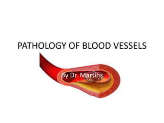
This document summarizes pathology of blood vessels. It begins by describing the normal structure of arteries, veins and capillaries. It then discusses the cells that make up blood vessel walls and their response to injury, which can lead to intimal thickening. It also briefly mentions some congenital vessel anomalies. The majority of the document focuses on arteriosclerosis and its subtype, atherosclerosis - describing the morphology, risk factors, pathogenesis, natural history and approaches for prevention. It concludes by outlining hypertensive vascular disease, its causes and pathogenesis.
Dear all, Pathologybasics is out with a new series of power point presentations on Systemic Pathology.. Following is link presentation on 12th chapter of robbins - the heart.This presentation includes valvular heart diseases, endocarditis, cardiomyopathies, pericardial diseases and tumors of the heart. Remaining topics will be uploaded as a separate presentation soon.
Dear all, Pathologybasics is out with a new series of power point presentations on Systemic Pathology.. Following is link presentation on 12th chapter of robbins - the heart.This presentation includes valvular heart diseases, endocarditis, cardiomyopathies, pericardial diseases and tumors of the heart. Remaining topics will be uploaded as a separate presentation soon.
Pathology of Cardiomyopathies
Literally means “disease of the heart muscle”.
Term “cardiomyopathy” is used to describe heart disease resulting from an abnormality in the myocardium.
Diseases of the myocardium usually produce:
>abnormalities in cardiac wall thickness and chamber size.
>mechanical or electrical dysfunction
>associated with significant morbidity and mortality.
Pathology of Central nervous system /certified fixed orthodontic courses by I...Indian dental academy
The Indian Dental Academy is the Leader in continuing dental education , training dentists in all aspects of dentistry and offering a wide range of dental certified courses in different formats.
Indian dental academy provides dental crown & Bridge,rotary endodontics,fixed orthodontics,
Dental implants courses.for details pls visit www.indiandentalacademy.com ,or call
0091-9248678078
Atherosclerosis - Definition - Risk Factors - Lesser and Non Quantitated risk factors - Arterial wall - The development of Atherosclerosis - Many Features of the injury Hypothesis - The process of Atherogenesis - Pathogenesis in short - Morphology of Atheroma - Components of Atheromatous Plaque (MP) - Complications and clinical significance - Cardiovascular risk and its assessment.
CAD is the commonest cause of deaths worldwide. Mortality rates have declined over the past four decades in western countries however this condition remains responsible for ~one-third of all deaths in individuals over age 35.
Mortality is on the rise in Low and middle income countries Tanzania being among. The 2016 Heart Disease and Stroke Statistics update of the AHA reported that 15.5 million people in the USA. have CHD.
The reported prevalence increases with age for both women and men. For those US people, the lifetime risk of developing CHD with ≥2 major risk factors is 37.5% for men and 18.3% for women.
Couples presenting to the infertility clinic- Do they really have infertility...Sujoy Dasgupta
Dr Sujoy Dasgupta presented the study on "Couples presenting to the infertility clinic- Do they really have infertility? – The unexplored stories of non-consummation" in the 13th Congress of the Asia Pacific Initiative on Reproduction (ASPIRE 2024) at Manila on 24 May, 2024.
- Video recording of this lecture in English language: https://youtu.be/lK81BzxMqdo
- Video recording of this lecture in Arabic language: https://youtu.be/Ve4P0COk9OI
- Link to download the book free: https://nephrotube.blogspot.com/p/nephrotube-nephrology-books.html
- Link to NephroTube website: www.NephroTube.com
- Link to NephroTube social media accounts: https://nephrotube.blogspot.com/p/join-nephrotube-on-social-media.html
These lecture slides, by Dr Sidra Arshad, offer a quick overview of physiological basis of a normal electrocardiogram.
Learning objectives:
1. Define an electrocardiogram (ECG) and electrocardiography
2. Describe how dipoles generated by the heart produce the waveforms of the ECG
3. Describe the components of a normal electrocardiogram of a typical bipolar leads (limb II)
4. Differentiate between intervals and segments
5. Enlist some common indications for obtaining an ECG
Study Resources:
1. Chapter 11, Guyton and Hall Textbook of Medical Physiology, 14th edition
2. Chapter 9, Human Physiology - From Cells to Systems, Lauralee Sherwood, 9th edition
3. Chapter 29, Ganong’s Review of Medical Physiology, 26th edition
4. Electrocardiogram, StatPearls - https://www.ncbi.nlm.nih.gov/books/NBK549803/
5. ECG in Medical Practice by ABM Abdullah, 4th edition
6. ECG Basics, http://www.nataliescasebook.com/tag/e-c-g-basics
New Directions in Targeted Therapeutic Approaches for Older Adults With Mantl...i3 Health
i3 Health is pleased to make the speaker slides from this activity available for use as a non-accredited self-study or teaching resource.
This slide deck presented by Dr. Kami Maddocks, Professor-Clinical in the Division of Hematology and
Associate Division Director for Ambulatory Operations
The Ohio State University Comprehensive Cancer Center, will provide insight into new directions in targeted therapeutic approaches for older adults with mantle cell lymphoma.
STATEMENT OF NEED
Mantle cell lymphoma (MCL) is a rare, aggressive B-cell non-Hodgkin lymphoma (NHL) accounting for 5% to 7% of all lymphomas. Its prognosis ranges from indolent disease that does not require treatment for years to very aggressive disease, which is associated with poor survival (Silkenstedt et al, 2021). Typically, MCL is diagnosed at advanced stage and in older patients who cannot tolerate intensive therapy (NCCN, 2022). Although recent advances have slightly increased remission rates, recurrence and relapse remain very common, leading to a median overall survival between 3 and 6 years (LLS, 2021). Though there are several effective options, progress is still needed towards establishing an accepted frontline approach for MCL (Castellino et al, 2022). Treatment selection and management of MCL are complicated by the heterogeneity of prognosis, advanced age and comorbidities of patients, and lack of an established standard approach for treatment, making it vital that clinicians be familiar with the latest research and advances in this area. In this activity chaired by Michael Wang, MD, Professor in the Department of Lymphoma & Myeloma at MD Anderson Cancer Center, expert faculty will discuss prognostic factors informing treatment, the promising results of recent trials in new therapeutic approaches, and the implications of treatment resistance in therapeutic selection for MCL.
Target Audience
Hematology/oncology fellows, attending faculty, and other health care professionals involved in the treatment of patients with mantle cell lymphoma (MCL).
Learning Objectives
1.) Identify clinical and biological prognostic factors that can guide treatment decision making for older adults with MCL
2.) Evaluate emerging data on targeted therapeutic approaches for treatment-naive and relapsed/refractory MCL and their applicability to older adults
3.) Assess mechanisms of resistance to targeted therapies for MCL and their implications for treatment selection
Pulmonary Thromboembolism - etilogy, types, medical- Surgical and nursing man...VarunMahajani
Disruption of blood supply to lung alveoli due to blockage of one or more pulmonary blood vessels is called as Pulmonary thromboembolism. In this presentation we will discuss its causes, types and its management in depth.
Lung Cancer: Artificial Intelligence, Synergetics, Complex System Analysis, S...Oleg Kshivets
RESULTS: Overall life span (LS) was 2252.1±1742.5 days and cumulative 5-year survival (5YS) reached 73.2%, 10 years – 64.8%, 20 years – 42.5%. 513 LCP lived more than 5 years (LS=3124.6±1525.6 days), 148 LCP – more than 10 years (LS=5054.4±1504.1 days).199 LCP died because of LC (LS=562.7±374.5 days). 5YS of LCP after bi/lobectomies was significantly superior in comparison with LCP after pneumonectomies (78.1% vs.63.7%, P=0.00001 by log-rank test). AT significantly improved 5YS (66.3% vs. 34.8%) (P=0.00000 by log-rank test) only for LCP with N1-2. Cox modeling displayed that 5YS of LCP significantly depended on: phase transition (PT) early-invasive LC in terms of synergetics, PT N0—N12, cell ratio factors (ratio between cancer cells- CC and blood cells subpopulations), G1-3, histology, glucose, AT, blood cell circuit, prothrombin index, heparin tolerance, recalcification time (P=0.000-0.038). Neural networks, genetic algorithm selection and bootstrap simulation revealed relationships between 5YS and PT early-invasive LC (rank=1), PT N0—N12 (rank=2), thrombocytes/CC (3), erythrocytes/CC (4), eosinophils/CC (5), healthy cells/CC (6), lymphocytes/CC (7), segmented neutrophils/CC (8), stick neutrophils/CC (9), monocytes/CC (10); leucocytes/CC (11). Correct prediction of 5YS was 100% by neural networks computing (area under ROC curve=1.0; error=0.0).
CONCLUSIONS: 5YS of LCP after radical procedures significantly depended on: 1) PT early-invasive cancer; 2) PT N0--N12; 3) cell ratio factors; 4) blood cell circuit; 5) biochemical factors; 6) hemostasis system; 7) AT; 8) LC characteristics; 9) LC cell dynamics; 10) surgery type: lobectomy/pneumonectomy; 11) anthropometric data. Optimal diagnosis and treatment strategies for LC are: 1) screening and early detection of LC; 2) availability of experienced thoracic surgeons because of complexity of radical procedures; 3) aggressive en block surgery and adequate lymph node dissection for completeness; 4) precise prediction; 5) adjuvant chemoimmunoradiotherapy for LCP with unfavorable prognosis.
Title: Sense of Smell
Presenter: Dr. Faiza, Assistant Professor of Physiology
Qualifications:
MBBS (Best Graduate, AIMC Lahore)
FCPS Physiology
ICMT, CHPE, DHPE (STMU)
MPH (GC University, Faisalabad)
MBA (Virtual University of Pakistan)
Learning Objectives:
Describe the primary categories of smells and the concept of odor blindness.
Explain the structure and location of the olfactory membrane and mucosa, including the types and roles of cells involved in olfaction.
Describe the pathway and mechanisms of olfactory signal transmission from the olfactory receptors to the brain.
Illustrate the biochemical cascade triggered by odorant binding to olfactory receptors, including the role of G-proteins and second messengers in generating an action potential.
Identify different types of olfactory disorders such as anosmia, hyposmia, hyperosmia, and dysosmia, including their potential causes.
Key Topics:
Olfactory Genes:
3% of the human genome accounts for olfactory genes.
400 genes for odorant receptors.
Olfactory Membrane:
Located in the superior part of the nasal cavity.
Medially: Folds downward along the superior septum.
Laterally: Folds over the superior turbinate and upper surface of the middle turbinate.
Total surface area: 5-10 square centimeters.
Olfactory Mucosa:
Olfactory Cells: Bipolar nerve cells derived from the CNS (100 million), with 4-25 olfactory cilia per cell.
Sustentacular Cells: Produce mucus and maintain ionic and molecular environment.
Basal Cells: Replace worn-out olfactory cells with an average lifespan of 1-2 months.
Bowman’s Gland: Secretes mucus.
Stimulation of Olfactory Cells:
Odorant dissolves in mucus and attaches to receptors on olfactory cilia.
Involves a cascade effect through G-proteins and second messengers, leading to depolarization and action potential generation in the olfactory nerve.
Quality of a Good Odorant:
Small (3-20 Carbon atoms), volatile, water-soluble, and lipid-soluble.
Facilitated by odorant-binding proteins in mucus.
Membrane Potential and Action Potential:
Resting membrane potential: -55mV.
Action potential frequency in the olfactory nerve increases with odorant strength.
Adaptation Towards the Sense of Smell:
Rapid adaptation within the first second, with further slow adaptation.
Psychological adaptation greater than receptor adaptation, involving feedback inhibition from the central nervous system.
Primary Sensations of Smell:
Camphoraceous, Musky, Floral, Pepperminty, Ethereal, Pungent, Putrid.
Odor Detection Threshold:
Examples: Hydrogen sulfide (0.0005 ppm), Methyl-mercaptan (0.002 ppm).
Some toxic substances are odorless at lethal concentrations.
Characteristics of Smell:
Odor blindness for single substances due to lack of appropriate receptor protein.
Behavioral and emotional influences of smell.
Transmission of Olfactory Signals:
From olfactory cells to glomeruli in the olfactory bulb, involving lateral inhibition.
Primitive, less old, and new olfactory systems with different path
ARTIFICIAL INTELLIGENCE IN HEALTHCARE.pdfAnujkumaranit
Artificial intelligence (AI) refers to the simulation of human intelligence processes by machines, especially computer systems. It encompasses tasks such as learning, reasoning, problem-solving, perception, and language understanding. AI technologies are revolutionizing various fields, from healthcare to finance, by enabling machines to perform tasks that typically require human intelligence.
Report Back from SGO 2024: What’s the Latest in Cervical Cancer?bkling
Are you curious about what’s new in cervical cancer research or unsure what the findings mean? Join Dr. Emily Ko, a gynecologic oncologist at Penn Medicine, to learn about the latest updates from the Society of Gynecologic Oncology (SGO) 2024 Annual Meeting on Women’s Cancer. Dr. Ko will discuss what the research presented at the conference means for you and answer your questions about the new developments.
micro teaching on communication m.sc nursing.pdfAnurag Sharma
Microteaching is a unique model of practice teaching. It is a viable instrument for the. desired change in the teaching behavior or the behavior potential which, in specified types of real. classroom situations, tends to facilitate the achievement of specified types of objectives.
New Drug Discovery and Development .....NEHA GUPTA
The "New Drug Discovery and Development" process involves the identification, design, testing, and manufacturing of novel pharmaceutical compounds with the aim of introducing new and improved treatments for various medical conditions. This comprehensive endeavor encompasses various stages, including target identification, preclinical studies, clinical trials, regulatory approval, and post-market surveillance. It involves multidisciplinary collaboration among scientists, researchers, clinicians, regulatory experts, and pharmaceutical companies to bring innovative therapies to market and address unmet medical needs.
Prix Galien International 2024 Forum ProgramLevi Shapiro
June 20, 2024, Prix Galien International and Jerusalem Ethics Forum in ROME. Detailed agenda including panels:
- ADVANCES IN CARDIOLOGY: A NEW PARADIGM IS COMING
- WOMEN’S HEALTH: FERTILITY PRESERVATION
- WHAT’S NEW IN THE TREATMENT OF INFECTIOUS,
ONCOLOGICAL AND INFLAMMATORY SKIN DISEASES?
- ARTIFICIAL INTELLIGENCE AND ETHICS
- GENE THERAPY
- BEYOND BORDERS: GLOBAL INITIATIVES FOR DEMOCRATIZING LIFE SCIENCE TECHNOLOGIES AND PROMOTING ACCESS TO HEALTHCARE
- ETHICAL CHALLENGES IN LIFE SCIENCES
- Prix Galien International Awards Ceremony
6. 2.VASCULAR WALL CELLS AND THEIR
RESPONSE TO INJURY
• Endothelial cells;
Elaboration of Anticoagulant, Antithrombotic,
Fibrinolytic Regulators
Elaboration of Prothrombotic Molecules
Extracellular Matrix Production (Collagen,
Proteoglycans)
Modulation of Blood Flow and Vascular Reactivity
Regulation of Inflammation and Immunity
Regulation of Cell Growth
Oxidation of LDL
7. Ctd
• Vascular Smooth Muscle Cells
proliferate when appropriately stimulated
synthesize ECM collagen, elastin, and proteoglycans
elaborate growth factors and cytokines
vasoconstriction or vasodilation
8. Response of Vascular Wall Cells to
Injury
• Endothelial injury contributes to a host of pathologies
including thrombosis, atherosclerosis, and
hypertensive vascular lesions.
• Injury to the vessel wall results in a healing response,
involving intimal expansion by proliferating SMCs and
newly synthesized ECM.
• The recruitment and activation of the SMCs in this
process involves signals from cells eg ECs, and
mediators derived from coagulation and complement
cascades.
• Therefore, intimal thickening is a stereotyped
Response to Vascular Injury
9.
10. 3.CONGENITAL ANOMALIES
• Rarely symptomatic;
Developmental (berry) aneurysms occur in cerebral
vessels.
Arteriovenous fistulas are direct connections between
arteries and veins that bypass the intervening
capillaries.
Fibromuscular dysplasia is a focal irregular thickening
of the walls of medium and large muscular arteries.
11. 4.ARTERIOSCLEROSIS
• Arterial wall thickening and loss of elasticity.
Arteriolosclerosis affects small arteries and arterioles with
two anatomic variants hyaline and hyperplastic.
Mönckeberg medial calcific sclerosis; calcific deposits in
muscular arteries.
Atherosclerosis.
12. 5.ATHEROSCLEROSIS
• General descriptions;
intimal lesions called atheromas (also called
atheromatous or atherosclerotic plaques), that
protrude into vascular lumina.
plaque consists of a raised lesion with a soft, yellow,
grumous core of cholesterol (esters) covered by a firm,
white fibrous cap.
obstructs blood flow & weaken the underlying media
and rupture, causing acute catastrophic vessel
thrombosis.
causes ischemic heart disease (IHD) .
13. Epidemiology
• Causes more morbidity and mortality (roughly
half of all deaths) in the Western world than any
other disorder.
• The mortality rate for IHD in the United States is
among the highest in the world and is
approximately five times higher than that in
Japan.
• Japanese who immigrate to the United States and
adopt American lifestyles and dietary customs
acquire the same predisposition to
atherosclerosis as the homegrown population.
14. Major Constitutional Risk Factors for
IHD
• Nonmodifiable factors;
Age; between ages 40 and 60, the incidence of myocardial
infarction in men increases fivefold.
Gender; premenopausal women are relatively protected
against atherosclerosis and its consequences compared with
age-matched men.
Genetics; well-established familial predisposition to
atherosclerosis and IHD is multifactorial:such as hypertension
or diabetes, familial hypercholesterolemia, that result in
excessively high blood lipid levels.
15. • Major Modifiable Factors;
Hyperlipidemia; esp. hypercholesteremia i.e. LDL cholesterol
has an essential physiologic role delivering cholesterol to
peripheral tissues while mobilizes cholesterol from developing
and existing atheromas.
Hypertension; increase the risk of IHD by approximately 60%
in comparison with normotensive populations.
Cigarette Smoking; Prolonged (years) smoking of one pack of
cigarettes or more daily increases the death rate from IHD by
200%.
Diabetes Mellitus; induces hypercholesterolemia, the
incidence of myocardial infarction is twice as high in diabetic
as in nondiabetic individuals.
16. • Additional Factors;
Inflammation as marked by C-reactive protein.
Hyperhomocystinemia.
Lipoprotein a.
Factors Affecting Hemostasis.
Other Factors; like lack of exercise; competitive, stressful
lifestyle ("type A" personality); and obesity.
17. Pathogenesis
• Response-to-injury hypothesis;
Atherosclerosis as a chronic inflammatory response of the
arterial wall to endothelial injury.
Lesion progression occurs through interactions of modified
lipoproteins, monocyte-derived macrophages, T lymphocytes,
and the normal cellular constituents of the arterial wall.
18. • Central tenets of the hypothesis;
Chronic endothelial injury, with resultant endothelial
dysfunction, causing (among other things) increased
permeability, leukocyte adhesion, and thrombosis.
Accumulation of lipoproteins (mainly LDL and its oxidized forms)
in the vessel wall.
Monocyte adhesion to the endothelium, followed by migration
into the intima and transformation into macrophages and foam
cells.
Platelet adhesionFactor release from activated platelets,
macrophages, and vascular wall cells, inducing
SMC recruitment, either from the media or from circulating
precursors.
SMC proliferation and ECM production.
Lipid accumulation both extracellularly and within cells
(macrophages and SMCs).
19.
20.
21. Morphology
• Fatty Streaks; composed of lipid-filled foam cells but are not significantly raised,
begin as multiple minute yellow, flat spots that can coalesce into elongated
streaks,
• Atherosclerotic Plaque; a.k.a fibrous or fibrofatty plaques impinge on the lumen of
the artery and grossly appear white to yellow; thrombosis superimposed over the
surface of ulcerated plaques is red-brown in color.
• Atherosclerotic plaques have three principal components: (1) cells, including
SMCs, macrophages, and T cells; (2) ECM, including collagen, elastic fibers, and
proteoglycans; and (3) intracellular and extracellular lipid.
• Parts of the plaque(on cross-section):
Superficial fibrous cap is composed of SMCs and relatively dense collagen.
“Shoulder”- beneath and to the side of the cap = more cellular area containing macrophages,
T cells, and SMCs.
Necrotic core; deep to the fibrous cap, containing cholesterol (esters), debris from dead cells,
foam cells (lipid-laden macrophages and SMCs), fibrin, variably organized thrombus, and other
plasma proteins; the cholesterol content is frequently present as crystalline aggregates that
are washed out during routine tissue processing and leave behind only empty "clefts.“
Neovascularization at the periphery of the lesions,
25. • Plaque changes;
Rupture, ulceration, or erosion; exposes the bloodstream to
highly thrombogenic substances and induces thrombus
formation and occlude the lumen and lead to downstream
ischemia.
Hemorrhage into a plaque
Atheroembolism; plaque rupture can discharge debris into
the bloodstream, producing microemboli composed of
plaque contents.
Aneurysm formation; Atherosclerosis-induced pressure or
ischemic atrophy of the underlying media, with loss of
elastic tissue, causes weakness of the vessel wall and
development of aneurysms that may rupture
27. Prevention of Atherosclerotic Vascular
Disease
• Primary prevention programs; cessation of cigarette
smoking, control of hypertension, weight loss, exercise,
and lowering total and LDL blood cholesterol levels
while increasing HDL (e.g., by diet or through statins)
• Secondary prevention programs; use of aspirin (anti-
platelet agent), statins, and beta blockers (to limit
cardiac demand), as well as surgical interventions (e.g.,
coronary artery bypass surgery, carotid
endarterectomy). These can successfully reduce
recurrent myocardial or cerebral events.
28. 6.HYPERTENSIVE VASCULAR DISEASE
• General description;
Elevated blood pressure is called hypertension.
Remains asymptomatic until late in its course.
Contributes to the pathogenesis of coronary heart
disease and cerebrovascular accidents, causes cardiac
hypertrophy and heart failure, aortic dissection, and
renal failure.
29. Regulation of Blood Pressure
• BP= CO X PR
• RAA System
• Vasodilators
• Adrenal aldosterone
• ANP
30. Pathogenesis of Hypertension
• 90% to 95% of hypertension is idiopathic (essential
hypertension), which is compatible with long life, unless a
myocardial infarction, cerebrovascular accident, or other
complication supervenes.
• Most of the remainder of "benign hypertension" is
secondary to renal disease or, less often, to narrowing of
the renal artery, usually by an atheromatous plaque
(renovascular hypertension).
• Accelerated or malignant hypertension, the clinical
syndrome is characterized by severe hypertension (diastolic
pressure over 120mmHg), renal failure, and retinal
hemorrhages and exudates, with or without papilledema
32. Vascular Pathology in Hypertension
• Accelerating atherogenesis
• Potentiate both aortic dissection and
cerebrovascular hemorrhage
• Two forms of small blood vessel disease
Hyaline Arteriolosclerosis: a homogeneous pink hyaline
thickening of the walls of arterioles with loss of underlying
structural detail and with narrowing of the lumen
Hyperplastic Arteriolosclerosis. Related to acute or severe
elevations of blood pressure, characteristic of malignant
hypertension , associated with "onion-skin," concentric,
laminated thickening of the walls of arterioles with luminal
narrowing ,the laminations consist of SMCs and thickened,
duplicated basement membrane.
33.
34. 7.ANEURYSMS AND DISSECTIONS
• General description;
aneurysm is a localized abnormal dilation of a blood vessel
or the heart
"true" aneurysm.
false aneurysm (pseudoaneurysm): pulsating hematoma.
arterial dissection arises when blood enters the wall of the
artery, as a hematoma dissecting between its layers.
Causes; atherosclerosis and cystic medial degeneration of
the arterial media, wall-weakening factors like include
trauma, congenital defects (e.g., berry aneurysms), infections
(mycotic aneurysms), or syphilis, and vasculitis.
Mycotic aneurysms can originate (1) from embolization of a
septic thrombus, usually as a complication of infective
endocarditis; (2) as an extension of an adjacent suppurative
process; or (3) by circulating organisms directly infecting the
arterial wall.
35.
36. Abdominal Aortic Aneurysm
• Definition
Out pouching of the aorta, most commonly in
abdominal aorta, following
atherosclerotic destruction of the aortic media,
leading to vessel wall weakness
37. • Morphology
Big fusiform or sacular bulge out the side of the
abdominal aorta of varying width and length below the
renal arteries and above the bifurcation of the aorta.
Often contains atheromatous ulcers with thrombi (source
of atherothrombotic origin)
May be so large that it compresses nearby vessels or the
thrombus causes occlusion syndromes
Special Variants
• Inflammatory AAA = Macrophages, Giant Cells, Lymphocytes,
Plasma cells, AAA
• Mycotic AAA = Supuritic lesions with organisms hiding inside
(salmonella)
39. • Pathogenesis
Atherosclerosis is number 1 cause
Cystic Medial Degeneration = Collagen. Aneurysms
come from abnormal collagen
(Marfan’s) or an abnormality of collagen remodeling
(increased degradation decreased
synthesis caused by an immune reaction) resulting in
an inherently weakened aortic wall
40. • Clinical Course
Rupture is often fatal. Hemorrhage occurs into the
peritoneum. ↑size = ↑risk
Obstruction of a vessel (mesenteric, renal, vertebral)
leading to ischemic injury
Direct Compression of the ureter or of the vertebrae
(casing vertebral erosion)
Embolism from the thrombus that’s sitting inside
Tumor = AAA is a palpable mass that may
41. Syphilitic Aneurysm
• Cause
obliterative endarteritis characteristic of the tertiary
stage of syphilis can involve small vessels in any part
of the body.
Syphilis (stage 3) has a vascular predilection for
small vessels, especially of the aorta
Infection with subsequent Inflammation of the vasa
vasorum leads to obstruction, inducing ischemia
and obliterative endarteritis of the aorta, causing
death of muscle and elastic tissue, called syphilitic
aortitis
42. • Morphology
Starts in the adventitia with vessels with
inflammation reactions in adventitia.
syphilitic aortitis.
Muscle that dies is replaced with fibrous scar
tissue in the media, contraction of which
causes the “tree‐barking” appearance
Effects thoracic aorta with possible dilation
of aortic valve leading to insufficiency
43. • Presentation
All causes by the encroachment on
mediastinal tissues
• Crush the lungs = Dyspnea Pain from
rib and vertebral erosions Death from
rupture
• Crush the esophagus = Dysphagia,
Cardiac Disease from valve
involvement Aortic Regurgitation
44. Aortic Dissection
• Definition
blood splays apart the laminar planes of the media to
form a blood-filled channel within the aortic wall
ruptures through the adventitia and into various
spaces, where it causes either massive hemorrhage or
cardiac tamponade (hemorrhage into the pericardial
sac).
men aged 40 to 60 years, with antecedent
hypertension (> 90% of cases ), and younger patients
with systemic or localized abnormalities of connective
tissue affecting the aorta ( Marfan’s )
45. • Morphology
A single intimal tear cuts into but not through the
media of the ascending aorta creating a blood filled
pocket within the aorta, between layers
Dissection can continue in both direction (towards the
heart and towards the femoral)
Usually rupture outwards, but can rupture back
inwards
– Outward = hemorrhage = fatal
– Inward = second intimal tear back into the normal lumen, forming
a new vascular channel (“double‐barrel Aorta”) which can, over
time, endothelize and become a permanent vessel.
47. • Pathogenesis
HTN is primary causative agent, if you see HTN pick
Dissecting Hematoma
Medial weakening is not required for dissection, but
cystic medial necrosis from
Marfan’s or Ehler’s Danlos (weak elastic layer)
predisposes dissection
If you have a dissection, pick HTN. If HTN isn’t there,
pick Marfans
Once dissection has happened, arterial blood pressure
favors hematoma
48. • Clinical Course
If the dissection is Proximal subclavian/carotid (type A
= bad), if distal (type B = better)
Pain in the chest radiating to the back is a tell tale sign
Death usually results from rupture
If dissection involves aortic root, you can get valve
problems (regurgitation, murmur
50. 8.VASCULITIS
• General description;
Inflammation of vessel walls.
2 mechanisms: immune-mediated inflammation and
direct invasion of vascular walls by infectious
pathogens.
Noninfectious Vasculitis.
Infectious Vasculitis.
Vasculitis Associated with Other Disorders .
52. • Pathogenesis;
immune complex deposition; Antibody and complement are
typically detected in vasculitic lesions, DNA-anti-DNA
complexes(SLE) and drug hypersensitivity.Can be 2ndary to
viral infections.
antineutrophil cytoplasmic antibodies (ANCAs); circulating
antibodies that react with neutrophil cytoplasmic antigens.
Cytoplasmic localization (c-ANCA) >>proteinase-3 (PR3) and
Perinuclear localization (p-ANCA) >>myeloperoxidase (MPO).
anti-endothelial cell antibodies; Antibodies to ECs may
predispose to certain vasculitides, for example Kawasaki
disease.
53. • Giant-Cell (Temporal) Arteritis
Most common form of Vasculitis, especially in Elderly Women
affects the arteries in the head-esp. the temporal arteries-but
also the vertebral and ophthalmic arteries & the aorta (giant-
cell aortitis).
T cell-mediated immune response to an unknown vessel wall
antigen.
nodular intimal thickening with reduction of the lumen and
occasional thrombosis.
granulomatous inflammation within the inner media
centered on the internal elastic membrane.
fragmentation of the internal elastic lamina
Head pain, vision disturbances /blindness(ophthalmic artery
involved)
Responds well to steroids.
54.
55. • Takayasu Arteritis
"pulseless disease“
transmural fibrous thickening of the aorta-
particularly the aortic arch and great vessels.
severe luminal narrowing of the major branch
vessels- loss of pulse.
in women younger than 40 years of
age(Japanese population)
intimal hyperplasia and irregular thickening of
the vessel wall
56.
57. adventitial mononuclear infiltrates >>
mononuclear inflammation in the media >>
granulomatous inflammation(giant cells and
patchy medial necrosis)
reduced blood pressure and weaker pulses in
the upper extremities with coldness or
numbness of the fingers; ocular disturbances,
and neurologic deficits. Claudication of the
legs(distal aorta involved); pulmonary
hypertension(PA involved). Narrowing of the
coronary ostia >>MI, systemic
hypertension(renal arteries involved)
58. • Polyarteritis Nodosa
Involves renal and visceral vessels but not the pulmonary
circulation.
segmental transmural necrotizing inflammation of small to
medium-sized arteries
part of the vessel circumference involved, with a
predilection for branch points.
weakens the arterial wall >>aneurysms or even rupture.
Impaired perfusion >>in the distribution of affected vessels
transmural inflammation >>fibrinoid necrosis >>fibrous
/nodular thickening of the vessel wall that can extend into
the adventitia(which may coexist)
59. • Kawasaki Disease
self-limited illness of infancy and childhood (80% of patients
are younger than 4 years)
coronary arteritis >>aneurysms that rupture or thrombose,
causing AMI.
leading cause of acquired heart disease in children.
delayed-type hypersensitivity -T cells to uncharacterized
vascular antigen.
pronounced inflammation affecting the entire thickness of the
vessel wall.
Clinical Course: mucocutaneous lymph node syndrome with
conjunctival & oral erythema and erosion, edema of the hands
and feet, erythema of the palms and soles, a desquamative
rash, and cervical lymph node enlargement
60. • Microscopic Polyangiitis
• necrotizing vasculitis that affects capillaries , arterioles and
venules.
• hypersensitivity vasculitis or leukocytoclastic vasculitis.
• necrotizing glomerulonephritis (90% of patients) and pulmonary
capillaritis present.
• an antibody response to antigens such as drugs (e.g., penicillin),
microorganisms (e.g., streptococci), heterologous proteins, or
tumor proteins.
• segmental fibrinoid necrosis of the media- with focal transmural
necrotizing lesions
• granulomatous inflammation is absent.
• infiltrating and fragmenting neutrophils- in postcapillary venules.
• little or no immunoglobulin can be seen in most lesions (so-called
"pauci-immune" injury).
61. • Wegener Granulomatosis
Triad
– Acute necrotizing granulomas of the upper respiratory tract
– Necrotizing or granulomatous vasculitis
– Renal disease in the form of focal necrotizing, often crescentic,
glomerulonephritis.
cell-mediated hypersensitivity response, possibly to an
inhaled infectious or other environmental agent.
The upper respiratory tract lesions range from
inflammatory sinusitis with mucosal granulomas to
ulcerative lesions of the nose, palate, or pharynx, rimmed
by granulomas with geographic patterns of central necrosis
and accompanying vasculitis
renal lesions
62. Clinical Features
• persistent pneumonitis with bilateral nodular and cavitary
infiltrates
• chronic sinusitis (90%),
• mucosal ulcerations of the nasopharynx (75%), and evidence
of renal disease (80%).
• Other features include rashes, muscle pains, articular
involvement, mononeuritis or polyneuritis, and fever
• Allergic granulomatosis and angiitis (Churg-Strauss
syndrome) has association with allergic rhinitis, bronchial
asthma, and peripheral eosinophilia; p-ANCAs are present in
roughly half the patients.
63. • Thromboangiitis Obliterans
Buerger Disease
segmental, thrombosing acute and chronic
inflammation(tibial and radial arteries)
exclusively in heavy smokers of cigarettes, usually beginning
before age 35.
direct toxicity to endothelium by some tobacco products, or
an idiosyncratic immune response to the same agents
sharply segmental acute and chronic vasculitis of medium-
sized and small arteries, predominantly of the extremities.
superficial nodular phlebitis, cold sensitivity of the Raynaud
type in the hands, and instep claudication.
64. Vasculitis Associated with Other
Disorders
• Disorders; rheumatoid arthritis, SLE,
malignancy, or systemic illnesses such as
mixed cryoglobulinemia, antiphospholipid
antibody syndrome and Henoch-Schönlein
purpura
• Rheumatoid vasculitis
• Lupus vasculitis
65. Infectious Vasculitis
• Direct invasion of infectious agents;
Aspergillus and Mucor species
mycotic aneurysms
can induce thrombosis and infarction.
66. 9.RAYNAUD PHENOMENON
• Results from an exaggerated vasoconstriction
of digital arteries and arterioles
• Two types;
Primary Raynaud phenomenon; exaggeration of
central and local vasomotor responses to cold or
emotion(common in young women)
Secondary Raynaud phenomenon; vascular
insufficiency of the extremities due to arterial disease
from other entities (SLE, scleroderma, Buerger disease
or atherosclerosis).
67. 10.VEINS AND LYMPHATICS
• Varicose Veins
Superficial veins (usually of the legs) become
distended, more cosmetic than anything else
Obesity, jobs with legs dependent (barber,
surgeon), and pregnancy are disposing factors
Stasis of blood, rupture of valves, or thinning of walls
allows distention
May cause ulceration, but are generally asymptomatic
(do not cause emboli)
Can also be in the esophagus (esopheal varices) and in
the anus (hemorrhoids).
68. • Thrombophlebitis and Phlebothrombosis
Aka Deep Vein Thrombosis, DVT.
The distinction between thrombosis of the vein
(phlebothrombosis) and inflammation of the
Vein with thrombosis (thrombophlebitis) is not
relevant, they are all DVTs
Presents with pain in the calf with red, tender lesions,
with a positive Homan’s Sign(dorsiflexion induces pain)
Risk increases with Estrogen, Birth Control, Smoking,
Age, Hypercoagulability
Can result in pulmonary embolism or edema
69. • Superior and Inferior Vena Caval Syndromes
Something blocks these large veins (invasive neoplasm,
mural thrombosis) that causes a back up distally,
without a failure of the heart
Massive edema inferiorly for IVC (ankle, ascites, pelvis)
or superiorly for SVC (face, neck, arms)
70. • Lymphangitis and Lymphedema
Infection gets into the lymph nodes.
Red streaks from site of penetration, follows along
lymph tract, finished at lymph node.
Lymphadenopathy is present with PMNs and
Lymphocytes infiltrating site of infection.
Caused by Cancer or Virulent Bacteria (Staph).
Type A (proximal) involves the ascending aorta, either in isolation (DeBakey I) or as part of a more extensive dissection (DeBakey II). Type B (distal, or DeBakey III) dissections arise after the take off of the great vessels. The serious complications predominantly occur in Type A dissections, which therefore mandate surgical intervention
c-ANCA is typical of Wegener granulomatosis and p-ANCA is found in most cases of microscopic polyangiitis and Churg-Strauss syndrome.
A plausible mechanism for ANCA vasculitis is:
Neutrophil release of PR3 and MPO (e.g., in the setting of infections) incites ANCA formation in a susceptible host.Some underlying disorder (e.g., infection, endotoxin exposure, etc.) elicits inflammatory cytokines, such as TNF, that result in surface expression of PR3 and MPO on neutrophils and other cell types.ANCAs react with these cytokine-primed cells and either cause direct injury (e.g., to endothelium) or induce activation (e.g., in neutrophils).ANCA-activated neutrophils degranulate and also cause injury by the release of reactive oxygen species, engendering EC toxicity and other direct tissue injury.
Thus, distinctions between active giant-cell lesions of the aorta are based largely on the age of the patient; most aortic giant-cell lesions in young patients (age 40 years and younger) are designated as Takayasu aortitis.
The most common manifestations are malaise, fever, and weight loss; hypertension, usually developing rapidly; abdominal pain and melena (bloody stool) caused by vascular GI lesions; diffuse muscular aches and pains; and peripheral neuritis, predominantly affecting motor nerves
Clinical Course
hemoptysis, hematuria, and proteinuria; bowel pain or bleeding; muscle pain or weakness; and palpable cutaneous purpura-depending on the vascular bed involved.
The renal lesions range over a spectrum. At one end, there is mild or early disease, where glomeruli show acute focal necrosis with thrombosis of isolated glomerular capillary loops (focal and segmental necrotizing glomerulonephritis). More advanced glomerular lesions are characterized by diffuse necrosis and parietal cell proliferation to form crescents (crescentic glomerulonephritis). Patients with focal lesions may have only hematuria and proteinuria responsive to therapy, whereas those with diffuse disease can develop rapidly progressive renal failure.