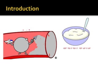
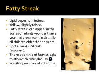
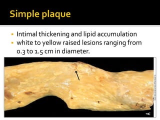
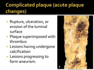
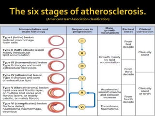
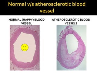
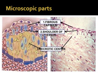
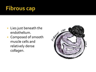
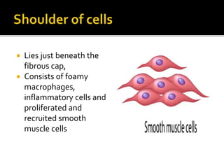
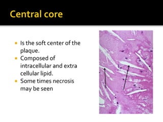
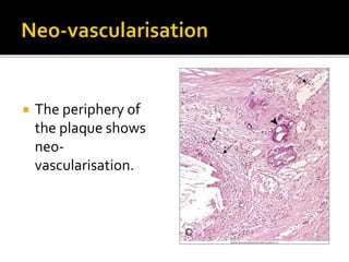
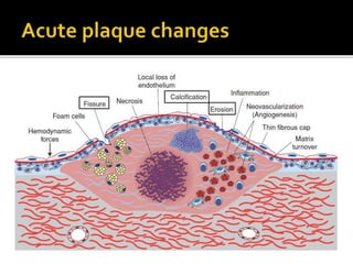
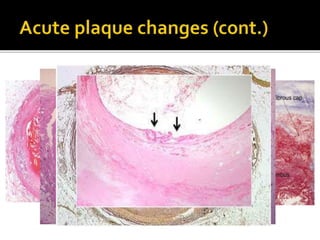
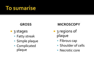
By- Sneha A (MBBS UG)
MORPHOLOGY
 Distribution: infrarenal abdominal aorta >
coronary arteries > popliteal arteries >
internal carotid arteries > the vessels of the
circle ofWillis.
 Spared: upper extremities, renal, mesenteric
(except ostia).
 Stages:
 Fatty streak
 Simple plaque
 Complicated plaque
 Lipid deposits in intima.
 Yellow, slightly raised.
 Fatty streaks can appear in the
aortas of infants younger than 1
year and are present in virtually
all children older than 10 years.
 Spot (1mm) -> Streak
(1x10mm).
 The relationship of fatty streaks
to atherosclerotic plaques
 Possible precursor of atheroma.
 Intimal thickening and lipid accumulation
 white to yellow raised lesions ranging from
0.3 to 1.5 cm in diameter.
 Rupture, ulceration, or
erosion of the luminal
surface
 Plaque superimposed with
thrombus
 Lesions having undergone
calcification
 Lesions progressing to
form aneurism.
NORMAL (HAPPY) BLOOD
VESSEL
ATHEROSCLEROTIC BLOOD
VESSELS
 3 parts
 Fibrous cap
 Shoulder of cells
 Central necrotic core
3.NECROTIC CENTER
2.SHOULDER OF
CELLS
1.FIBROUS
CAPSULE
 Lies just beneath the
endothelium.
 Composed of smooth
muscle cells and
relatively dense
collagen.
 Lies just beneath the
fibrous cap,
 Consists of foamy
macrophages,
inflammatory cells and
proliferated and
recruited smooth
muscle cells
 Is the soft center of the
plaque.
 Composed of
intracellular and extra
cellular lipid.
 Some times necrosis
may be seen
 The periphery of
the plaque shows
neo-
vascularisation.
GROSS
 3 stages
 Fatty streak
 Simple plaque
 Complicated
plaque
MICROSCOPY
 3 regions of
plaque
 Fibrous cap
 Shoulder of cells
 Necrotic core
Atherosclerosis  morphology

Atherosclerosis morphology

  • 1.
    By- Sneha A(MBBS UG) MORPHOLOGY
  • 3.
     Distribution: infrarenalabdominal aorta > coronary arteries > popliteal arteries > internal carotid arteries > the vessels of the circle ofWillis.  Spared: upper extremities, renal, mesenteric (except ostia).
  • 5.
     Stages:  Fattystreak  Simple plaque  Complicated plaque
  • 6.
     Lipid depositsin intima.  Yellow, slightly raised.  Fatty streaks can appear in the aortas of infants younger than 1 year and are present in virtually all children older than 10 years.  Spot (1mm) -> Streak (1x10mm).  The relationship of fatty streaks to atherosclerotic plaques  Possible precursor of atheroma.
  • 7.
     Intimal thickeningand lipid accumulation  white to yellow raised lesions ranging from 0.3 to 1.5 cm in diameter.
  • 8.
     Rupture, ulceration,or erosion of the luminal surface  Plaque superimposed with thrombus  Lesions having undergone calcification  Lesions progressing to form aneurism.
  • 11.
  • 12.
     3 parts Fibrous cap  Shoulder of cells  Central necrotic core 3.NECROTIC CENTER 2.SHOULDER OF CELLS 1.FIBROUS CAPSULE
  • 13.
     Lies justbeneath the endothelium.  Composed of smooth muscle cells and relatively dense collagen.
  • 14.
     Lies justbeneath the fibrous cap,  Consists of foamy macrophages, inflammatory cells and proliferated and recruited smooth muscle cells
  • 15.
     Is thesoft center of the plaque.  Composed of intracellular and extra cellular lipid.  Some times necrosis may be seen
  • 16.
     The peripheryof the plaque shows neo- vascularisation.
  • 19.
    GROSS  3 stages Fatty streak  Simple plaque  Complicated plaque MICROSCOPY  3 regions of plaque  Fibrous cap  Shoulder of cells  Necrotic core