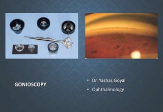
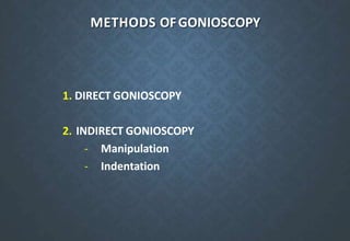
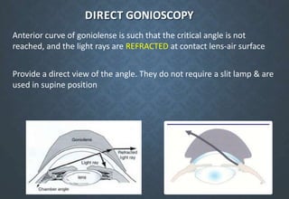
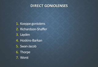
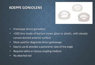
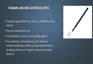
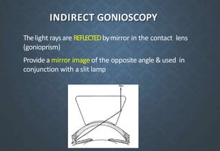
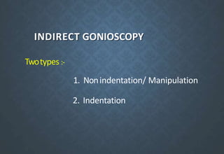
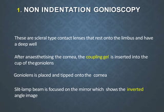
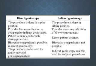
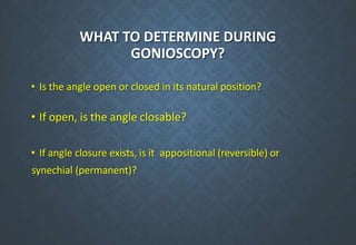
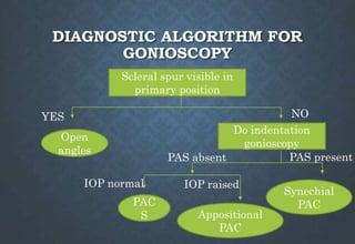
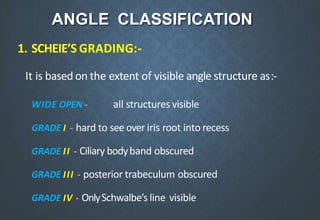
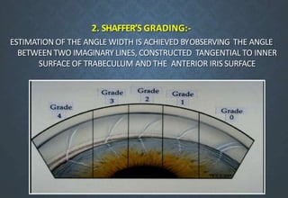
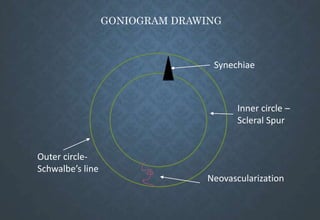
GONIOSCOPY
• Dr. Yashas Goyal
• Ophthalmology
• Biomicroscopic examination of the anterior chamber angle of
the eye
• Diagnostic
• Prognostic
• Therapeutic
HISTORY
Alexios Trantas (1907)
- first to examineangle in keratoglobus eyeusingdirect
ophthalmoscopewithdigitalscleralindentation
- introduced the term ā€œgonioscopyā€(Greek: angle
observation)
Maximilian Salzmann (1914)
- recognized that normal angle is not visible d/t total
internal reflection
- first to view the angle through a contact lens
- stressed the importance of gonioscopic examination in
patients with history of angle closure
- known as ā€œfatherof gonioscopyā€
Koeppe (1919)
- used the Zeiss slit lamp to examine the angle with his
newly developed lens
- thicker and more convex than Salzmann’s lens
Otto Barkan (1936)
- used a slit lamp suspended from the ceiling and a
hand-held illuminator to view the angle through a
Koeppe lens
- brought gonioscopy into practical clinical application
- made the distinction between ā€œdeep- chamberā€ and ā€œ
shallow-chamberā€ glaucoma
Goldmann (1938)
- introduced the gonioprism
- illustrated method of indirect gonioscopy
CONCEPT OF GONIOSCOPY
Lightrayscomingfromanglestructures
undergototalinternalreflection
Theangleatwhichtheystrikethecorneal
epitheliumisgreaterthanthecriticalangle
forcornea-airinterface(46degrees)
Goniolens/ Gonioprism
Eliminates total internal reflection by
replacing the cornea-air interface bya new
interface
Refractive indexis slightlygreater than that of
corneaand tears so that light rays undergo
minimal refraction
Reach the observer’s eye either directly or
after getting reflected in a mirror
METHODS OFGONIOSCOPY
1. DIRECT GONIOSCOPY
2. INDIRECT GONIOSCOPY
- Manipulation
- Indentation
DIRECT GONIOSCOPY
Anterior curve of goniolense is such that the critical angle is not
reached, and the light rays are REFRACTED at contact lens-air surface
Provide a direct view of the angle. They do not require a slit lamp & are
used in supine position
DIRECT GONIOLENSES
1. Koeppegoniolens
2. Richardson-Shaffer
3. Layden
4. Hoskins-Barkan
5. Swan-Jacob
6. Thorpe
7. Worst
KOEPPE GONIOLENS
• Prototype direct goniolens
• +50D lens made of barium crown glass or plastic, with steeply
convex domed anterior surface
• Most used for diagnosticdirect gonioscopy
• Easyto use & provides a panoramic viewof the angle
• Requiressalineorviscouscouplingmedium
• No attachedrod
SWAN JACOB GONIOLENS
• Surgical goniolens for use in children and
adults
• Has an attached rod
• Viscoelastic used as coupling agent
• Procedures- Goniotomy, Ab interno
trabeculotomy, while using trabectome,
putting iStent or Cypass suprachoroidal
device
Richardson-Shaffer’s goniolens –
small Koeppe lens used for
infants.
Layden goniolens –
for premature infants
Hoskins-Barkan lens –
prototype surgical goniolens
used for goniotomy
Thorpe goniolens –
surgical and diagnostic
lens for ORs
Worst goniolens –
surgical goniolens for
children
TECHNIQUE
Patientlies supine with examiner seated on
the sideof the eyeto beexamined
After topical anesthesia, goniolens is
positioned on the cornea, usingbalanced
salt solution or methyl cellulose
Withthebiomicroscope in onehand & the
Barkan light sourcein other, examiner
scansthe anterior chamber angle by
shifting his or her position until all 360
degreeshavebeen studied
Mag of Koeppe lens: 1.5x
Mag of biomicroscope: 16x
Total magnification: 24x
ADVANTAGES
Greaterflexibility asposition of observercan
be changed
Panoramicviewis obtained soonepart of the angle
can becompared with other
Angledeepin supine position soit is easytosee the
angle
Canbeused in sedated patient and in infants
Providesa straight viewrather than invertedview
makingsurgicalprocedureseasier
DISADVANTAGES
• Annoyinglight reflexes from cornea
• Timeconsuming
• Benefits of slit-lamp likevariable light &
better clarity are not available
INDIRECT GONIOSCOPY
Thelight rays are REFLECTEDbymirror in the contact lens
(gonioprism)
Providea mirror image of the opposite angle & used in
conjunction with a slit lamp
INDIRECT GONIOSCOPY
Twotypes :-
1. Nonindentation/ Manipulation
2. Indentation
1. NON INDENTATION GONIOSCOPY
These are scleral type contact lenses that rest onto the limbus and have
a deep well
After anaesthetising the cornea,the couplinggel is inserted into the
cup of thegoniolens
Goniolensis placed and tipped ontothe cornea
Slit-lamp beam is focused on the mirrorwhich showsthe inverted
angleimage
GONIOPRISMS (MANIPULATION) -
Surface is slightly larger than cornea& requirea coupling gel:
1. Goldmann single mirror: single mirror inclined at 62°
2. Goldmann double mirror: two mirrors aligned at 62 ° each
3. Goldmann 3 mirror: mirror for gonioscopy at 59°, for pars plana at 67°,
and for equator 73°
4. Allen-Thorpe: 4 mirrors inclined at 62° each
Goldmann 3 mirror lens
GOLDMANN MIRRORS
GOLDMANN SINGLEMIRROR GOLDMANN 2 MIRROR GOLDMANN 3 MIRROR
2. INDENTATION GONIOSCOPY
Cornealtypegoniolens–curvaturesimilartocornea
Nocouplingfluidrequired,tearfilmactsascouplingmedium
Varyingamount of pressure applied to the cornea
Examinerobservesthe effect on angle width
Increased pressure indents the central cornea &displaces fluid into the angle,
opening itwider
Differentiates between appositional and synechial angle closure
GONIOPRISMS (INDENTATION):
1. Zeiss four mirror
2. Sussmmanfour mirror
3. Tokel single mirror
4. Posner four mirror
1. ZEISS -4 MIRROR GONIOPRISM:
It has four identical mirrors angled at 64° which allow examination of the four
quadrants without rotation of the lens
Thecontact surface of lens has a 9 mm diameter
2. SUSSMMAN LENS :
It is also similar toZeiss lens but it has nohandle
3.TOKEL GONIOLENS:
It is a single mirrorgoniolens & has got a wider field ofview
4.POSNER GONIOLENS:
It is likeZeiss-goniolens but it is made of plastic &also has a fixed handle
ANGLE VIEWING
• Adjust slit to a thin beam of height to 2-3 mm
• Switch on slit lamp only after slit is directed onto mirror, without crossing
the pupil with the beam
• Image seen in the mirror is of opposite angle but NOT laterally inverted
• View inferior angle first (generally wider and more pigmented) followed
by the superior angle
• Turn beam horizontally to view nasal and temporal angle
• For manipulation, patient asked to look slightly towards the mirror and
angle becomes visible over the steep iris
• For eyes with angle closure in the primary position, indentation done
with corneal type lens to differentiate type of closure (PACS;
appositional PAC or synechial PAC)
• Each quadrant examined separately
ADVANTAGES OF INDIRECT GONIOSCOPY:
Easy to perform when mastered
Allfour quadrants can be seenwhen four mirror gonioprism used
With slit lamp, variable magnification and illumination can
beachieved
Indentation Gonioscopy can be performed, which allows
differentiation of appositional andsynechial angle closure
Couplingagent is not used, sovisualization of fundus &
photography ispossible
DISADVANTAGES
Difficult to master
Mirror image can be confusing
Mayopen the angle artefactually if pressureapplied
GONIOSCOPIC VIEW OF ANGLE STRUCTURES
1. SCHWALBE’S LINE:-
Most anterior structure
Irregular elevation of 50 to 100µm width
Posterior part of zone S (anterior part is the termination of
corneal Descemet membrane)
Marked only bya slight changein colorfrom trabecular meshwork or by
a faint white line.
Best identified bylocating the ā€œCorneal Wedgeā€.
Prominent and anteriorly
displaced Schwalbe’s line in
Axenfeld anomaly (posterior
embryotoxon)
Pigmentation anterior to
Schwalbe’s line maybe seen
(Sampaolesi’s line) in Pigment
Dispersion syndrome and PXF
Corneal wedge localization helps
differentiate it from PTM
2. TRABECULAR MESHWORK :-
- It lies between the Schwalbe’s line
anteriorly &scleral spur posteriorly and
has an averagewidth of 600 µm
- Anterior non pigmented part (non-
functional) and posterior pigmented
part that overlies the canal of Schlemm
- Smoothin infants but becomes coarseandpigmented with age
- Inferior TM more pigmented than superior, due to gravity and
aqueous currents
3. SCLERAL SPUR :-
-Anterior most projection of thesclera into which the ciliary body is
inserted
- Situated just posterior to the TMand appears as a narrow, dense,
white band
-Maybe obscured byiris process,and pigments.
- Visibility of scleral spur in primary position denotes an open angle
4. CILIARY BODY BAND:-
- Liesjust behind the scleral spur asa dull brownor slate greyband
- Portion of ciliary bodywhich is visible in the angle asa result of insertion
of the iris into the ciliary body
- Maybewideinmyopes andnarrow inhyperopes.
5. ROOT OF IRIS :-
Iris contour is normally slightly convexorflat
Iris processes- fine finger like extensions arising fromiris and
attaching atvariable position in angle, not crossing TM
Color varies in differentindividuals
Radial marking and crypts arepresent
Normal blood vessels – loops of vessels near iris root, run radially
towards pupil; never cross scleral spur
IRIS PROCESSES VS. PERIPHERAL ANTERIOR
SYNECHIAE
Iris processes PAS
Fine, finger like Broad based
Do not cross trabecular meshwork Attach on and anterior to TM
Follow concavity of iris Bridge the angle structures
Allow free movement of iris
posteriorly upon indentation
Do not allow free movement of iris
OTHER STRUCTURES
IRIS PROCESSES:-
Normal variants
Appear as fine strands extending from iris to thescleral
spur
Thesedo not inhibit the
movement of the irisduring
indentation gonioscopy
WHAT TO DETERMINE DURING
GONIOSCOPY?
• Is the angle open or closed in its natural position?
• If open, is the angle closable?
• If angle closure exists, is it appositional (reversible) or
synechial (permanent)?
DIAGNOSTIC ALGORITHM FOR
GONIOSCOPY
Scleral spur visible in
primary position
Open
angles
YES NO
Do indentation
gonioscopy
PAS present
PAS absent
Synechial
PAC
PAC
S
IOP normal IOP raised
Appositional
PAC
ANGLE CLASSIFICATION
1. SCHEIE’S GRADING:-
It is based on the extent of visible angle structure as:-
WIDE OPEN - all structures visible
GRADE I - hard to see over iris root into recess
GRADE II - Ciliary bodyband obscured
GRADE III - posterior trabeculum obscured
GRADE IV - OnlySchwalbe’s line visible
2. SHAFFER’S GRADING:-
ESTIMATION OF THE ANGLE WIDTH IS ACHIEVED BYOBSERVING THE ANGLE
BETWEEN TWO IMAGINARY LINES, CONSTRUCTED TANGENTIAL TO INNER
SURFACE OF TRABECULUM AND THE ANTERIOR IRIS SURFACE
Grade Angle
width
Configuration Chances
of Closure
Structures visible
on gonioscopy
IV 35-45° Wide open Nil
III 20-35° Open angle Nil
From Schwalbe’s line
to ciliary body
From Schwalbe’s line
to scleral spur
II 20° possible
10°
Moderately
narrow
Very narrow probable
I
O 0° closed Closed
From Schwalbe’s line
to TM
Schwalbe’s line only
No angle structure
visible
3. SPAETH CLASSIFICATION:-
This classification elaborated a complex grading system that
captures a detailed three dimensional information in coded
form
or flat
DOCUMENTATION OF GONIOSCOPIC FINDINGS
- Posterior most structure visible in
primary gaze with thin short slit beam
- Posterior most structure on indentation
or manipulation
- Iris configuration – concave, regular or
convex/steep
- Any specific angle abnormality
• Goniosynechiae
• Pigmentation
• Angle recession
• Anterior insertion of iris
• Iridodialysis/cyclodialysis
S
L
TM
+2
TM
+1
TM
+1
Diagrammatic representation
ATM
TO SS
ATM
TO SS
ATM
PTM
TO SS
PTM
TO SS
PTM
GONIOGRAM DRAWING
Inner circle –
Scleral Spur
Outer circle-
Schwalbe’s line
Synechiae
Neovascularization
CLINICAL USES OF GONIOSCOPY
DIAGNOSTIC
Differentiation between POAG and PACG
To diagnosesecondary glaucomas-anglerecession,uveitic,
pigmentary,pseudoexfoliative,neovascular,lens induced
Todiagnose tumors of anterior segment
Earlydetection of KFRing,neovascularization (NVA)
THERAPEUTIC
Toperform argon laser trabeculoplasty
Laser iridoplasty
Laser sclerostomy
Goniotomy & trabeculotomy
LIMITATIONS OFGONIOSCOPY
Cannot be performed in painful inflamedeyes
Indirect gonioscopy- requires skill for interpretation
Compromisedcornea – may obscure view
Cannot be performed in lacerated or perforated globe
COMPLICATIONS- Corneal abrasion, hyphema
Contact procedure
GONIOSCOPY: PATHOLOGICAL CONDITIONS
ANGLE CLOSUREGLAUCOMA
The angle is first occluded appositionally, followed by
a sticky adhesion & later by peripheral anterior
synechiae over wide area
.
PERIPHERAL ANTERIOR
SYNECHIAE
Not pathognomonic of any single disease entity
Tend to form first:
Superiorly -Angle closure glaucoma
Inferiorly – Uveitis
Anywhere –post traumatic cases
PLATEAU IRIS CONFIGURATION
Iris appears to be in plane to intersect withSchwalbes line, a steep
drop off occurs immediately beforeTM
Potential for causing angle closure glaucoma
Anteriorly positioned ciliaryprocess
On indentation gonioscopy, TM becomes visible; peripheral hump of iris
draped over ciliary processes gives a ā€œsine wave appearance"
PLATEAU IRIS CONFIGURATION
UBM showing obliteration of
iridociliary sulcus in plateau iris
configuration
Sine wave appearance
PIGMENTARY GLAUCOMA
Heavypigment dispersion in leadingto occlusionof trabecularmeshwork
Typical patients are youngmyopic males
Iris configuration is anteriorly concave due to posterior bowing of peripheral iris
contact with zonules leads to dispersion of pigments from posterior pigmented iris
epithelium
NEOVASCULAR GLAUCOMA
New vessel formatiom occurs on iris
known as RUBIOSIS IRIDIS, which is
frequently associated with severe form of
glaucomad/t neovascularizationatangle.
Normal Blood vessels-
Branches of major circle of iris
Unlike neovascularization, they
are either arranged radially or form
loops
Broad, do not cross scleral spur,
do not arborize
Fine, arborizing blood vessels
crossing over scleral spur up to
SL  NVA
ANGLE RECESSION
- Irregular widening of ciliary bodyband occurs sometimes following blunt
trauma to the eye
- It occurs due to tearbetween longitudenal and circular muscles of ciliary
body
- Indirectindicatorofsevereinjurytothetrabecularmeshworkleadingtorise
inIOPovertime
MISCELLENIOUS GONIOSCOPIC FINDINGS
Blood in Schlemm’s canal
STERILIZATION OF GONIOSCOPES
• Cleaning - Rinse with cold water and mild soap immediately after use
• Disinfection - soak in 2% glutaraldehyde for 20 minutes OR in 10% bleach
solution (1 part bleach to 9 parts water) for 10 minutes
• Rinse with water thoroughly and air dry after disinfection
• Can also wipe with sterile swab (soaked in 70% isopropyl alcohol) or clean
with 1:1000 Merthiolate solution
• Sterilization – Ethylene Oxide (ETO) exposure - at 56° C for 1 hour
• AVOID steam autoclave / alcohol soaking
NEWER WAYSTO EVALUATE ANTERIOR CHAMBER
ANGLE
THANK YOU!

Gonioscopy

  • 1.
    GONIOSCOPY • Dr. YashasGoyal • Ophthalmology
  • 2.
    • Biomicroscopic examinationof the anterior chamber angle of the eye • Diagnostic • Prognostic • Therapeutic
  • 3.
    HISTORY Alexios Trantas (1907) -first to examineangle in keratoglobus eyeusingdirect ophthalmoscopewithdigitalscleralindentation - introduced the term ā€œgonioscopyā€(Greek: angle observation)
  • 4.
    Maximilian Salzmann (1914) -recognized that normal angle is not visible d/t total internal reflection - first to view the angle through a contact lens - stressed the importance of gonioscopic examination in patients with history of angle closure - known as ā€œfatherof gonioscopyā€ Koeppe (1919) - used the Zeiss slit lamp to examine the angle with his newly developed lens - thicker and more convex than Salzmann’s lens
  • 5.
    Otto Barkan (1936) -used a slit lamp suspended from the ceiling and a hand-held illuminator to view the angle through a Koeppe lens - brought gonioscopy into practical clinical application - made the distinction between ā€œdeep- chamberā€ and ā€œ shallow-chamberā€ glaucoma Goldmann (1938) - introduced the gonioprism - illustrated method of indirect gonioscopy
  • 6.
  • 7.
    Goniolens/ Gonioprism Eliminates totalinternal reflection by replacing the cornea-air interface bya new interface Refractive indexis slightlygreater than that of corneaand tears so that light rays undergo minimal refraction Reach the observer’s eye either directly or after getting reflected in a mirror
  • 8.
    METHODS OFGONIOSCOPY 1. DIRECTGONIOSCOPY 2. INDIRECT GONIOSCOPY - Manipulation - Indentation
  • 9.
    DIRECT GONIOSCOPY Anterior curveof goniolense is such that the critical angle is not reached, and the light rays are REFRACTED at contact lens-air surface Provide a direct view of the angle. They do not require a slit lamp & are used in supine position
  • 10.
    DIRECT GONIOLENSES 1. Koeppegoniolens 2.Richardson-Shaffer 3. Layden 4. Hoskins-Barkan 5. Swan-Jacob 6. Thorpe 7. Worst
  • 11.
    KOEPPE GONIOLENS • Prototypedirect goniolens • +50D lens made of barium crown glass or plastic, with steeply convex domed anterior surface • Most used for diagnosticdirect gonioscopy • Easyto use & provides a panoramic viewof the angle • Requiressalineorviscouscouplingmedium • No attachedrod
  • 12.
    SWAN JACOB GONIOLENS •Surgical goniolens for use in children and adults • Has an attached rod • Viscoelastic used as coupling agent • Procedures- Goniotomy, Ab interno trabeculotomy, while using trabectome, putting iStent or Cypass suprachoroidal device
  • 13.
    Richardson-Shaffer’s goniolens – smallKoeppe lens used for infants. Layden goniolens – for premature infants Hoskins-Barkan lens – prototype surgical goniolens used for goniotomy Thorpe goniolens – surgical and diagnostic lens for ORs Worst goniolens – surgical goniolens for children
  • 14.
    TECHNIQUE Patientlies supine withexaminer seated on the sideof the eyeto beexamined After topical anesthesia, goniolens is positioned on the cornea, usingbalanced salt solution or methyl cellulose Withthebiomicroscope in onehand & the Barkan light sourcein other, examiner scansthe anterior chamber angle by shifting his or her position until all 360 degreeshavebeen studied Mag of Koeppe lens: 1.5x Mag of biomicroscope: 16x Total magnification: 24x
  • 15.
    ADVANTAGES Greaterflexibility asposition ofobservercan be changed Panoramicviewis obtained soonepart of the angle can becompared with other Angledeepin supine position soit is easytosee the angle Canbeused in sedated patient and in infants Providesa straight viewrather than invertedview makingsurgicalprocedureseasier
  • 16.
    DISADVANTAGES • Annoyinglight reflexesfrom cornea • Timeconsuming • Benefits of slit-lamp likevariable light & better clarity are not available
  • 17.
    INDIRECT GONIOSCOPY Thelight raysare REFLECTEDbymirror in the contact lens (gonioprism) Providea mirror image of the opposite angle & used in conjunction with a slit lamp
  • 18.
    INDIRECT GONIOSCOPY Twotypes :- 1.Nonindentation/ Manipulation 2. Indentation
  • 19.
    1. NON INDENTATIONGONIOSCOPY These are scleral type contact lenses that rest onto the limbus and have a deep well After anaesthetising the cornea,the couplinggel is inserted into the cup of thegoniolens Goniolensis placed and tipped ontothe cornea Slit-lamp beam is focused on the mirrorwhich showsthe inverted angleimage
  • 21.
    GONIOPRISMS (MANIPULATION) - Surfaceis slightly larger than cornea& requirea coupling gel: 1. Goldmann single mirror: single mirror inclined at 62° 2. Goldmann double mirror: two mirrors aligned at 62 ° each 3. Goldmann 3 mirror: mirror for gonioscopy at 59°, for pars plana at 67°, and for equator 73° 4. Allen-Thorpe: 4 mirrors inclined at 62° each
  • 22.
  • 23.
    GOLDMANN MIRRORS GOLDMANN SINGLEMIRRORGOLDMANN 2 MIRROR GOLDMANN 3 MIRROR
  • 24.
    2. INDENTATION GONIOSCOPY Cornealtypegoniolens–curvaturesimilartocornea Nocouplingfluidrequired,tearfilmactsascouplingmedium Varyingamountof pressure applied to the cornea Examinerobservesthe effect on angle width Increased pressure indents the central cornea &displaces fluid into the angle, opening itwider Differentiates between appositional and synechial angle closure
  • 25.
    GONIOPRISMS (INDENTATION): 1. Zeissfour mirror 2. Sussmmanfour mirror 3. Tokel single mirror 4. Posner four mirror
  • 26.
    1. ZEISS -4MIRROR GONIOPRISM: It has four identical mirrors angled at 64° which allow examination of the four quadrants without rotation of the lens Thecontact surface of lens has a 9 mm diameter
  • 27.
    2. SUSSMMAN LENS: It is also similar toZeiss lens but it has nohandle
  • 28.
    3.TOKEL GONIOLENS: It isa single mirrorgoniolens & has got a wider field ofview 4.POSNER GONIOLENS: It is likeZeiss-goniolens but it is made of plastic &also has a fixed handle
  • 29.
    ANGLE VIEWING • Adjustslit to a thin beam of height to 2-3 mm • Switch on slit lamp only after slit is directed onto mirror, without crossing the pupil with the beam • Image seen in the mirror is of opposite angle but NOT laterally inverted • View inferior angle first (generally wider and more pigmented) followed by the superior angle
  • 30.
    • Turn beamhorizontally to view nasal and temporal angle • For manipulation, patient asked to look slightly towards the mirror and angle becomes visible over the steep iris • For eyes with angle closure in the primary position, indentation done with corneal type lens to differentiate type of closure (PACS; appositional PAC or synechial PAC) • Each quadrant examined separately
  • 31.
    ADVANTAGES OF INDIRECTGONIOSCOPY: Easy to perform when mastered Allfour quadrants can be seenwhen four mirror gonioprism used With slit lamp, variable magnification and illumination can beachieved Indentation Gonioscopy can be performed, which allows differentiation of appositional andsynechial angle closure Couplingagent is not used, sovisualization of fundus & photography ispossible
  • 32.
    DISADVANTAGES Difficult to master Mirrorimage can be confusing Mayopen the angle artefactually if pressureapplied
  • 34.
    GONIOSCOPIC VIEW OFANGLE STRUCTURES
  • 35.
    1. SCHWALBE’S LINE:- Mostanterior structure Irregular elevation of 50 to 100µm width Posterior part of zone S (anterior part is the termination of corneal Descemet membrane)
  • 36.
    Marked only byaslight changein colorfrom trabecular meshwork or by a faint white line. Best identified bylocating the ā€œCorneal Wedgeā€.
  • 37.
    Prominent and anteriorly displacedSchwalbe’s line in Axenfeld anomaly (posterior embryotoxon) Pigmentation anterior to Schwalbe’s line maybe seen (Sampaolesi’s line) in Pigment Dispersion syndrome and PXF Corneal wedge localization helps differentiate it from PTM
  • 38.
    2. TRABECULAR MESHWORK:- - It lies between the Schwalbe’s line anteriorly &scleral spur posteriorly and has an averagewidth of 600 µm - Anterior non pigmented part (non- functional) and posterior pigmented part that overlies the canal of Schlemm - Smoothin infants but becomes coarseandpigmented with age - Inferior TM more pigmented than superior, due to gravity and aqueous currents
  • 39.
    3. SCLERAL SPUR:- -Anterior most projection of thesclera into which the ciliary body is inserted - Situated just posterior to the TMand appears as a narrow, dense, white band -Maybe obscured byiris process,and pigments. - Visibility of scleral spur in primary position denotes an open angle
  • 40.
    4. CILIARY BODYBAND:- - Liesjust behind the scleral spur asa dull brownor slate greyband - Portion of ciliary bodywhich is visible in the angle asa result of insertion of the iris into the ciliary body - Maybewideinmyopes andnarrow inhyperopes.
  • 41.
    5. ROOT OFIRIS :- Iris contour is normally slightly convexorflat Iris processes- fine finger like extensions arising fromiris and attaching atvariable position in angle, not crossing TM Color varies in differentindividuals Radial marking and crypts arepresent Normal blood vessels – loops of vessels near iris root, run radially towards pupil; never cross scleral spur
  • 42.
    IRIS PROCESSES VS.PERIPHERAL ANTERIOR SYNECHIAE Iris processes PAS Fine, finger like Broad based Do not cross trabecular meshwork Attach on and anterior to TM Follow concavity of iris Bridge the angle structures Allow free movement of iris posteriorly upon indentation Do not allow free movement of iris
  • 44.
    OTHER STRUCTURES IRIS PROCESSES:- Normalvariants Appear as fine strands extending from iris to thescleral spur Thesedo not inhibit the movement of the irisduring indentation gonioscopy
  • 45.
    WHAT TO DETERMINEDURING GONIOSCOPY? • Is the angle open or closed in its natural position? • If open, is the angle closable? • If angle closure exists, is it appositional (reversible) or synechial (permanent)?
  • 46.
    DIAGNOSTIC ALGORITHM FOR GONIOSCOPY Scleralspur visible in primary position Open angles YES NO Do indentation gonioscopy PAS present PAS absent Synechial PAC PAC S IOP normal IOP raised Appositional PAC
  • 47.
    ANGLE CLASSIFICATION 1. SCHEIE’SGRADING:- It is based on the extent of visible angle structure as:- WIDE OPEN - all structures visible GRADE I - hard to see over iris root into recess GRADE II - Ciliary bodyband obscured GRADE III - posterior trabeculum obscured GRADE IV - OnlySchwalbe’s line visible
  • 48.
    2. SHAFFER’S GRADING:- ESTIMATIONOF THE ANGLE WIDTH IS ACHIEVED BYOBSERVING THE ANGLE BETWEEN TWO IMAGINARY LINES, CONSTRUCTED TANGENTIAL TO INNER SURFACE OF TRABECULUM AND THE ANTERIOR IRIS SURFACE
  • 49.
    Grade Angle width Configuration Chances ofClosure Structures visible on gonioscopy IV 35-45° Wide open Nil III 20-35° Open angle Nil From Schwalbe’s line to ciliary body From Schwalbe’s line to scleral spur II 20° possible 10° Moderately narrow Very narrow probable I O 0° closed Closed From Schwalbe’s line to TM Schwalbe’s line only No angle structure visible
  • 50.
    3. SPAETH CLASSIFICATION:- Thisclassification elaborated a complex grading system that captures a detailed three dimensional information in coded form
  • 51.
  • 52.
    DOCUMENTATION OF GONIOSCOPICFINDINGS - Posterior most structure visible in primary gaze with thin short slit beam - Posterior most structure on indentation or manipulation - Iris configuration – concave, regular or convex/steep - Any specific angle abnormality • Goniosynechiae • Pigmentation • Angle recession • Anterior insertion of iris • Iridodialysis/cyclodialysis S L TM +2 TM +1 TM +1 Diagrammatic representation ATM TO SS ATM TO SS ATM PTM TO SS PTM TO SS PTM
  • 53.
    GONIOGRAM DRAWING Inner circle– Scleral Spur Outer circle- Schwalbe’s line Synechiae Neovascularization
  • 54.
    CLINICAL USES OFGONIOSCOPY DIAGNOSTIC Differentiation between POAG and PACG To diagnosesecondary glaucomas-anglerecession,uveitic, pigmentary,pseudoexfoliative,neovascular,lens induced Todiagnose tumors of anterior segment Earlydetection of KFRing,neovascularization (NVA)
  • 55.
    THERAPEUTIC Toperform argon lasertrabeculoplasty Laser iridoplasty Laser sclerostomy Goniotomy & trabeculotomy
  • 56.
    LIMITATIONS OFGONIOSCOPY Cannot beperformed in painful inflamedeyes Indirect gonioscopy- requires skill for interpretation Compromisedcornea – may obscure view Cannot be performed in lacerated or perforated globe COMPLICATIONS- Corneal abrasion, hyphema Contact procedure
  • 57.
    GONIOSCOPY: PATHOLOGICAL CONDITIONS ANGLECLOSUREGLAUCOMA The angle is first occluded appositionally, followed by a sticky adhesion & later by peripheral anterior synechiae over wide area .
  • 58.
    PERIPHERAL ANTERIOR SYNECHIAE Not pathognomonicof any single disease entity Tend to form first: Superiorly -Angle closure glaucoma Inferiorly – Uveitis Anywhere –post traumatic cases
  • 59.
    PLATEAU IRIS CONFIGURATION Irisappears to be in plane to intersect withSchwalbes line, a steep drop off occurs immediately beforeTM Potential for causing angle closure glaucoma Anteriorly positioned ciliaryprocess
  • 60.
    On indentation gonioscopy,TM becomes visible; peripheral hump of iris draped over ciliary processes gives a ā€œsine wave appearance" PLATEAU IRIS CONFIGURATION UBM showing obliteration of iridociliary sulcus in plateau iris configuration Sine wave appearance
  • 61.
    PIGMENTARY GLAUCOMA Heavypigment dispersionin leadingto occlusionof trabecularmeshwork Typical patients are youngmyopic males Iris configuration is anteriorly concave due to posterior bowing of peripheral iris contact with zonules leads to dispersion of pigments from posterior pigmented iris epithelium
  • 62.
    NEOVASCULAR GLAUCOMA New vesselformatiom occurs on iris known as RUBIOSIS IRIDIS, which is frequently associated with severe form of glaucomad/t neovascularizationatangle. Normal Blood vessels- Branches of major circle of iris Unlike neovascularization, they are either arranged radially or form loops Broad, do not cross scleral spur, do not arborize Fine, arborizing blood vessels crossing over scleral spur up to SL  NVA
  • 63.
    ANGLE RECESSION - Irregularwidening of ciliary bodyband occurs sometimes following blunt trauma to the eye - It occurs due to tearbetween longitudenal and circular muscles of ciliary body - Indirectindicatorofsevereinjurytothetrabecularmeshworkleadingtorise inIOPovertime
  • 64.
  • 65.
    STERILIZATION OF GONIOSCOPES •Cleaning - Rinse with cold water and mild soap immediately after use • Disinfection - soak in 2% glutaraldehyde for 20 minutes OR in 10% bleach solution (1 part bleach to 9 parts water) for 10 minutes • Rinse with water thoroughly and air dry after disinfection • Can also wipe with sterile swab (soaked in 70% isopropyl alcohol) or clean with 1:1000 Merthiolate solution • Sterilization – Ethylene Oxide (ETO) exposure - at 56° C for 1 hour • AVOID steam autoclave / alcohol soaking
  • 66.
    NEWER WAYSTO EVALUATEANTERIOR CHAMBER ANGLE
  • 67.