Examination of theanterior chamber angle is a critical part of the eye examination, especially in glaucoma. Gonioscopy remains the gold standard technique for visualising the angle structures
and for devising an appropriate management. This reference is designed to provide a guide to identifying the structures, but the clinician should practice this technique frequently to become
expert at its deployment.
CHAIR-SIDE REFERENCE: GONIOSCOPY AND THE ANTERIOR CHAMBER ANGLE
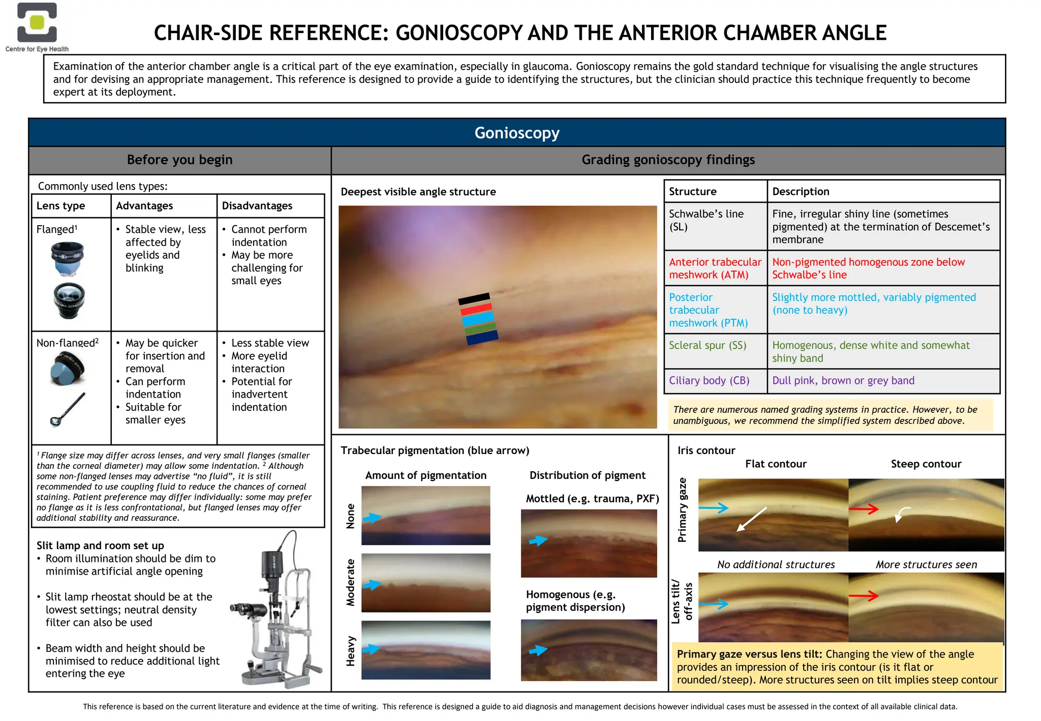
Gonioscopy
Before you begin Grading gonioscopy findings
Commonly used lens types:
Lens type Advantages Disadvantages
Flanged1 • Stable view, less
affected by
eyelids and
blinking
• Cannot perform
indentation
• May be more
challenging for
small eyes
Non-flanged2 • May be quicker
for insertion and
removal
• Can perform
indentation
• Suitable for
smaller eyes
• Less stable view
• More eyelid
interaction
• Potential for
inadvertent
indentation
1 Flange size may differ across lenses, and very small flanges (smaller
than the corneal diameter) may allow some indentation. 2 Although
some non-flanged lenses may advertise “no fluid”, it is still
recommended to use coupling fluid to reduce the chances of corneal
staining. Patient preference may differ individually: some may prefer
no flange as it is less confrontational, but flanged lenses may offer
additional stability and reassurance.
Slit lamp and room set up
• Room illumination should be dim to
minimise artificial angle opening
• Slit lamp rheostat should be at the
lowest settings; neutral density
filter can also be used
• Beam width and height should be
minimised to reduce additional light
entering the eye
Deepest visible angle structure Structure Description
Schwalbe’s line
(SL)
Fine, irregular shiny line (sometimes
pigmented) at the termination of Descemet’s
membrane
Anterior trabecular
meshwork (ATM)
Non-pigmented homogenous zone below
Schwalbe’s line
Posterior
trabecular
meshwork (PTM)
Slightly more mottled, variably pigmented
(none to heavy)
Scleral spur (SS) Homogenous, dense white and somewhat
shiny band
Ciliary body (CB) Dull pink, brown or grey band
Trabecular pigmentation (blue arrow)
Amount of pigmentation Distribution of pigment
None
Moderate
Heavy
Mottled (e.g. trauma, PXF)
Homogenous (e.g.
pigment dispersion)
Iris contour
Flat contour Steep contour
Primary
gaze
Lens
tilt/
off-axis
Primary gaze versus lens tilt: Changing the view of the angle
provides an impression of the iris contour (is it flat or
rounded/steep). More structures seen on tilt implies steep contour
More structures seen
No additional structures
Note: These structures form
“zones” and may widen or
thin throughout the angle. There are numerous named grading systems in practice. However, to be
unambiguous, we recommend the simplified system described above.
This reference is based on the current literature and evidence at the time of writing. This reference is designed a guide to aid diagnosis and management decisions however individual cases must be assessed in the context of all available clinical data.
2.
CHAIR-SIDE REFERENCE: GONIOSCOPYAND THE ANTERIOR CHAMBER ANGLE
Imaging modalities for assessing the anterior chamber angle
Optical coherence tomography
Key advantages
• Quick, non-invasive, high
resolution
• Can be repurposed from posterior
segment imaging devices
• Many quantitative parameters
become available [note: no
parameter cut-off exists for
identifying angle closure]
• Can visualise iris contour, lens-iris
interaction and lens vault
• Relatively well-controlled
background lighting
Key disadvantages
• Requires visualisation of key landmarks such as scleral spur,
Schlemm’s canal (not possible in around 20% of patients)
• Cannot visualise key anatomical structures in en face manner
(e.g. trabecular meshwork)
• Most commercially available instruments only give one slice
(not sufficient for describing entirety of the anterior chamber
angle)
• Specialised (e.g. swept-source) devices more appropriate than
repurposed posterior segment devices
• Affected by anterior segment pathologies (e.g. conjunctiva)
and corneal compensation protocols required to adjust for
image magnification – see examples below
Examples of challenges with anterior segment imaging
• The lateral angles are easier to image compared to
superior/inferior angles, which are susceptible to
distortions due to instrument-specific image scaling
(A, example of superior angle imaging, where primary
gaze imaging is not possible)
• Opacities on the conjunctiva and cornea can obscure
the angle structures (B, example of pterygium)
Scheimpflug imaging Ultrasound biomicroscopy
Key features
• Allows quantification of anterior
chamber parameters (A)
• Allows visualisation of the angle
across the anterior chamber width
• Cannot visualise anterior chamber
angle itself (B); few normative data
available for quantitative
information
Example images Key features
• Allows visualisation of the retroiridal
space by penetrating the pigment
epithelium (e.g. iridociliary cysts,
ciliary body position and lesions)
• Resolution much lower than that of
optical coherence tomography
• Requires contact with ocular surface
Example case
Legend:
White: ciliary body
Red: scleral spur
Blue: Schlemm’s
canal
Yellow: iris
Open angle Closed angle
Iris
contour
Angle
opening
Plateau configuration Posteriorly bowed Regular
A B
This reference is based on the current literature and evidence at the time of writing. This reference is designed a guide to aid diagnosis and management decisions however individual cases must be assessed in the context of all available clinical data.
A
B
3.
This reference isbased on the current literature and evidence at the time of writing. This reference is designed a guide to aid diagnosis and management decisions however individual cases must be assessed in the context of all available clinical data.
CHAIR-SIDE REFERENCE: GONIOSCOPY AND THE ANTERIOR CHAMBER ANGLE
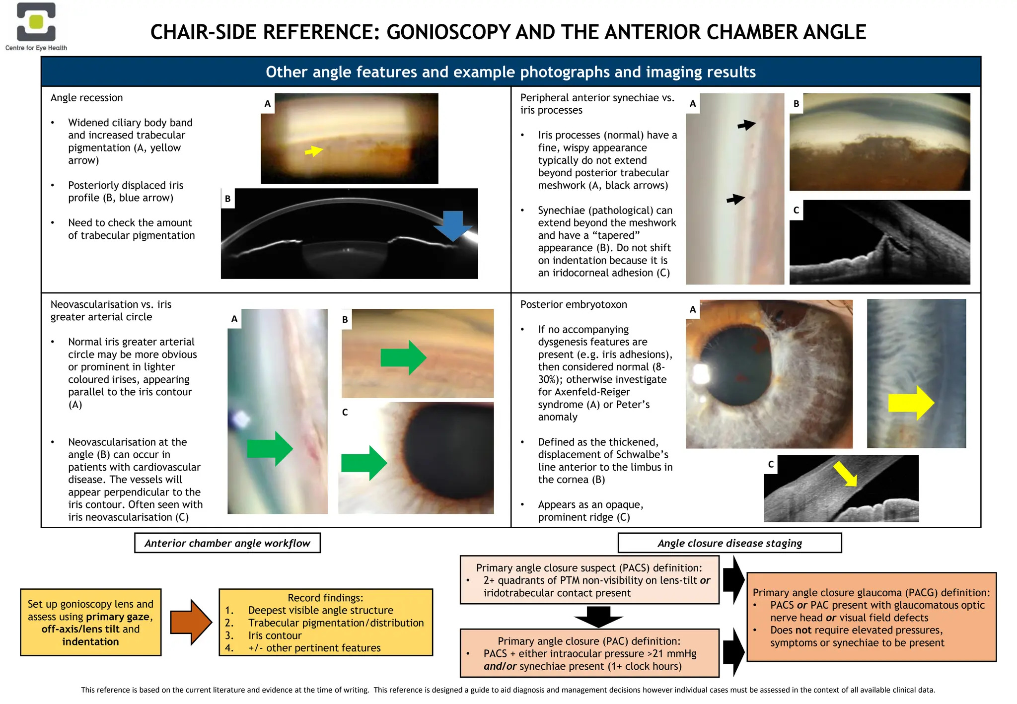
Other angle features and example photographs and imaging results
Angle recession
• Widened ciliary body band
and increased trabecular
pigmentation (A, yellow
arrow)
• Posteriorly displaced iris
profile (B, blue arrow)
• Need to check the amount
of trabecular pigmentation
Peripheral anterior synechiae vs.
iris processes
• Iris processes (normal) have a
fine, wispy appearance
typically do not extend
beyond posterior trabecular
meshwork (A, black arrows)
• Synechiae (pathological) can
extend beyond the meshwork
and have a “tapered”
appearance (B). Do not shift
on indentation because it is
an iridocorneal adhesion (C)
Neovascularisation vs. iris
greater arterial circle
• Normal iris greater arterial
circle may be more obvious
or prominent in lighter
coloured irises, appearing
parallel to the iris contour
(A)
• Neovascularisation at the
angle (B) can occur in
patients with cardiovascular
disease. The vessels will
appear perpendicular to the
iris contour. Often seen with
iris neovascularisation (C)
Posterior embryotoxon
• If no accompanying
dysgenesis features are
present (e.g. iris adhesions),
then considered normal (8-
30%); otherwise investigate
for Axenfeld-Reiger
syndrome (A) or Peter’s
anomaly
• Defined as the thickened,
displacement of Schwalbe’s
line anterior to the limbus in
the cornea (B)
• Appears as an opaque,
prominent ridge (C)
A
B
B
C
A
A
C
B
Set up gonioscopy lens and
assess using primary gaze,
off-axis/lens tilt and
indentation
Record findings:
1. Deepest visible angle structure
2. Trabecular pigmentation/distribution
3. Iris contour
4. +/- other pertinent features
Primary angle closure suspect (PACS) definition:
• 2+ quadrants of PTM non-visibility on lens-tilt or
iridotrabecular contact present
Primary angle closure (PAC) definition:
• PACS + either intraocular pressure >21 mmHg
and/or synechiae present (1+ clock hours)
Primary angle closure glaucoma (PACG) definition:
• PACS or PAC present with glaucomatous optic
nerve head or visual field defects
• Does not require elevated pressures,
symptoms or synechiae to be present
Anterior chamber angle workflow
A B
C
Angle closure disease staging
4.
CHAIR-SIDE REFERENCE: GONIOSCOPYAND THE ANTERIOR CHAMBER ANGLE
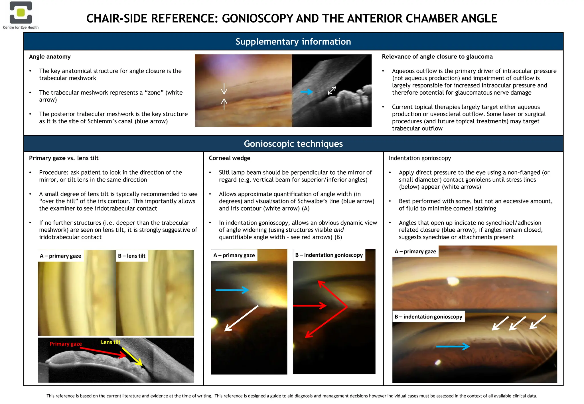
Supplementary information
Angle anatomy
• The key anatomical structure for angle closure is the
trabecular meshwork
• The trabecular meshwork represents a “zone” (white
arrow)
• The posterior trabecular meshwork is the key structure
as it is the site of Schlemm’s canal (blue arrow)
Relevance of angle closure to glaucoma
• Aqueous outflow is the primary driver of intraocular pressure
(not aqueous production) and impairment of outflow is
largely responsible for increased intraocular pressure and
therefore potential for glaucomatous nerve damage
• Current topical therapies largely target either aqueous
production or uveoscleral outflow. Some laser or surgical
procedures (and future topical treatments) may target
trabecular outflow
Gonioscopic techniques
Primary gaze vs. lens tilt
• Procedure: ask patient to look in the direction of the
mirror, or tilt lens in the same direction
• A small degree of lens tilt is typically recommended to see
“over the hill” of the iris contour. This importantly allows
the examiner to see iridotrabecular contact
• If no further structures (i.e. deeper than the trabecular
meshwork) are seen on lens tilt, it is strongly suggestive of
iridotrabecular contact
Corneal wedge
• Slitl lamp beam should be perpendicular to the mirror of
regard (e.g. vertical beam for superior/inferior angles)
• Allows approximate quantification of angle width (in
degrees) and visualisation of Schwalbe’s line (blue arrow)
and iris contour (white arrow) (A)
• In indentation gonioscopy, allows an obvious dynamic view
of angle widening (using structures visible and
quantifiable angle width – see red arrows) (B)
Indentation gonioscopy
• Apply direct pressure to the eye using a non-flanged (or
small diameter) contact goniolens until stress lines
(below) appear (white arrows)
• Best performed with some, but not an excessive amount,
of fluid to minimise corneal staining
• Angles that open up indicate no synechiael/adhesion
related closure (blue arrow); if angles remain closed,
suggests synechiae or attachments present
A – primary gaze B – lens tilt
Lens tilt
Primary gaze
A – primary gaze B – indentation gonioscopy
B – indentation gonioscopy
A – primary gaze
This reference is based on the current literature and evidence at the time of writing. This reference is designed a guide to aid diagnosis and management decisions however individual cases must be assessed in the context of all available clinical data.
![CHAIR-SIDE REFERENCE: GONIOSCOPY AND THE ANTERIOR CHAMBER ANGLE
Imaging modalities for assessing the anterior chamber angle
Optical coherence tomography
Key advantages
• Quick, non-invasive, high
resolution
• Can be repurposed from posterior
segment imaging devices
• Many quantitative parameters
become available [note: no
parameter cut-off exists for
identifying angle closure]
• Can visualise iris contour, lens-iris
interaction and lens vault
• Relatively well-controlled
background lighting
Key disadvantages
• Requires visualisation of key landmarks such as scleral spur,
Schlemm’s canal (not possible in around 20% of patients)
• Cannot visualise key anatomical structures in en face manner
(e.g. trabecular meshwork)
• Most commercially available instruments only give one slice
(not sufficient for describing entirety of the anterior chamber
angle)
• Specialised (e.g. swept-source) devices more appropriate than
repurposed posterior segment devices
• Affected by anterior segment pathologies (e.g. conjunctiva)
and corneal compensation protocols required to adjust for
image magnification – see examples below
Examples of challenges with anterior segment imaging
• The lateral angles are easier to image compared to
superior/inferior angles, which are susceptible to
distortions due to instrument-specific image scaling
(A, example of superior angle imaging, where primary
gaze imaging is not possible)
• Opacities on the conjunctiva and cornea can obscure
the angle structures (B, example of pterygium)
Scheimpflug imaging Ultrasound biomicroscopy
Key features
• Allows quantification of anterior
chamber parameters (A)
• Allows visualisation of the angle
across the anterior chamber width
• Cannot visualise anterior chamber
angle itself (B); few normative data
available for quantitative
information
Example images Key features
• Allows visualisation of the retroiridal
space by penetrating the pigment
epithelium (e.g. iridociliary cysts,
ciliary body position and lesions)
• Resolution much lower than that of
optical coherence tomography
• Requires contact with ocular surface
Example case
Legend:
White: ciliary body
Red: scleral spur
Blue: Schlemm’s
canal
Yellow: iris
Open angle Closed angle
Iris
contour
Angle
opening
Plateau configuration Posteriorly bowed Regular
A B
This reference is based on the current literature and evidence at the time of writing. This reference is designed a guide to aid diagnosis and management decisions however individual cases must be assessed in the context of all available clinical data.
A
B](https://image.slidesharecdn.com/13-250918133428-12a6dbef/75/13-Gonioscopy-and-anterior-chamber-angle-pdf-2-2048.jpg)