Downloaded 762 times


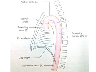
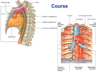
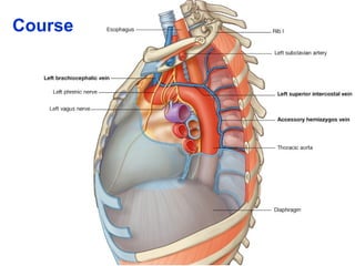
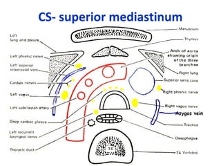

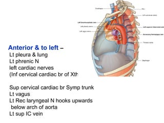
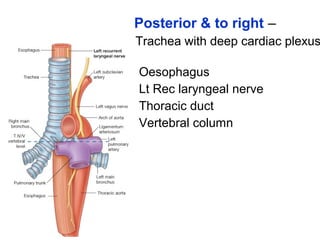
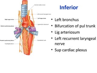
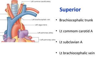
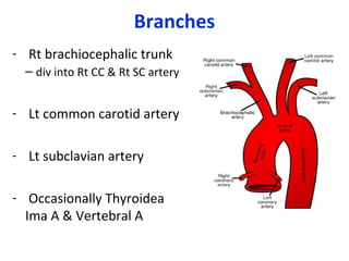
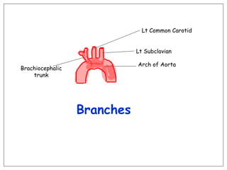

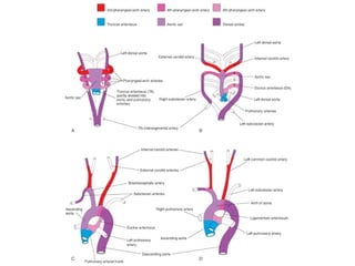
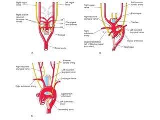

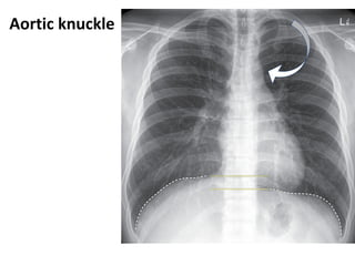
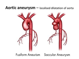


The arch of the aorta begins at the level of the sternal angle and arches over the root of the left lung in the superior mediastinum. It begins as the continuation of the ascending aorta and passes up, back, and left before turning backwards and downwards to become the descending aorta at the level of T4. It has anterior relations to the left lung and pleura and posterior relations to the trachea, esophagus, and thoracic duct. Its branches include the brachiocephalic trunk, left common carotid artery, and left subclavian artery.


![ONFH[AVN HIP] -TRIPLE REGIME -A NOVAL SURGICAL CONCEPT .pptx](https://cdn.slidesharecdn.com/ss_thumbnails/onfhavnhip2026koaconcalicutdrgokuldevdrmashraf-260210064517-213ec005-thumbnail.jpg?width=640&height=640&fit=bounds)


![PERI-PROSTHETIC FRACTURE NAIL-PLATE CONSTRUCT [NPC].pptx](https://cdn.slidesharecdn.com/ss_thumbnails/drarunkumardrmohamedashrafperiprostheticfrasturenail-plateconstructnpc-260209164459-7e9d15a1-thumbnail.jpg?width=640&height=640&fit=bounds)