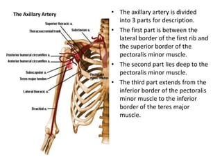
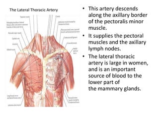
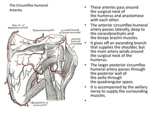
The fascial compartments of thigh are the three fascial compartments that divide and contain the thigh muscles. The fascia lata is the strong and deep fascia of the thigh that surrounds the thigh muscles and forms the outer limits of the compartments. Internally the muscle compartments are divided by the lateral and medial intermuscular septa.
The Transradial technique is the true minimally invasive "Drive-through" approach to perform percutaneous coronary and peripheral angiograms and interventions.
ARTERIAL SUPPLY OF UPPER LIMBThe arterial supply to the .docxrossskuddershamus
ARTERIAL SUPPLY
OF UPPER LIMB
The arterial supply to the upper limb begins in the chest as the subclavian artery. The right subclavian artery arises from the brachiocephalic trunk, while the left subclavian branches directly off the arch of aorta.
When the subclavian arteries cross the lateral edge of the 1st rib, they enter the axilla, and are called axillary arteries.
IN THE AXILLA
The axillary artery passes through the axilla, just underneath the pectoralis minor muscle, enclosed in the axillary sheath.
At the level of the humeral surgical neck, the posterior and anterior circumflex humeral arteries arise. They circle posteriorly around the humerus to supply the shoulder region. The largest branch of the axillary artery also arises here – the subscapular artery.
The axillary artery becomes the brachial artery at the level of the teres major muscle.
Anteriorly
(i) Skin.
(ii) Superficial fascia
(iii) Deep fascia.
(iv) Clavicular part of the pectoralis major.
(v) Clavipectoral fascia with cephalic vein, lateral pectoral nerve, and thoracoacromial vessels.
Posteriorly
(i) First intercostal space with the external intercostal muscle.
(ii) First and second digitations of the serratus anterior with the nerve to serratus anterior.
(iii) Medial cord of brachial plexus with its medial pectoral
branch
laterally
Lateral and posterior cords of the brachial plexus.
Medially
Axillary vein
The first part of the axillary artery is enclosed (together with the brachial plexus) in the axillary sheath, derived from the prevertebral layer of deep cervical fascia.
Anteriorly
(i) Skin.
(ii) Superficial fascia.
(iii) Deep fascia.
(iv) Pectoralis major.
(v) Pectoralis minor
Posteriorly
(i) Posterior cord of brachial plexus.
(ii) subscapularis
Medially
(i) Medial cord of brachial plexus,
(ii) Medial pectoral nerve,
(iii) Axillary vein.
Laterally
Lateral cord of brachial plexus.
Anteriorly
(i) Skin.
(ii) Superficial fascia,
(iii) Deep fascia.
(iv) In the upper part there are the pectoralis major and medial root of the median nerve.
Posteriorly
(i) Radial nerve.
(ii) Axillary nerve in the upper part,
(iii) Subscapularis in the upper part,
(iv) Tendons of the latissimus dorsi and the teres major in the lower part.
Laterally
Coracobrachialis.
Musculocutaneous nerve in the upper part,
Lateral root of median nerve in the upper part,
Trunk of median nerve in the lower part.
Medially
(i) Axillary vein,
(ii) Medial cutaneous nerve of the forearm and ulnar nerve
(iii) Medial cutaneous nerve of arm
1st part:
Superior Thoracic artery
2nd part:
Acromiothoracic artery
Lateral Thoracic artery
3rd part:
Subscapular artery
Ant circumflex humeral artery
Post circumflex humeral artery
CLINICAL RELEVANCE: AXILLARY ARTERY ANEURYSM
An aneurysm is dilation of a blood vessel to more than twice its original size. Although rare, axillary artery aneurysms can occur as a result of atherosclerosis, thoracic out.
Title: Sense of Smell
Presenter: Dr. Faiza, Assistant Professor of Physiology
Qualifications:
MBBS (Best Graduate, AIMC Lahore)
FCPS Physiology
ICMT, CHPE, DHPE (STMU)
MPH (GC University, Faisalabad)
MBA (Virtual University of Pakistan)
Learning Objectives:
Describe the primary categories of smells and the concept of odor blindness.
Explain the structure and location of the olfactory membrane and mucosa, including the types and roles of cells involved in olfaction.
Describe the pathway and mechanisms of olfactory signal transmission from the olfactory receptors to the brain.
Illustrate the biochemical cascade triggered by odorant binding to olfactory receptors, including the role of G-proteins and second messengers in generating an action potential.
Identify different types of olfactory disorders such as anosmia, hyposmia, hyperosmia, and dysosmia, including their potential causes.
Key Topics:
Olfactory Genes:
3% of the human genome accounts for olfactory genes.
400 genes for odorant receptors.
Olfactory Membrane:
Located in the superior part of the nasal cavity.
Medially: Folds downward along the superior septum.
Laterally: Folds over the superior turbinate and upper surface of the middle turbinate.
Total surface area: 5-10 square centimeters.
Olfactory Mucosa:
Olfactory Cells: Bipolar nerve cells derived from the CNS (100 million), with 4-25 olfactory cilia per cell.
Sustentacular Cells: Produce mucus and maintain ionic and molecular environment.
Basal Cells: Replace worn-out olfactory cells with an average lifespan of 1-2 months.
Bowman’s Gland: Secretes mucus.
Stimulation of Olfactory Cells:
Odorant dissolves in mucus and attaches to receptors on olfactory cilia.
Involves a cascade effect through G-proteins and second messengers, leading to depolarization and action potential generation in the olfactory nerve.
Quality of a Good Odorant:
Small (3-20 Carbon atoms), volatile, water-soluble, and lipid-soluble.
Facilitated by odorant-binding proteins in mucus.
Membrane Potential and Action Potential:
Resting membrane potential: -55mV.
Action potential frequency in the olfactory nerve increases with odorant strength.
Adaptation Towards the Sense of Smell:
Rapid adaptation within the first second, with further slow adaptation.
Psychological adaptation greater than receptor adaptation, involving feedback inhibition from the central nervous system.
Primary Sensations of Smell:
Camphoraceous, Musky, Floral, Pepperminty, Ethereal, Pungent, Putrid.
Odor Detection Threshold:
Examples: Hydrogen sulfide (0.0005 ppm), Methyl-mercaptan (0.002 ppm).
Some toxic substances are odorless at lethal concentrations.
Characteristics of Smell:
Odor blindness for single substances due to lack of appropriate receptor protein.
Behavioral and emotional influences of smell.
Transmission of Olfactory Signals:
From olfactory cells to glomeruli in the olfactory bulb, involving lateral inhibition.
Primitive, less old, and new olfactory systems with different path
New Directions in Targeted Therapeutic Approaches for Older Adults With Mantl...i3 Health
i3 Health is pleased to make the speaker slides from this activity available for use as a non-accredited self-study or teaching resource.
This slide deck presented by Dr. Kami Maddocks, Professor-Clinical in the Division of Hematology and
Associate Division Director for Ambulatory Operations
The Ohio State University Comprehensive Cancer Center, will provide insight into new directions in targeted therapeutic approaches for older adults with mantle cell lymphoma.
STATEMENT OF NEED
Mantle cell lymphoma (MCL) is a rare, aggressive B-cell non-Hodgkin lymphoma (NHL) accounting for 5% to 7% of all lymphomas. Its prognosis ranges from indolent disease that does not require treatment for years to very aggressive disease, which is associated with poor survival (Silkenstedt et al, 2021). Typically, MCL is diagnosed at advanced stage and in older patients who cannot tolerate intensive therapy (NCCN, 2022). Although recent advances have slightly increased remission rates, recurrence and relapse remain very common, leading to a median overall survival between 3 and 6 years (LLS, 2021). Though there are several effective options, progress is still needed towards establishing an accepted frontline approach for MCL (Castellino et al, 2022). Treatment selection and management of MCL are complicated by the heterogeneity of prognosis, advanced age and comorbidities of patients, and lack of an established standard approach for treatment, making it vital that clinicians be familiar with the latest research and advances in this area. In this activity chaired by Michael Wang, MD, Professor in the Department of Lymphoma & Myeloma at MD Anderson Cancer Center, expert faculty will discuss prognostic factors informing treatment, the promising results of recent trials in new therapeutic approaches, and the implications of treatment resistance in therapeutic selection for MCL.
Target Audience
Hematology/oncology fellows, attending faculty, and other health care professionals involved in the treatment of patients with mantle cell lymphoma (MCL).
Learning Objectives
1.) Identify clinical and biological prognostic factors that can guide treatment decision making for older adults with MCL
2.) Evaluate emerging data on targeted therapeutic approaches for treatment-naive and relapsed/refractory MCL and their applicability to older adults
3.) Assess mechanisms of resistance to targeted therapies for MCL and their implications for treatment selection
Couples presenting to the infertility clinic- Do they really have infertility...Sujoy Dasgupta
Dr Sujoy Dasgupta presented the study on "Couples presenting to the infertility clinic- Do they really have infertility? – The unexplored stories of non-consummation" in the 13th Congress of the Asia Pacific Initiative on Reproduction (ASPIRE 2024) at Manila on 24 May, 2024.
Report Back from SGO 2024: What’s the Latest in Cervical Cancer?bkling
Are you curious about what’s new in cervical cancer research or unsure what the findings mean? Join Dr. Emily Ko, a gynecologic oncologist at Penn Medicine, to learn about the latest updates from the Society of Gynecologic Oncology (SGO) 2024 Annual Meeting on Women’s Cancer. Dr. Ko will discuss what the research presented at the conference means for you and answer your questions about the new developments.
ARTIFICIAL INTELLIGENCE IN HEALTHCARE.pdfAnujkumaranit
Artificial intelligence (AI) refers to the simulation of human intelligence processes by machines, especially computer systems. It encompasses tasks such as learning, reasoning, problem-solving, perception, and language understanding. AI technologies are revolutionizing various fields, from healthcare to finance, by enabling machines to perform tasks that typically require human intelligence.
Pulmonary Thromboembolism - etilogy, types, medical- Surgical and nursing man...VarunMahajani
Disruption of blood supply to lung alveoli due to blockage of one or more pulmonary blood vessels is called as Pulmonary thromboembolism. In this presentation we will discuss its causes, types and its management in depth.
Ethanol (CH3CH2OH), or beverage alcohol, is a two-carbon alcohol
that is rapidly distributed in the body and brain. Ethanol alters many
neurochemical systems and has rewarding and addictive properties. It
is the oldest recreational drug and likely contributes to more morbidity,
mortality, and public health costs than all illicit drugs combined. The
5th edition of the Diagnostic and Statistical Manual of Mental Disorders
(DSM-5) integrates alcohol abuse and alcohol dependence into a single
disorder called alcohol use disorder (AUD), with mild, moderate,
and severe subclassifications (American Psychiatric Association, 2013).
In the DSM-5, all types of substance abuse and dependence have been
combined into a single substance use disorder (SUD) on a continuum
from mild to severe. A diagnosis of AUD requires that at least two of
the 11 DSM-5 behaviors be present within a 12-month period (mild
AUD: 2–3 criteria; moderate AUD: 4–5 criteria; severe AUD: 6–11 criteria).
The four main behavioral effects of AUD are impaired control over
drinking, negative social consequences, risky use, and altered physiological
effects (tolerance, withdrawal). This chapter presents an overview
of the prevalence and harmful consequences of AUD in the U.S.,
the systemic nature of the disease, neurocircuitry and stages of AUD,
comorbidities, fetal alcohol spectrum disorders, genetic risk factors, and
pharmacotherapies for AUD.
Tom Selleck Health: A Comprehensive Look at the Iconic Actor’s Wellness Journeygreendigital
Tom Selleck, an enduring figure in Hollywood. has captivated audiences for decades with his rugged charm, iconic moustache. and memorable roles in television and film. From his breakout role as Thomas Magnum in Magnum P.I. to his current portrayal of Frank Reagan in Blue Bloods. Selleck's career has spanned over 50 years. But beyond his professional achievements. fans have often been curious about Tom Selleck Health. especially as he has aged in the public eye.
Follow us on: Pinterest
Introduction
Many have been interested in Tom Selleck health. not only because of his enduring presence on screen but also because of the challenges. and lifestyle choices he has faced and made over the years. This article delves into the various aspects of Tom Selleck health. exploring his fitness regimen, diet, mental health. and the challenges he has encountered as he ages. We'll look at how he maintains his well-being. the health issues he has faced, and his approach to ageing .
Early Life and Career
Childhood and Athletic Beginnings
Tom Selleck was born on January 29, 1945, in Detroit, Michigan, and grew up in Sherman Oaks, California. From an early age, he was involved in sports, particularly basketball. which played a significant role in his physical development. His athletic pursuits continued into college. where he attended the University of Southern California (USC) on a basketball scholarship. This early involvement in sports laid a strong foundation for his physical health and disciplined lifestyle.
Transition to Acting
Selleck's transition from an athlete to an actor came with its physical demands. His first significant role in "Magnum P.I." required him to perform various stunts and maintain a fit appearance. This role, which he played from 1980 to 1988. necessitated a rigorous fitness routine to meet the show's demands. setting the stage for his long-term commitment to health and wellness.
Fitness Regimen
Workout Routine
Tom Selleck health and fitness regimen has evolved. adapting to his changing roles and age. During his "Magnum, P.I." days. Selleck's workouts were intense and focused on building and maintaining muscle mass. His routine included weightlifting, cardiovascular exercises. and specific training for the stunts he performed on the show.
Selleck adjusted his fitness routine as he aged to suit his body's needs. Today, his workouts focus on maintaining flexibility, strength, and cardiovascular health. He incorporates low-impact exercises such as swimming, walking, and light weightlifting. This balanced approach helps him stay fit without putting undue strain on his joints and muscles.
Importance of Flexibility and Mobility
In recent years, Selleck has emphasized the importance of flexibility and mobility in his fitness regimen. Understanding the natural decline in muscle mass and joint flexibility with age. he includes stretching and yoga in his routine. These practices help prevent injuries, improve posture, and maintain mobilit
- Video recording of this lecture in English language: https://youtu.be/lK81BzxMqdo
- Video recording of this lecture in Arabic language: https://youtu.be/Ve4P0COk9OI
- Link to download the book free: https://nephrotube.blogspot.com/p/nephrotube-nephrology-books.html
- Link to NephroTube website: www.NephroTube.com
- Link to NephroTube social media accounts: https://nephrotube.blogspot.com/p/join-nephrotube-on-social-media.html
These lecture slides, by Dr Sidra Arshad, offer a quick overview of physiological basis of a normal electrocardiogram.
Learning objectives:
1. Define an electrocardiogram (ECG) and electrocardiography
2. Describe how dipoles generated by the heart produce the waveforms of the ECG
3. Describe the components of a normal electrocardiogram of a typical bipolar leads (limb II)
4. Differentiate between intervals and segments
5. Enlist some common indications for obtaining an ECG
Study Resources:
1. Chapter 11, Guyton and Hall Textbook of Medical Physiology, 14th edition
2. Chapter 9, Human Physiology - From Cells to Systems, Lauralee Sherwood, 9th edition
3. Chapter 29, Ganong’s Review of Medical Physiology, 26th edition
4. Electrocardiogram, StatPearls - https://www.ncbi.nlm.nih.gov/books/NBK549803/
5. ECG in Medical Practice by ABM Abdullah, 4th edition
6. ECG Basics, http://www.nataliescasebook.com/tag/e-c-g-basics
micro teaching on communication m.sc nursing.pdfAnurag Sharma
Microteaching is a unique model of practice teaching. It is a viable instrument for the. desired change in the teaching behavior or the behavior potential which, in specified types of real. classroom situations, tends to facilitate the achievement of specified types of objectives.
4. The axillary artery is divided into 3 parts for description. The first part is between the lateral border of the first rib and the superior border of the pectoralis minor muscle. The second part lies deep to the pectoralis minor muscle. The third part extends from the inferior border of the pectoralis minor muscle to the inferior border of the teres major muscle. The Axillary Artery
5. First Part of the Axillary Artery This part is enclosed in the axillary sheath along with the axillary vein. This part of the axillary artery has only one branch, the supreme thoracic artery. This artery helps to supply the first two intercostal spaces and the superior part of the serratus anterior muscle.
6. Second Part of the Axillary Artery This part of the artery lies deep to the pectoralis minor muscle. The lateral, medial and posterior cords of the brachial plexus are so named respective to this part of the axillary artery. This part of the axillary artery has two branches, the thoracoacromial and lateral thoracic arteries.
7. This is a short, wide trunk that arises from the axillary artery deep to the pectoralis minor muscle. It pierces the costocoracoid membrane, which is part of the clavipectoral fascia. It then divides into 4 branches (acromial, deltoid, pectoral, and clavicular) deep to the clavicular head of the pectoralis major muscle. The Thoracoacromial Artery
8. The Lateral Thoracic Artery This artery descends along the axillary border of the pectoralis minor muscle. It supplies the pectoral muscles and the axillary lymph nodes. The lateral thoracic artery is large in women, and is an important source of blood to the lower part of the mammary glands.
9. Third Part of the Axillary Artery This part extends from the inferior border of the pectoralis minor muscle to the inferior border of the teres major muscle. It has three branches, the anterior and posterior circumflex humeral arteries and the subscapular artery.
10. The Circumflex Humeral Arteries These arteries pass around the surgical neck of the humerus and anastomose with each other. The anterior circumflex humeral artery passes laterally, deep to the coracobrachialis and the biceps brachii muscles. It gives off an ascending branch that supplies the shoulder, but the main artery winds around the surgical neck of the humerus. The larger posterior circumflex humeral artery passes through the posterior wall of the axilla through the quadrangular space. It is accompanied by the axillary nerve to supply the surrounding muscles.
11. The Subscapular Artery This is the largest branch of the axillary artery. It descends along the lateral border of the subscapularis muscle and divides into the circumflex scapular artery and thethoracodorsal artery. The circumflex scapular artery passes around the lateral border of the scapula to supply muscles on the dorsum of the scapula. The thoracodorsal artery continues the general course of the subscapular artery to supply adjacent muscles, principally, the latissimusdorsi muscle.
13. This artery begins at the inferior border of the teres major muscle as the continuation of the axillary artery. Runs inferiorly and slightly laterally on the medial side of the biceps brachii muscle to the cubitalfossa, where it ends opposite the neck of the radius. At first it lies medial to the humerus and then anterior to it. It lies anterior to the triceps brachii and brachialis muscles, and is overlapped by the biceps brachii and coracobrachialis muscles. It is superficial and palpable throughout its whole length ( brachial pulse). It accompanies the median nerve, which crosses it anteriorly at the middle of the arm. Divisions Under the cover of the bicipitalaponeurosis, the brachial artery divides into the radial and ulnar arteries. The brachial artery gives rise to many unnamed muscular branches, mainly from its lateral side. The named branches of the brachial artery are the profundabrachii artery, the nutrient humeral artery, and the ulnar collateral arteries (superior and inferior).
14. The ProfundaBrachii Artery This is the largest branch of the brachial artery, and has the most superior origin. It accompanies the radial nerve in its posterior course in the radial groove. Posterior to the humerus, the profundabrachii artery (or deep brachial artery) divides into anterior and posterior ascending branches. These help form the arterial anastomoses of the elbow region.
15. The Superior Ulnar Collateral Artery This arises from the brachial artery near the middle of the arm. It accompanies the ulnar nerve posterior to the medial epicondyle of the humerus. Here, it anastomoses with the posterior ulnar recurrent branch of the ulnar artery and the inferior ulnar collateral artery.
16. The Inferior Ulnar Collateral Artery This arises from the brachial artery about 5 cm proximal to the elbow crease. It then passes inferomedially, anterior to the medial epicondyle of the humerus. Here, it joins the anastomoses of the elbow region.
18. It begins in the cubitalfossa, opposite the neck of the radius and leaves the forearm by winding around the lateral aspect of the radius. The proximal part of the radial artery is overlapped by the fleshy belly of the brachioradialis muscle. The radial artery lies on muscle until it comes into contact with the distal end of the radius, where it is covered only by superficial and deep fascia and skin. It passes posteriorly between the lateral collateral ligament of the wrist joint and the tendons of the abductor pollicislongus and extensor pollicisbrevis muscles. It crosses the floor of the anatomical snuffbox formed by the scaphoid and trapezium bones. It enters the palm of the hand by passing between the heads of the first dorsal interosseous muscle. It divides into the princepspollicis artery and the radialisindicis artery (arteries to the thumb and index fingers). It then passes between the two heads of adductor pollicis muscle and ends by completing the deep palmar arterial arch in conjunction with the ulnar artery. Muscular branches of the radial artery supply muscles on the lateral side of the forearm
22. The Dorsal Carpal Branch This runs medially across the dorsal surface of the wrist, deep to the extensor tendons. Here it anastomoses with the dorsal carpal branch of the ulnar artery and with the terminations of the anterior and posterior interosseous arteries to form the dorsal carpal arch.
23. Palmar Metacarpal Arteries There are three of these arteries and they arise from the deep palmar arch and run distally, where they joint the common palmar digital arteries, which arise from the superficial palmar arterial arch.
25. It is the larger of the two terminal branches of the brachial artery and begins near the neck of the radius, just medial to the biceps brachii tendon. It passes inferomedially deep to the pronatorteres muscle. With the median nerve, the ulnar artery passes between the ulnar and radial heads of the flexor digitorumsuperficialis muscle. About midway between the elbow and the wrist, it crosses posterior to the median nerve to reach the medial side of the forearm, where it lies on the flexor digitorumsuperficialis muscle. In the distal 2/3 of the forearm, the ulnar artery lies lateral to the ulnarnerve.It leaves the forearm by passing superficial to the flexor retinaculum on the lateral side of the pisiform bone. At the wrist, the ulnar artery and nerve lie lateral to the tendon of flexor carpiulnarismuscle.Here, they are covered only by fascia and skin.
26. Ulnar Recurrent Arteries Anterior Ulnar Recurrent Artery This arises from the ulnar artery, just inferior to the elbow joint. It runs superiorly between the brachialis and pronatorteres muscles. It supplies these muscles and anastomoses with the inferior ulnar collateral artery. Posterior Ulnar Recurrent Artery This arises from the ulnar artery distal to the anterior ulnar recurrent artery. It passes superiorly, posterior to the medial epicondyle of the humerus, where it lies deep to the tendon of the flexor carpiulnaris muscle. It supplies the adjacent muscles and then takes part in the arterial anastomosis around the elbow.
27. Interosseous Arteries Common Interosseous Artery This is a short branch of the ulnar artery. It arises from the distal part of the cubitalfossa and divides into the anterior and posterior interosseous arteries. Anterior Interosseous Artery This artery passes distally on the interosseous membrane to the proximal border of the pronatorquadratus muscles. Here, it pierces the membrane and continues distally to joint the dorsal carpal arch. Posterior InterosseousArtery This artery passes posterior between the bones of the forearm, just proximal to the interosseous membrane. It supplies the adjacent muscles and then gives off the posterior interosseous recurrent artery, which passes superiorly, posterior to the lateral epicondyle of the humerus and participates in the arterial anastomosis around the elbow.
28. Carpal Branches of the Ulnar Artery Palmar Carpal Branch of the Ulnar Artery This arises just proximal to the pisiform bone. It passes across the anterior aspect of the wrist, deep to the tendon of flexor digitorumprofundus muscle. This branch anastomoses with the palmar carpal branch of the radial artery and forms the palmar carpal arch. Dorsal Carpal Branch of the Ulnar Artery This arises just proximal to the pisiform bone. It passes across the dorsal surface of the wrist, deep to the extensor tendons, where it anastomoses with the dorsal carpal branch of the radial artery to form the dorsal carpal arch.
29. Superficial and Palmar Branches of Ulnar Artery The Superficial Branch of the Ulnar Artery This artery continues into the palm as the superficial palmar arch. The Deep Palmar Branch of the Ulnar Artery This branch passes deeply in the hand, where it anastomoses with the radial artery and completes the deep palmar arch.
30. PalmarArtieral Arches Superficial Palmar Arterial Arch It is formed mainly by the ulnar artery. It gives rise to 3 common palmar digital arteries that anastomoses with the palmar metacarpal arteries from the deep palmar arch. Each common palmar digital artery divides into a pair of proper digital arteries, which runs along the sides of the 2nd to 4th digits. The Deep Palmar Arterial Arch This lies across the metacarpal bones just distal to their bases. This if formed mainly by the radial artery. It is about a fingerbreadth closer to the wrist than the superficial palmar arterial arch. It gives rise to 3 common palmar digital arteries.