Downloaded 638 times


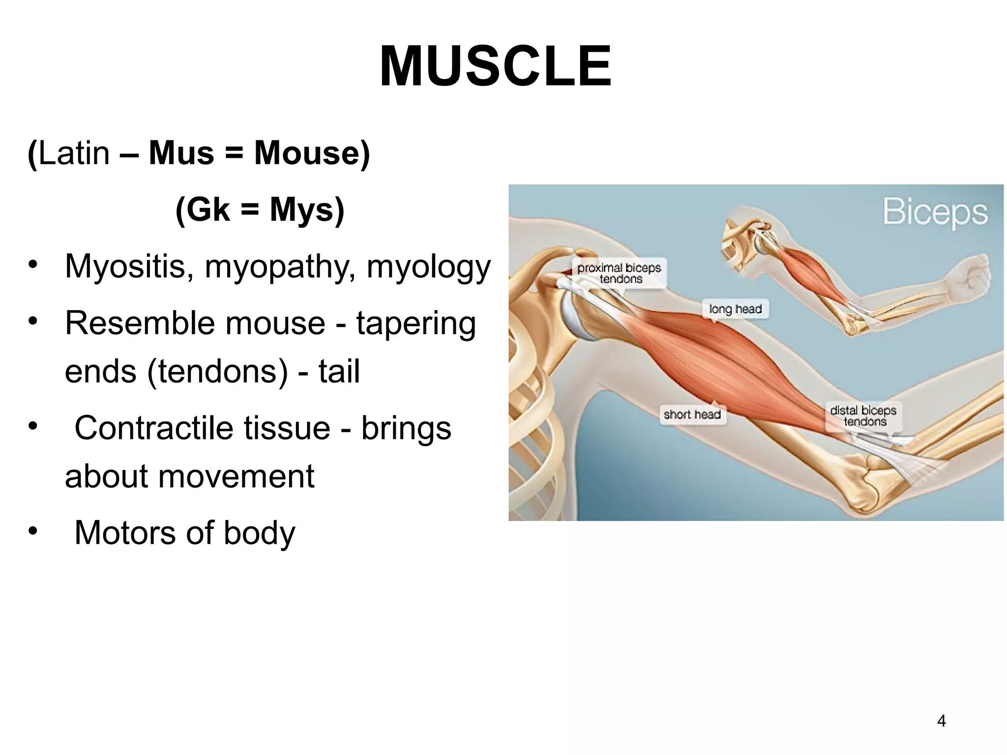
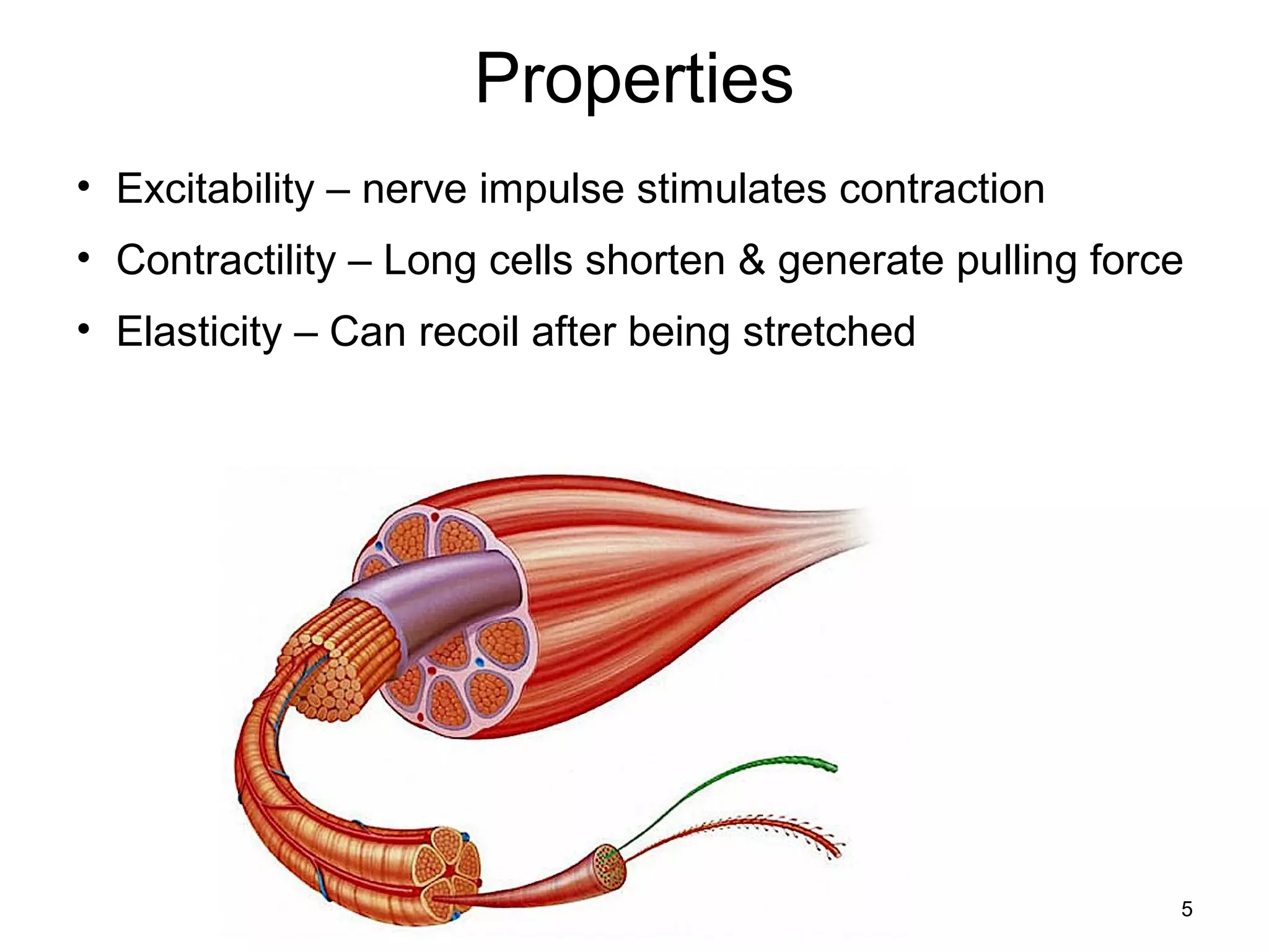


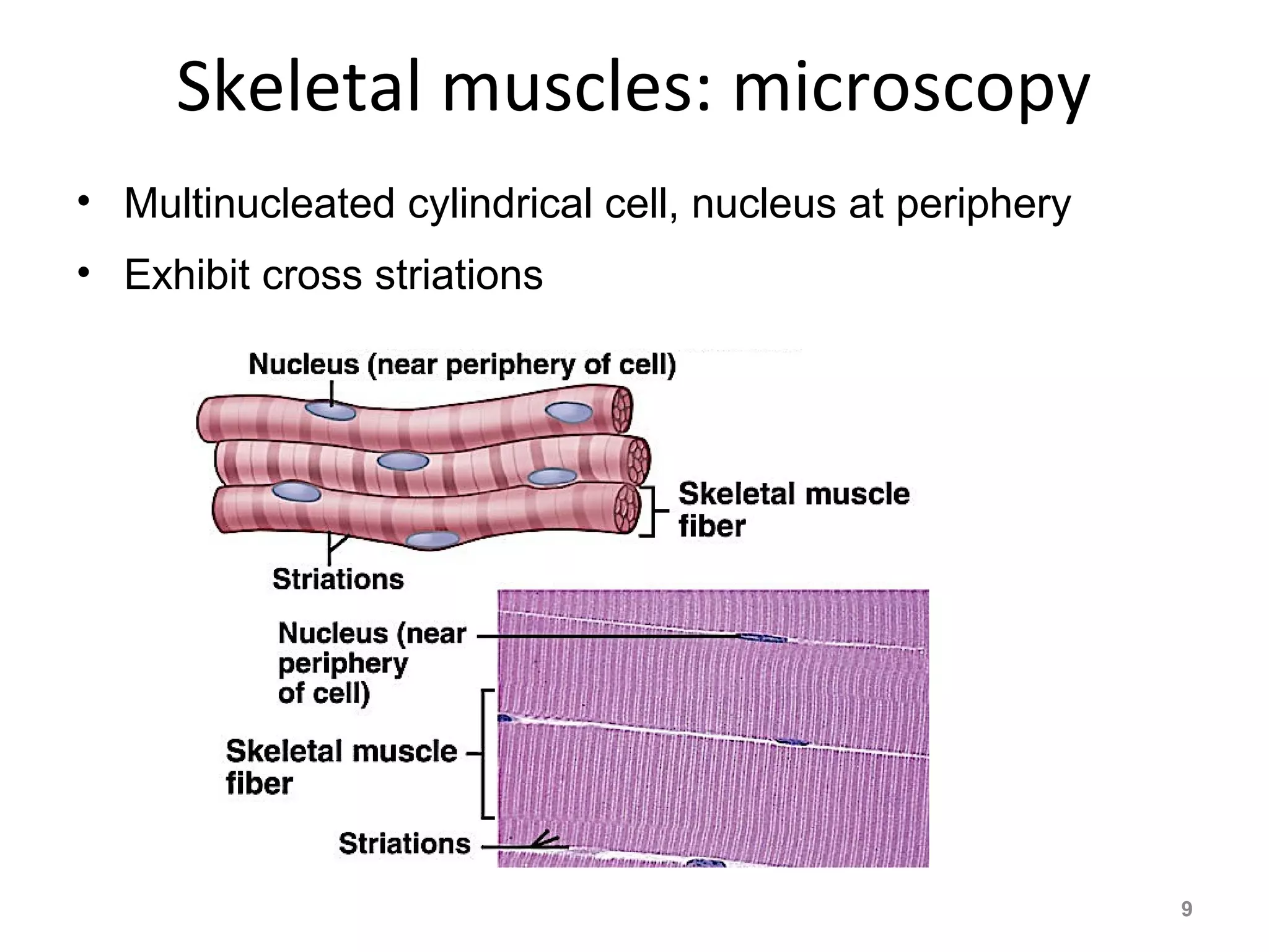

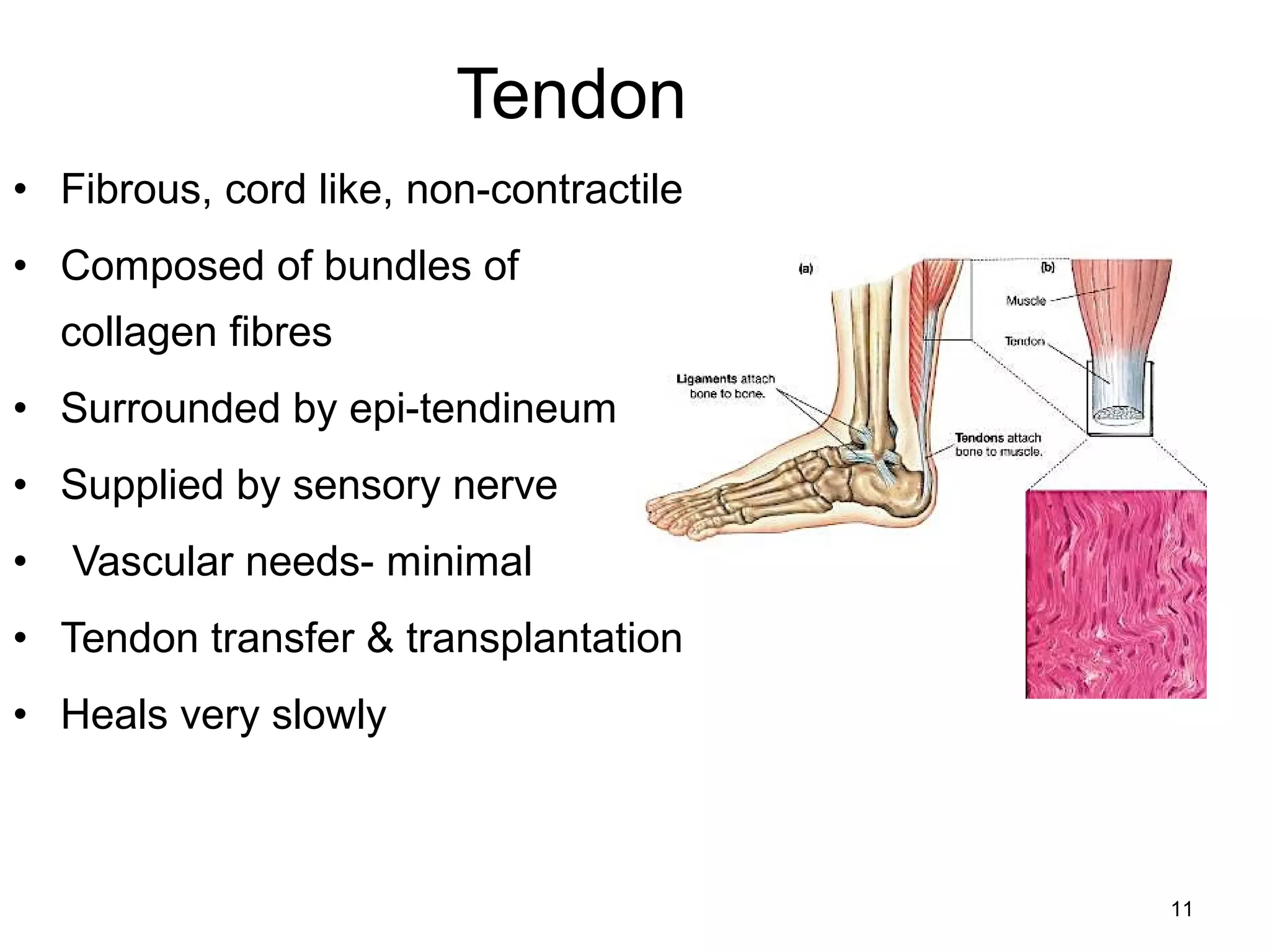

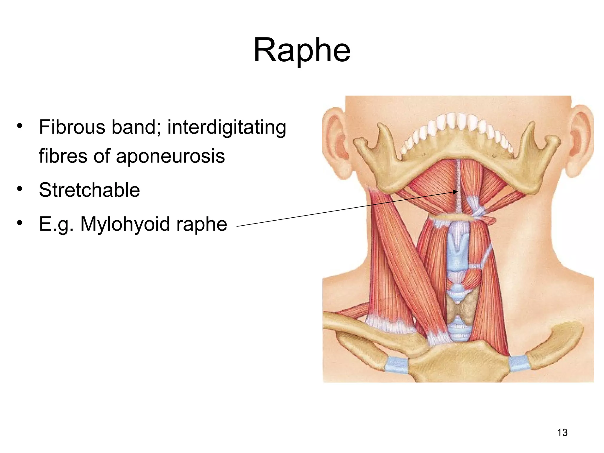
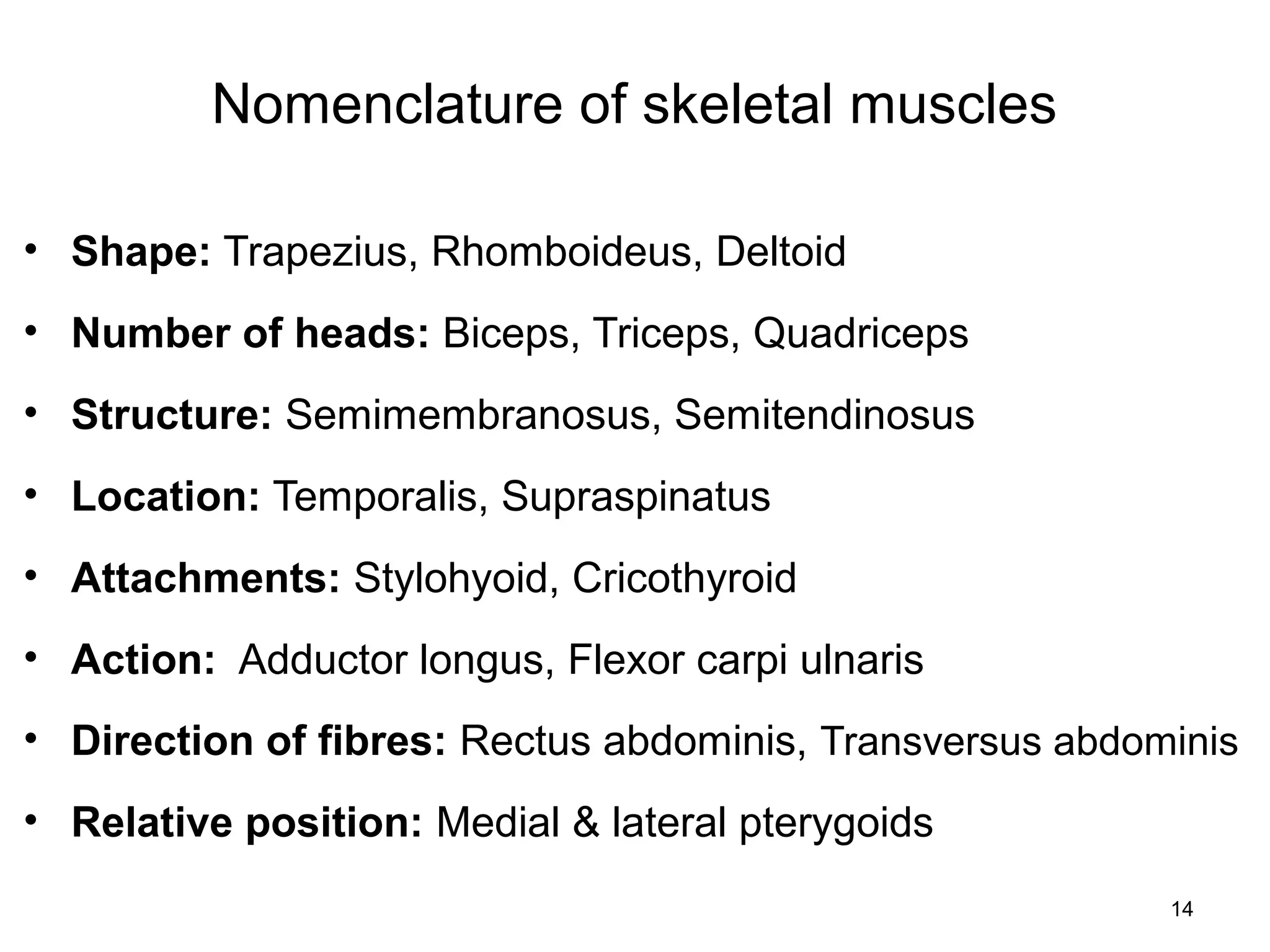
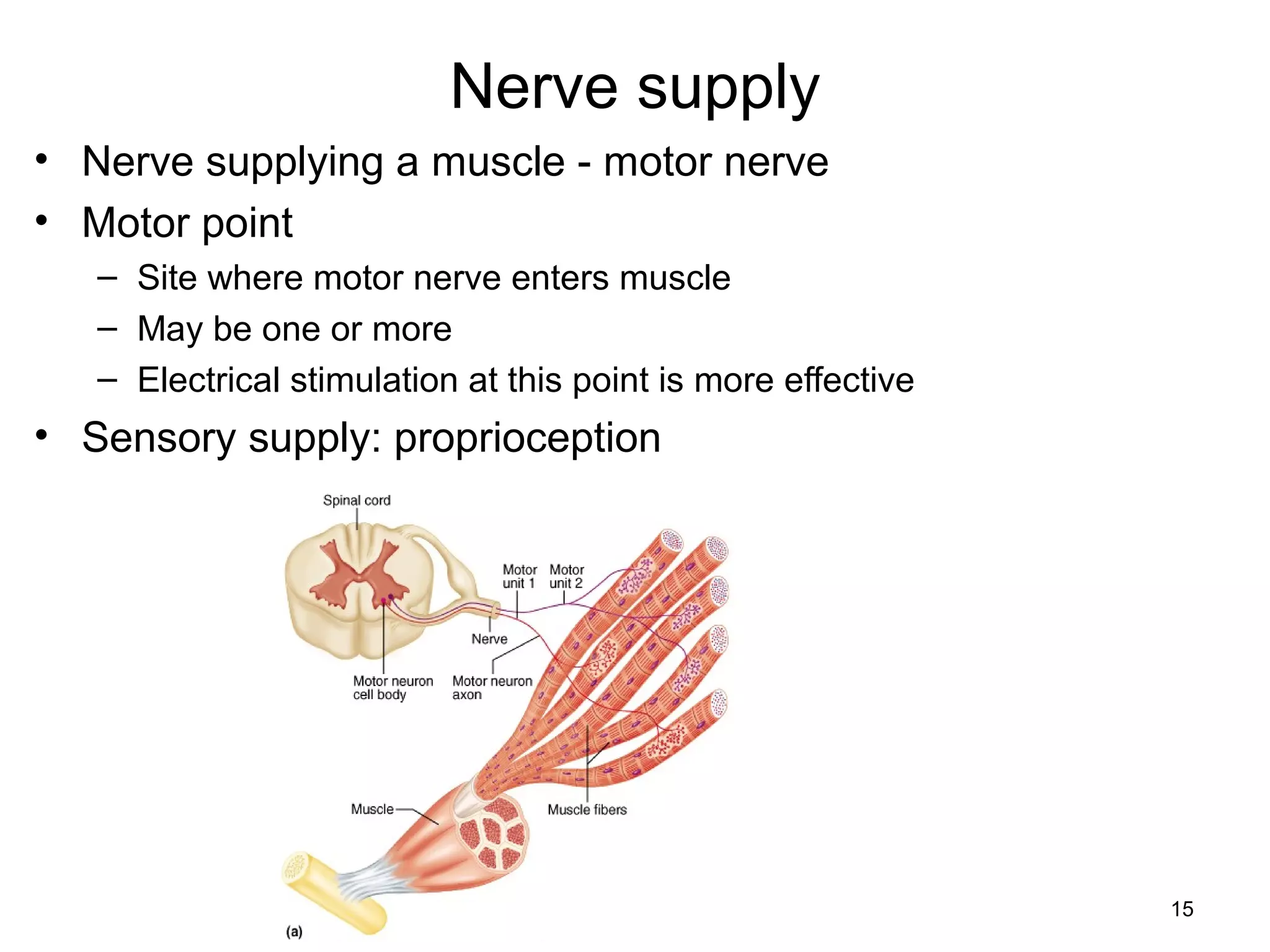


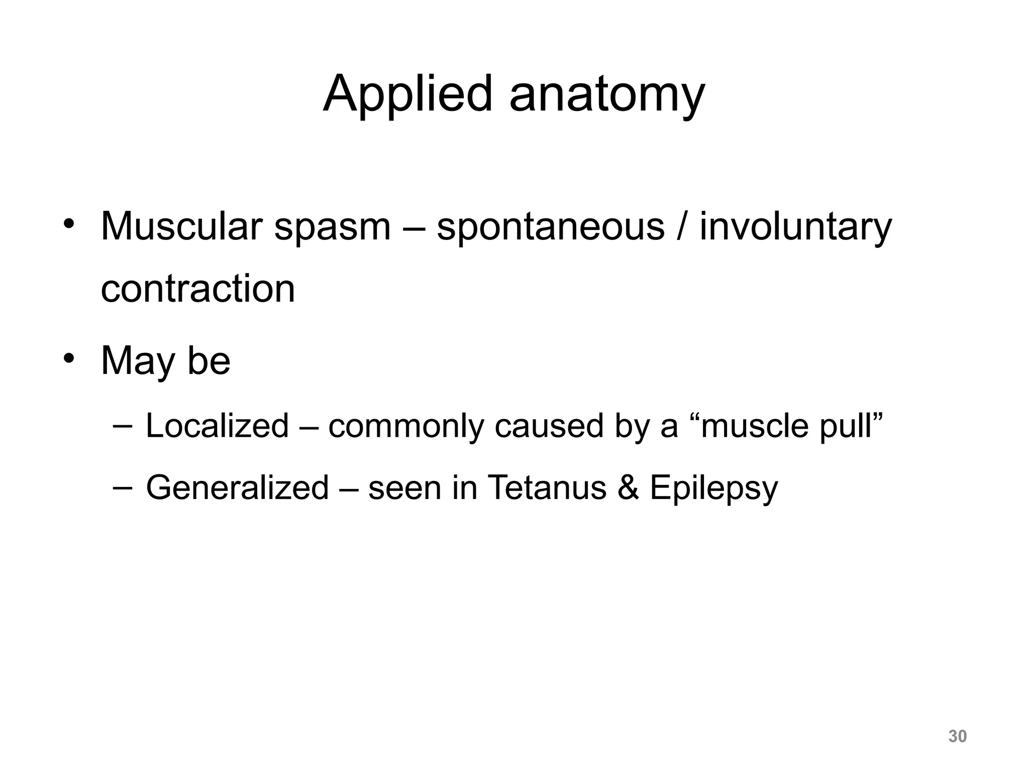
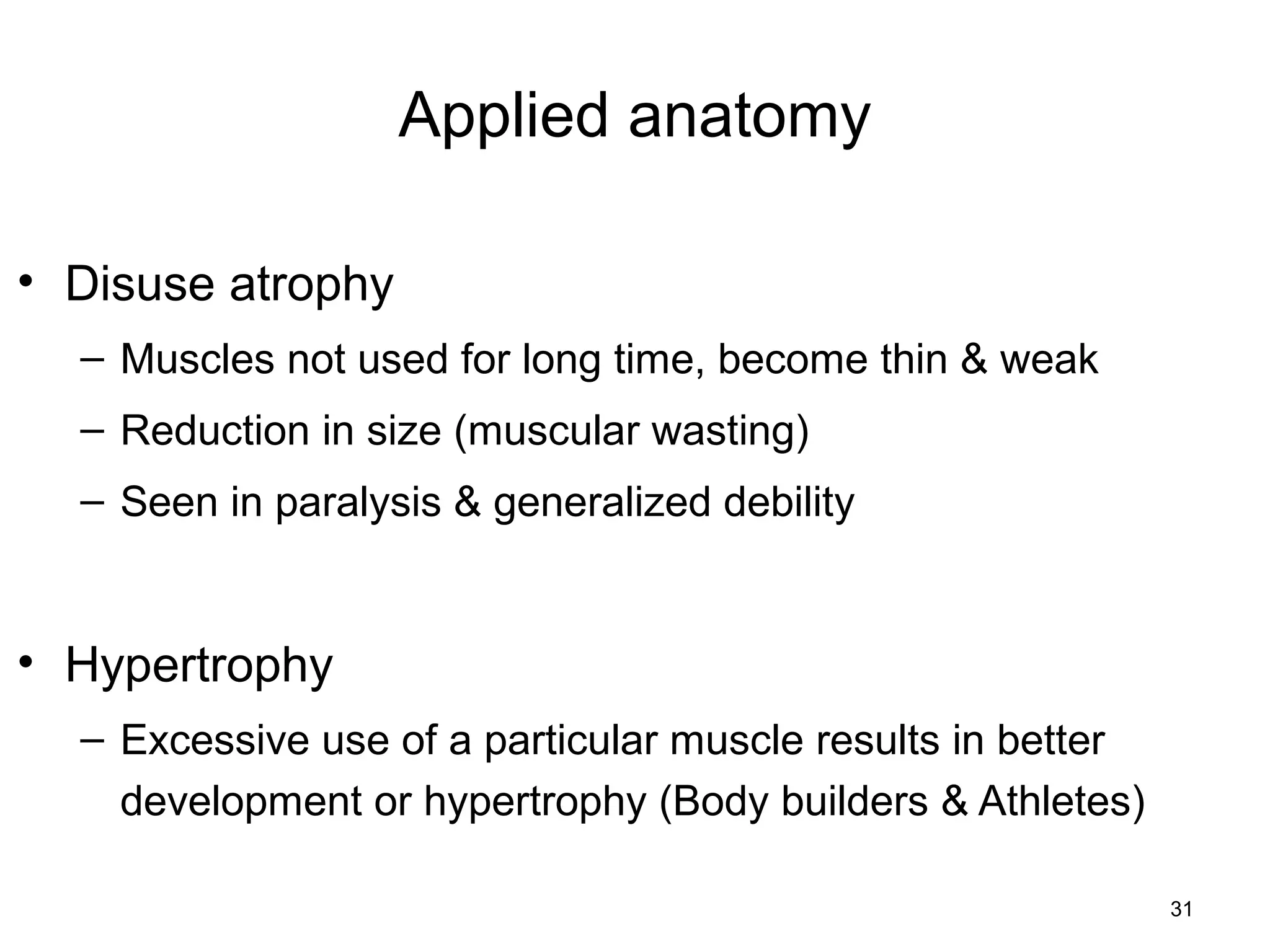
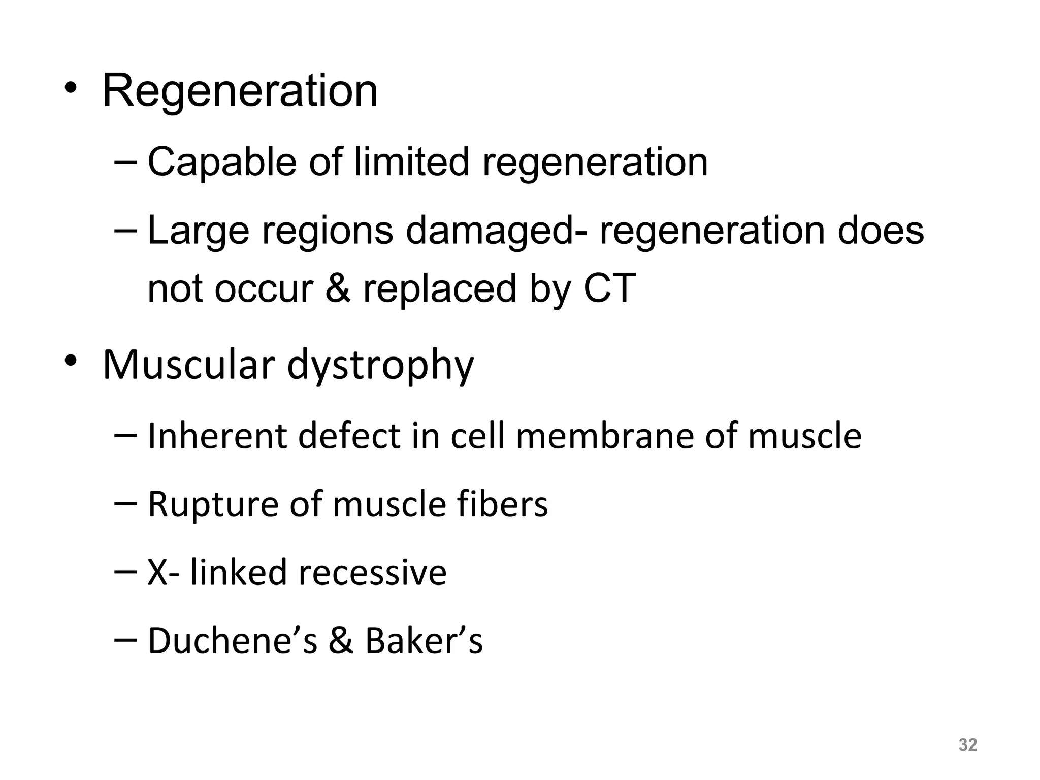


1) The document discusses skeletal muscle anatomy and physiology, describing the different types of skeletal muscle fibers and their classifications based on architecture and function. 2) It also covers motor units, nerve supply, and applied concepts like muscle paralysis, spasm, atrophy, hypertrophy, and regeneration. 3) Key points include the structures of skeletal muscle like tendons and fascicles, fiber types, and how muscles are classified based on their prime mover, antagonist, fixator, or synergist actions.


