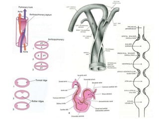
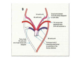
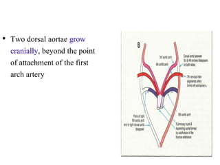
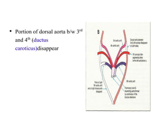
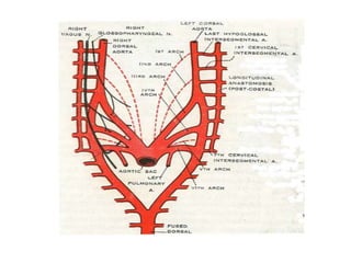
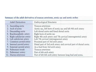
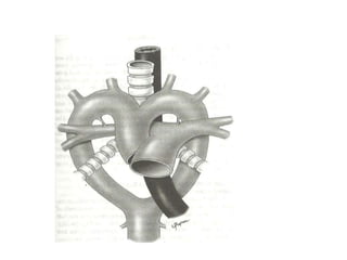
1. The aortic arches develop from the primitive aorta and pharyngeal arch arteries during embryonic development.
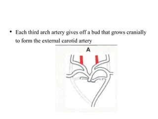
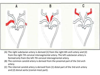
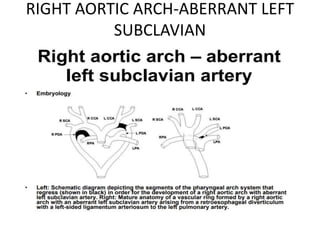
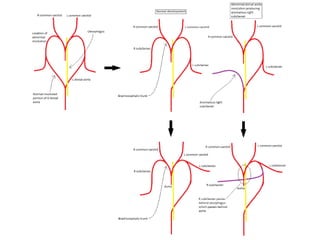
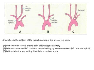
2. The right and left 3rd aortic arches normally form the common carotid arteries, while the 4th arches form the subclavian arteries.
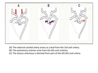
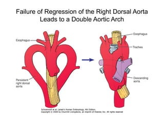
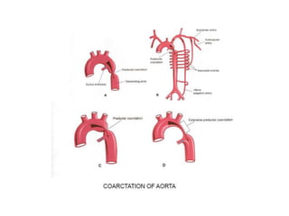
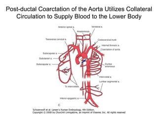
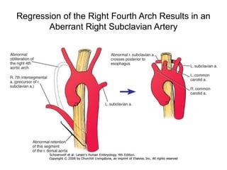
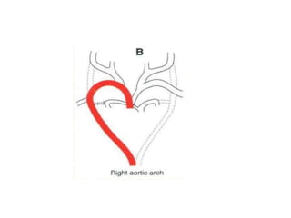
3. A number of anomalies can occur if parts of the arches fail to regress as normal or persist that typically regress. This includes double aortic arch, right aortic arch, patent ductus arteriosus, and interrupted aortic arch.
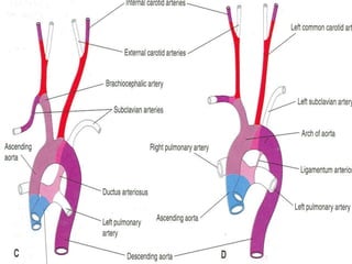
4. Proper development and regression of the aortic arches is required for normal formation of the branches of the aortic arch.
Cardiac Surgery
Internal thoracic ( mammary ) artery
ORIGIN and Course Of IMA
BRANCHES
Thymic Artery
The Xiphoid branch
The sternal branches
The Pericardiacophrenic branch
Anterior intercostal branches
Perforating branches
Musculophrenic artery
Superior Epigastric Artery
Clinical significance
The conotruncus comprises collectively two myocardial subsegments, the conus and the truncus.
Conus is the myocardial segment between ventricle and semi lunar valves which gives rise to sub arterial coni.
Truncus is the fibrous segment between semi lunar valves and aortic sac which gives rise to great arteries.
An overview of the normal embryological process of development of the Aortic arch and the clinically relevant anomalies of the aortic arch development. Ideal for Cardiology Fellows.
NVBDCP.pptx Nation vector borne disease control programSapna Thakur
NVBDCP was launched in 2003-2004 . Vector-Borne Disease: Disease that results from an infection transmitted to humans and other animals by blood-feeding arthropods, such as mosquitoes, ticks, and fleas. Examples of vector-borne diseases include Dengue fever, West Nile Virus, Lyme disease, and malaria.
Prix Galien International 2024 Forum ProgramLevi Shapiro
June 20, 2024, Prix Galien International and Jerusalem Ethics Forum in ROME. Detailed agenda including panels:
- ADVANCES IN CARDIOLOGY: A NEW PARADIGM IS COMING
- WOMEN’S HEALTH: FERTILITY PRESERVATION
- WHAT’S NEW IN THE TREATMENT OF INFECTIOUS,
ONCOLOGICAL AND INFLAMMATORY SKIN DISEASES?
- ARTIFICIAL INTELLIGENCE AND ETHICS
- GENE THERAPY
- BEYOND BORDERS: GLOBAL INITIATIVES FOR DEMOCRATIZING LIFE SCIENCE TECHNOLOGIES AND PROMOTING ACCESS TO HEALTHCARE
- ETHICAL CHALLENGES IN LIFE SCIENCES
- Prix Galien International Awards Ceremony
Flu Vaccine Alert in Bangalore Karnatakaaddon Scans
As flu season approaches, health officials in Bangalore, Karnataka, are urging residents to get their flu vaccinations. The seasonal flu, while common, can lead to severe health complications, particularly for vulnerable populations such as young children, the elderly, and those with underlying health conditions.
Dr. Vidisha Kumari, a leading epidemiologist in Bangalore, emphasizes the importance of getting vaccinated. "The flu vaccine is our best defense against the influenza virus. It not only protects individuals but also helps prevent the spread of the virus in our communities," he says.
This year, the flu season is expected to coincide with a potential increase in other respiratory illnesses. The Karnataka Health Department has launched an awareness campaign highlighting the significance of flu vaccinations. They have set up multiple vaccination centers across Bangalore, making it convenient for residents to receive their shots.
To encourage widespread vaccination, the government is also collaborating with local schools, workplaces, and community centers to facilitate vaccination drives. Special attention is being given to ensuring that the vaccine is accessible to all, including marginalized communities who may have limited access to healthcare.
Residents are reminded that the flu vaccine is safe and effective. Common side effects are mild and may include soreness at the injection site, mild fever, or muscle aches. These side effects are generally short-lived and far less severe than the flu itself.
Healthcare providers are also stressing the importance of continuing COVID-19 precautions. Wearing masks, practicing good hand hygiene, and maintaining social distancing are still crucial, especially in crowded places.
Protect yourself and your loved ones by getting vaccinated. Together, we can help keep Bangalore healthy and safe this flu season. For more information on vaccination centers and schedules, residents can visit the Karnataka Health Department’s official website or follow their social media pages.
Stay informed, stay safe, and get your flu shot today!
Knee anatomy and clinical tests 2024.pdfvimalpl1234
This includes all relevant anatomy and clinical tests compiled from standard textbooks, Campbell,netter etc..It is comprehensive and best suited for orthopaedicians and orthopaedic residents.
Title: Sense of Smell
Presenter: Dr. Faiza, Assistant Professor of Physiology
Qualifications:
MBBS (Best Graduate, AIMC Lahore)
FCPS Physiology
ICMT, CHPE, DHPE (STMU)
MPH (GC University, Faisalabad)
MBA (Virtual University of Pakistan)
Learning Objectives:
Describe the primary categories of smells and the concept of odor blindness.
Explain the structure and location of the olfactory membrane and mucosa, including the types and roles of cells involved in olfaction.
Describe the pathway and mechanisms of olfactory signal transmission from the olfactory receptors to the brain.
Illustrate the biochemical cascade triggered by odorant binding to olfactory receptors, including the role of G-proteins and second messengers in generating an action potential.
Identify different types of olfactory disorders such as anosmia, hyposmia, hyperosmia, and dysosmia, including their potential causes.
Key Topics:
Olfactory Genes:
3% of the human genome accounts for olfactory genes.
400 genes for odorant receptors.
Olfactory Membrane:
Located in the superior part of the nasal cavity.
Medially: Folds downward along the superior septum.
Laterally: Folds over the superior turbinate and upper surface of the middle turbinate.
Total surface area: 5-10 square centimeters.
Olfactory Mucosa:
Olfactory Cells: Bipolar nerve cells derived from the CNS (100 million), with 4-25 olfactory cilia per cell.
Sustentacular Cells: Produce mucus and maintain ionic and molecular environment.
Basal Cells: Replace worn-out olfactory cells with an average lifespan of 1-2 months.
Bowman’s Gland: Secretes mucus.
Stimulation of Olfactory Cells:
Odorant dissolves in mucus and attaches to receptors on olfactory cilia.
Involves a cascade effect through G-proteins and second messengers, leading to depolarization and action potential generation in the olfactory nerve.
Quality of a Good Odorant:
Small (3-20 Carbon atoms), volatile, water-soluble, and lipid-soluble.
Facilitated by odorant-binding proteins in mucus.
Membrane Potential and Action Potential:
Resting membrane potential: -55mV.
Action potential frequency in the olfactory nerve increases with odorant strength.
Adaptation Towards the Sense of Smell:
Rapid adaptation within the first second, with further slow adaptation.
Psychological adaptation greater than receptor adaptation, involving feedback inhibition from the central nervous system.
Primary Sensations of Smell:
Camphoraceous, Musky, Floral, Pepperminty, Ethereal, Pungent, Putrid.
Odor Detection Threshold:
Examples: Hydrogen sulfide (0.0005 ppm), Methyl-mercaptan (0.002 ppm).
Some toxic substances are odorless at lethal concentrations.
Characteristics of Smell:
Odor blindness for single substances due to lack of appropriate receptor protein.
Behavioral and emotional influences of smell.
Transmission of Olfactory Signals:
From olfactory cells to glomeruli in the olfactory bulb, involving lateral inhibition.
Primitive, less old, and new olfactory systems with different path
Title: Sense of Taste
Presenter: Dr. Faiza, Assistant Professor of Physiology
Qualifications:
MBBS (Best Graduate, AIMC Lahore)
FCPS Physiology
ICMT, CHPE, DHPE (STMU)
MPH (GC University, Faisalabad)
MBA (Virtual University of Pakistan)
Learning Objectives:
Describe the structure and function of taste buds.
Describe the relationship between the taste threshold and taste index of common substances.
Explain the chemical basis and signal transduction of taste perception for each type of primary taste sensation.
Recognize different abnormalities of taste perception and their causes.
Key Topics:
Significance of Taste Sensation:
Differentiation between pleasant and harmful food
Influence on behavior
Selection of food based on metabolic needs
Receptors of Taste:
Taste buds on the tongue
Influence of sense of smell, texture of food, and pain stimulation (e.g., by pepper)
Primary and Secondary Taste Sensations:
Primary taste sensations: Sweet, Sour, Salty, Bitter, Umami
Chemical basis and signal transduction mechanisms for each taste
Taste Threshold and Index:
Taste threshold values for Sweet (sucrose), Salty (NaCl), Sour (HCl), and Bitter (Quinine)
Taste index relationship: Inversely proportional to taste threshold
Taste Blindness:
Inability to taste certain substances, particularly thiourea compounds
Example: Phenylthiocarbamide
Structure and Function of Taste Buds:
Composition: Epithelial cells, Sustentacular/Supporting cells, Taste cells, Basal cells
Features: Taste pores, Taste hairs/microvilli, and Taste nerve fibers
Location of Taste Buds:
Found in papillae of the tongue (Fungiform, Circumvallate, Foliate)
Also present on the palate, tonsillar pillars, epiglottis, and proximal esophagus
Mechanism of Taste Stimulation:
Interaction of taste substances with receptors on microvilli
Signal transduction pathways for Umami, Sweet, Bitter, Sour, and Salty tastes
Taste Sensitivity and Adaptation:
Decrease in sensitivity with age
Rapid adaptation of taste sensation
Role of Saliva in Taste:
Dissolution of tastants to reach receptors
Washing away the stimulus
Taste Preferences and Aversions:
Mechanisms behind taste preference and aversion
Influence of receptors and neural pathways
Impact of Sensory Nerve Damage:
Degeneration of taste buds if the sensory nerve fiber is cut
Abnormalities of Taste Detection:
Conditions: Ageusia, Hypogeusia, Dysgeusia (parageusia)
Causes: Nerve damage, neurological disorders, infections, poor oral hygiene, adverse drug effects, deficiencies, aging, tobacco use, altered neurotransmitter levels
Neurotransmitters and Taste Threshold:
Effects of serotonin (5-HT) and norepinephrine (NE) on taste sensitivity
Supertasters:
25% of the population with heightened sensitivity to taste, especially bitterness
Increased number of fungiform papillae
Tom Selleck Health: A Comprehensive Look at the Iconic Actor’s Wellness Journeygreendigital
Tom Selleck, an enduring figure in Hollywood. has captivated audiences for decades with his rugged charm, iconic moustache. and memorable roles in television and film. From his breakout role as Thomas Magnum in Magnum P.I. to his current portrayal of Frank Reagan in Blue Bloods. Selleck's career has spanned over 50 years. But beyond his professional achievements. fans have often been curious about Tom Selleck Health. especially as he has aged in the public eye.
Follow us on: Pinterest
Introduction
Many have been interested in Tom Selleck health. not only because of his enduring presence on screen but also because of the challenges. and lifestyle choices he has faced and made over the years. This article delves into the various aspects of Tom Selleck health. exploring his fitness regimen, diet, mental health. and the challenges he has encountered as he ages. We'll look at how he maintains his well-being. the health issues he has faced, and his approach to ageing .
Early Life and Career
Childhood and Athletic Beginnings
Tom Selleck was born on January 29, 1945, in Detroit, Michigan, and grew up in Sherman Oaks, California. From an early age, he was involved in sports, particularly basketball. which played a significant role in his physical development. His athletic pursuits continued into college. where he attended the University of Southern California (USC) on a basketball scholarship. This early involvement in sports laid a strong foundation for his physical health and disciplined lifestyle.
Transition to Acting
Selleck's transition from an athlete to an actor came with its physical demands. His first significant role in "Magnum P.I." required him to perform various stunts and maintain a fit appearance. This role, which he played from 1980 to 1988. necessitated a rigorous fitness routine to meet the show's demands. setting the stage for his long-term commitment to health and wellness.
Fitness Regimen
Workout Routine
Tom Selleck health and fitness regimen has evolved. adapting to his changing roles and age. During his "Magnum, P.I." days. Selleck's workouts were intense and focused on building and maintaining muscle mass. His routine included weightlifting, cardiovascular exercises. and specific training for the stunts he performed on the show.
Selleck adjusted his fitness routine as he aged to suit his body's needs. Today, his workouts focus on maintaining flexibility, strength, and cardiovascular health. He incorporates low-impact exercises such as swimming, walking, and light weightlifting. This balanced approach helps him stay fit without putting undue strain on his joints and muscles.
Importance of Flexibility and Mobility
In recent years, Selleck has emphasized the importance of flexibility and mobility in his fitness regimen. Understanding the natural decline in muscle mass and joint flexibility with age. he includes stretching and yoga in his routine. These practices help prevent injuries, improve posture, and maintain mobilit
Explore natural remedies for syphilis treatment in Singapore. Discover alternative therapies, herbal remedies, and lifestyle changes that may complement conventional treatments. Learn about holistic approaches to managing syphilis symptoms and supporting overall health.
Lung Cancer: Artificial Intelligence, Synergetics, Complex System Analysis, S...Oleg Kshivets
RESULTS: Overall life span (LS) was 2252.1±1742.5 days and cumulative 5-year survival (5YS) reached 73.2%, 10 years – 64.8%, 20 years – 42.5%. 513 LCP lived more than 5 years (LS=3124.6±1525.6 days), 148 LCP – more than 10 years (LS=5054.4±1504.1 days).199 LCP died because of LC (LS=562.7±374.5 days). 5YS of LCP after bi/lobectomies was significantly superior in comparison with LCP after pneumonectomies (78.1% vs.63.7%, P=0.00001 by log-rank test). AT significantly improved 5YS (66.3% vs. 34.8%) (P=0.00000 by log-rank test) only for LCP with N1-2. Cox modeling displayed that 5YS of LCP significantly depended on: phase transition (PT) early-invasive LC in terms of synergetics, PT N0—N12, cell ratio factors (ratio between cancer cells- CC and blood cells subpopulations), G1-3, histology, glucose, AT, blood cell circuit, prothrombin index, heparin tolerance, recalcification time (P=0.000-0.038). Neural networks, genetic algorithm selection and bootstrap simulation revealed relationships between 5YS and PT early-invasive LC (rank=1), PT N0—N12 (rank=2), thrombocytes/CC (3), erythrocytes/CC (4), eosinophils/CC (5), healthy cells/CC (6), lymphocytes/CC (7), segmented neutrophils/CC (8), stick neutrophils/CC (9), monocytes/CC (10); leucocytes/CC (11). Correct prediction of 5YS was 100% by neural networks computing (area under ROC curve=1.0; error=0.0).
CONCLUSIONS: 5YS of LCP after radical procedures significantly depended on: 1) PT early-invasive cancer; 2) PT N0--N12; 3) cell ratio factors; 4) blood cell circuit; 5) biochemical factors; 6) hemostasis system; 7) AT; 8) LC characteristics; 9) LC cell dynamics; 10) surgery type: lobectomy/pneumonectomy; 11) anthropometric data. Optimal diagnosis and treatment strategies for LC are: 1) screening and early detection of LC; 2) availability of experienced thoracic surgeons because of complexity of radical procedures; 3) aggressive en block surgery and adequate lymph node dissection for completeness; 4) precise prediction; 5) adjuvant chemoimmunoradiotherapy for LCP with unfavorable prognosis.
- Video recording of this lecture in English language: https://youtu.be/lK81BzxMqdo
- Video recording of this lecture in Arabic language: https://youtu.be/Ve4P0COk9OI
- Link to download the book free: https://nephrotube.blogspot.com/p/nephrotube-nephrology-books.html
- Link to NephroTube website: www.NephroTube.com
- Link to NephroTube social media accounts: https://nephrotube.blogspot.com/p/join-nephrotube-on-social-media.html
2. Embryology
• Heart is first seen in the form of two endothelial heart tubes-
18th day of foetal life
• Fusion results in a single tube with a series of
dilatations(Bulbus cordis, Ventricle, Atrium,Sinus venosus)
and begins to beat by 22nd day
3. • Ventricle and atrium
are connected by a
narrow atrio-
ventricular canal.
• The sinus venosus has
prolongations that are
referred to as its right
and left horns.
4. • Bulbus cordis represents
arterial end of the tube-
Proximal -- No special name
Middle -- Conus
Distal -- Truncus arteriosus
5. • Truncus continues with the aortic sac from which right
and left pharyngeal arch arteries arises
• They arch backward on lateral side of foregut –continues
as right and left dorsal aorta-fuse to form descending
aorta
6. • Sinus Venosus
1. Lies at the venous end of
the heart
1. Right and Left horns-
- Vitelline vein
- Umbilical vein
- Common cardinal vein
8. • First arteries to appear are right and left Primitive Aorta connected to the two
endocardial heart tubes
Parts of Primitive Aorta-
1. Ventral aorta-Ventral to foregut
2. Arched portion-connected to first pharyngeal arch
3. Dorsal aorta-Dorsal to foregut
9. • After the fusion of endocardial heart tubes,ventral aorta fuse
to form Aortic Sac
• Unfused parts remaining as the right and left horns of the sac
10. • During 4th and 5th
week,successive arterial arches
appear in 2nd to 6th pharyngeal
arches
• Each connects ventrally to aortic
sac & dorsally to dorsal aorta
12. • 3rd and 4th open to ventral part of aortic sac
• 6th open to dorsal part of aortic sac
• Spiral septum formed in truncus arteriousus in the 5th
week extends to aortic sac, fuses with posterior wall-
-Blood from pulmonary artery goes to 6th arch artery
-while from ascending aorta passes into 3rd &4th arch
arteries.
13.
14.
15. • Two dorsal aortae grow
cranially, beyond the point
of attachment of the first
arch artery
16. • Portion of dorsal aorta b/w 3rd
and 4th (ductus
caroticus)disappear
17. • Portion of the right dorsal
aorta, which lies between
the point of attachment of
the fourth arch artery and
the point of fusion of the
two dorsal aortae,
disappears
18. • Each 6th arch artery connects
to the pulmonary vascular
tree
• Portion between this
connection and dorsal
aorta(Ductus
Arteriosus)regresses on right
side
• Ductus Arteriosus is
obliterated after birth and is
then seen as the ligamentutn
arteriosum.
19. • Each third arch artery gives off a bud that grows cranially
to form the external carotid artery
20. • Dorsal aorta gives lateral intersegmental
branches to body wall.7th cervical
intersegmental supplies upper limb bud
21. • The ascending aorta and the pulmonary trunk are formed
from the truncus arteriosus
22. • The arch of the aorta is derived from the ventral part of
the aortic sac (1), its left horn (2), and the left fourth arch
artery (3)
23. • The descending aorta is derived from the left dorsal aorta,
below the attachment of fourth arch artery (1), along with
the fused median vessel (2)
25. (A) The right subclavian artery is derived (1) from the right 4th arch artery and {2)
from the right 7th cervical intersegmental artery. The left subclavian artery is
formed only from the left 7th cervical intersegmental artery.
(B) The common carotid artery is derived from the proximal part of the 3rd arch
artery.
(C) The internal carotid artery is derived from (1) distal part of the 3rd arch artery
and (2) dorsal aorta (cranial-most part).
26. (A) The external carotid artery arises as a bud from the 3rd arch artery.
(B) The pulmonary arteries arise from the 6th arch arteries.
(C) The ductus arteriosus is derived from part of the left 6th arch artery.
27. • As the right third and fourth arch arteries arise from the
right horn of the aortic sac, the common carotid and
subclavian arteries become branches of the
brachiocephalic artery.
• With the formation of the neck , and the descent
of the heart into the thoracic cavity ,
• The point of origin of the subclavian artery from
the aorta gradually shifts upwards and comes to
lie close to the origin of the Lt common carotid
artery
28.
29.
30. -The nerve of the sixth arch (recurrent laryngeal), is at first caudal to the artery of this arch
-on the right side, the nerve moves cranially and comes into relationship with the right
fourth arch artery (subclavian)
-On the left side, it retains its relationship to that part of the sixth arch which forms the
ductus arteriosus
31.
32.
33. AORTIC ARCH ANOMALIES
• DOUBLE ARCH
• RIGHT ARCH
• PATENT DUCTUS ARTERIOSIS
• ABERRANT RIGHT SUBCLAVIAN ARTERY
• INTERRUPTED AORTIC ARCH
• COARCTATION
• ANOMALOUS ORIGIN OF PULMONARY ARTERY
• ABNORMAL LEFT ARCH
• ABNORMAL RIGHT ARCH
• CERVICAL ARCH
34. EDWARD’S DOUBLE AORTIC ARCH
MODE
• Anomalies of aortic arch to be conceptualized as variations in
regression of different segments from a “hypothetical double arch”.
• The concept of “hypothetical double aortic arch” emphasises the
potential contribution of nearly all embryonic arches to components
of definitive arch system.
• Demonstrate possible embryologic explanations for each arch
anomaly.
35.
36. SIDEDNESS OF THE AORTA
• LEFT AND RIGHT ARCH REFERS TO WHICH
BRONCHUS IS CROSSED BY THE ARCH
37. • Occurs due to-
1. Some parts that normally disappear may persist
2. Some parts that normally persist may disappear.
38. Double Aortic arch
• Both right and left arches present.
• Both arches can be patent or one hypoplastic or atretic(usually
left)
• Persistence of both right and left 4th arch which join Truncus
arteriosus sac to their respective dorsal aortae
• Only one 6th remain.
• Form complete vascular rings.
• When both arches are patent, rings typically tight and present
with sridor in first week of life.
40. TRUNCUS ARTERIOSUS
• A single trunk arising from the heart
• 4 truncus and 2 conal cushions develop.
• Dextro- sinistro cushions of both conus and truncus fuse
to form Conotruncal septum
41. • Because the cushions
are dextro-superior and
sinistro inferior in
truncus and dextro-
dorsal and sinistro-
ventral in conus union
forms a spiral septum
than true lineal relation.
42. • Failure of aortopulmonary septum to Septation give rise to
persistent truncus arteriosus
43. CLASSIFICATION OF TRUNCUS
ARTERIOSUS
I) Edward & Collett classification-
• Type I – main pulmonary trunk arises from truncus arteriosus
and gives rise to RPA & LPA.
• Type II- RPA & LPA arteries arises directly and lying close
to one another.
• Type III- RPA & LPA arises from separate ostium lying at
some distance from one another.
• Type IV-absence of branch of PA ,pulmonary blood flow is
derived from aortopulmonary collaterals.
45. Aortopulmonary Window
-opening between ascending aorta and
pulmonary artery
-resulting from abnormal septation of the
truncus arteriosus into the aorta and
pulmonary artery
46. • . Classification
Type I : Between posteromedial wall of ascending
aorta and lateral wall of MPA
Type II : Between posterior wall of ascending aorta
and origin of RPA
Type III : Anomalous origin of RPA from postero-
lateral wall of ascending aorta
47. PATENT DUCTUS ARTERIOSIS
• The ductus arteriosus, which is normally occluded soon
after birth, may remain patent
• Communication between the pulmonary artery and the
aorta
48.
49. Type A - conical duct with well defined aortic ampulla and constriction near the
pulmonary artery end.
Type B - large duct with window like structure which is very short in length.
Type C - tubular duct without any constriction.
Type D - complex duct with multiple constrictions.
Type E - elongated duct with constriction remote from the edge of the trachea (as
viewed on lateral angiography)
50. COARCTATION OF THE AORTA
• Aorta may show a localized narrowing of its lumen
• May be distal to the attachment of the ductus (postductal)
• proximal to the attachment (preductal)- right ventricle
supplies the distal part of the body throug h the ductus
arteriosus
53. RIGHT AORTIC ARCH
MOST COMMON TYPES
• MIRROR IMAGE
• ABERRANT LEFT SUBCLAVIAN
• ISOLATED LEFT SUBCLAVIAN
54.
55. RIGHT AORTIC ARCH-MIRROR IMAGE
TYPE
• Sequence of arch vessels-left innominate,right
carotid.
• Ligamentum left sided.
• No vascular ring.
• Almost always associated with CHD (48% TOF)
57. Regression of the Right Fourth Arch Results in an
Aberrant Right Subclavian Artery
58.
59. • The ductus caroticus may persist. As a result, the left
internal carotid arises directly from the aortic arch, and the
right internal carotid from the subclavian
60. Anomalies in the pattern of the main branches of the arch of the aorta.
(A) Left common carotid arising from brachiocephalic artery.
(B) Left subclavian and left common carotid arising by a common stem (left brachiocephalic).
(C) Left vertebral artery arising directly from arch of aorta.
61. RIGHT AORTIC ARCH
MOST COMMON TYPES
• MIRROR IMAGE
• ABERRANT LEFT SUBCLAVIAN
• ISOLATED LEFT SUBCLAVIAN
62.
63. RIGHT AORTIC ARCH-MIRROR IMAGE
TYPE
• Sequence of arch vessels-left innominate,right
carotid.
• Ligamentum left sided.
• No vascular ring.
• Almost always associated with CHD (48% TOF)
65. INTERRUPTED AORTIC ARCH
Obliteration of the right and left fourth aortic arches
• Defined as complete separation of ascending
and descending aorta
• A segment of the aortic arch, may be missing
• ascending aorta ends by supplying the left
common carotid artery
• Left subclavian artery arises from the distal
segment which receives blood through a
patent ductus arteriosus.
66. Obliteration of the Right and Left Fourth Aortic
Arches Leads to Interruption of the Aorta
67. • Celoria and Patton classification(1959)
– Type A-interruption distal to SCA that is ipsilateral to 2nd carotid
artery
Type A-involution of both dorsal aorta distal to 4th
arch,prox to persistent 6th arch
– Type B-interruption b/w 2nd carotid and ipsilateral subclavian
Type b-involution of one 4th arch and one dorsal aorta b/w
4th and 6th
– Type C-interruption b/w carotids
Type C-involution of one limb of truncoaortic sac
68. Anomalous origin of pulmonary artery
from Ascending Aorta
• Anomalous pulmonary artery branch arising from ascending aorta in
presence of a MPA arising separately
• Anomalous RPA-
◦ More common
◦ Embryonic branch pulmonary artery joins right side of TA
sac, but fails the leftward migration to join MPA
before septation
• Anomalous LPA
◦ a/w TOF in 74%
◦ Embryonic branch pulmonary artery fails to join TA sac
69. Anomalous origin of LPA from RPA
• LPA arises from RPA and passes b/w trachea and esophagus-
Pulmonary artery sling.
70. LEFT AORTIC ARCH AND RETROESOPHAGEAL
DIVERTICULUM OF KOMMERELL
• First vascular ring to be diagnosed during life
• Similar to previous except for persistent 6th arch-
ligamentum which completes a vascular Ring
• Proximal RSCA dilated to form diverticulum
71. RIGHT AORTIC ARCH WITH RETROESOPHAGEAL
DIVERTICULUM OF KOMMERELL
• Sequence –left carotid, right carotid ,RSCA, a large
retroesophageal vessel( diverticulum) from which LSCA
arises
• Left ligamentum completes the ring
• Disappearance of Lt 4th arch and persistence of 6th arch
72. Abnormal Left Arch
• a) Left arch with retroesophageal RSCA
• b) Left aortic arch and retroesophageal
diverticulum of Kommerell
• c) Left aortic arch,right descending aorta,
right.ductus(circumflex aortic arch)
• d) Left aortic arch & isolated RSCA
• e) Left aortic arch with cervical origin of Rt subclavian
73. LEFT AORTIC ARCH,RIGHT DESCENDING AORTA,
RIGHT.DUCTUS(CIRCUMFLEX AORTIC ARCH
• Disappearance of right 4th arch but with left distal dorsal
aorta forming definitive distal arch and passing
retroesophageally to a descending aorta beginning to right
of vertebral column.
74. LEFT AORTIC ARCH & ISOLATED RSCA
• RSCA arises only from righ` t ductus.
• Right 6th arch persists with dossolution of right 4
th arch and right dorsal aorta.
75. LEFT AORTIC ARCH WITH CERVICAL ORIGIN OF
RIGHT SUBCLAVIAN
• Innominate trifurcates in the neck-RSCA travels back to
thorax
• Subclavian artery arises from 3rd arch in stead of 4 th .
• 3 rd arch being more cephalad gives origin to RSCA in
the neck.
76. • Dissolution of left dorsal aorta distal to the origin of left 7 th
intersegmental artery
• Left 4th arch becomes proximal left subclavian artery.
• Left Sixth arch persists- left ductus arises from underside of
left innominate artery and passes to LPA .
• Alternatively right ductus persists giving true mirror image
of normal
77. Persistent 5th Arch
• Double lumen aortic arch in which both arches appear on
same side of trachea.
78. RIGHT ARCH WITH LEFT DECENDING
AORTA & LEFT LIGAMENTUM
• Also known as circumflex right aortic arch.
• Embryology : Dissolution of either left dorsal aorta distal to
takeoff of the left subclavian artery or the left fourth arch.
Persistent left sixth arch connects to the left sided dorsal
aorta completing vascular ring .
• Aortic arch itself crosses midline to the left at the level of
T4 vertebral body -connects to left ductus to form vascular
ring
• Aortic arch is retroesophageal and not the subclavian artery.
79. RIGHT AORTIC ARCH WITH RETROESOPHAGEAL
INNOMINATE ARTEY
• Dissolution of left branch of truncoaortic sac and left 4th arch
• The left dorsal aorta supplies left 7th IS artery and left 3rd arch.