Downloaded 1,455 times


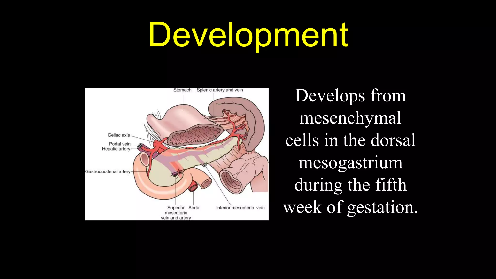


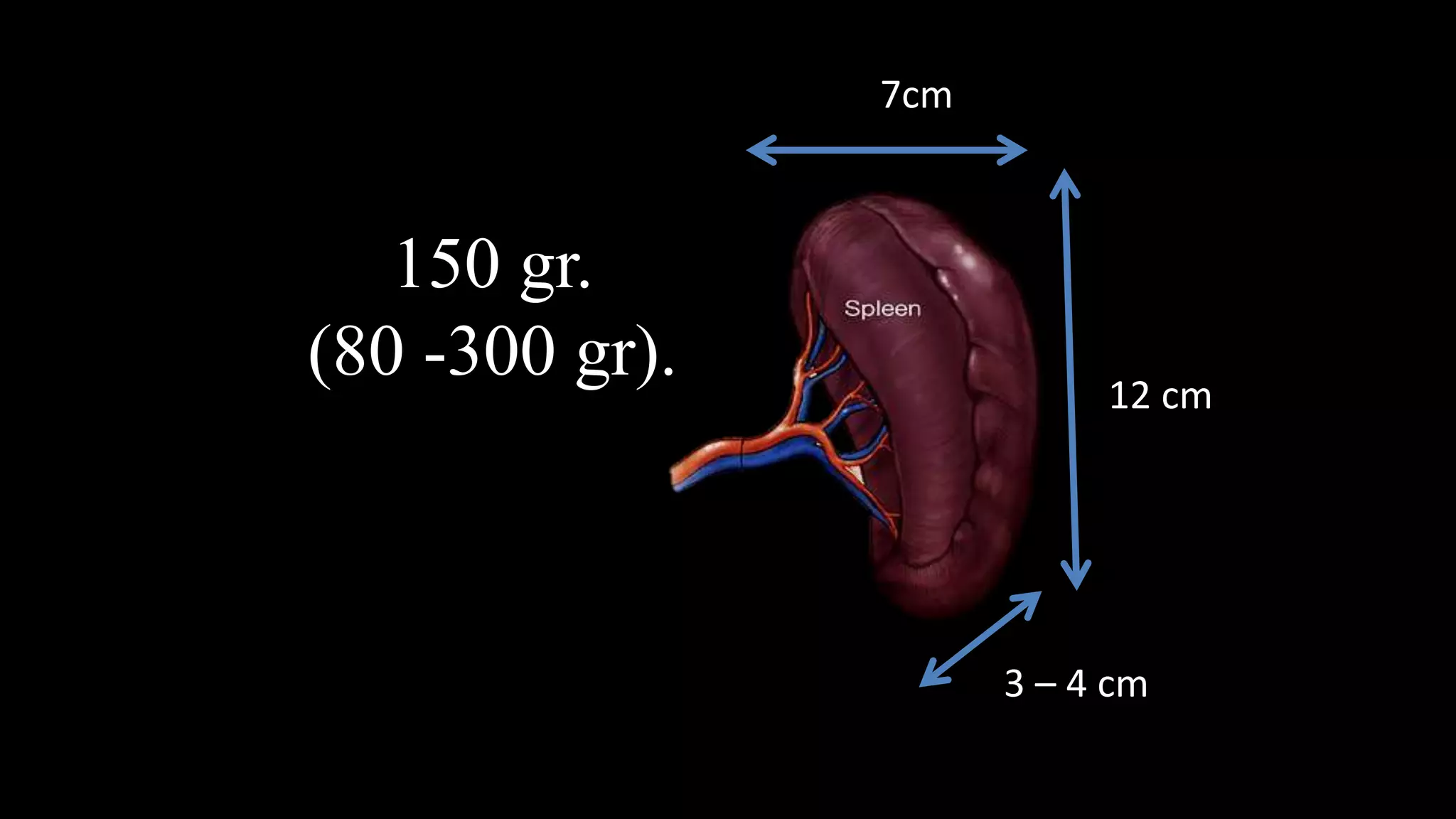


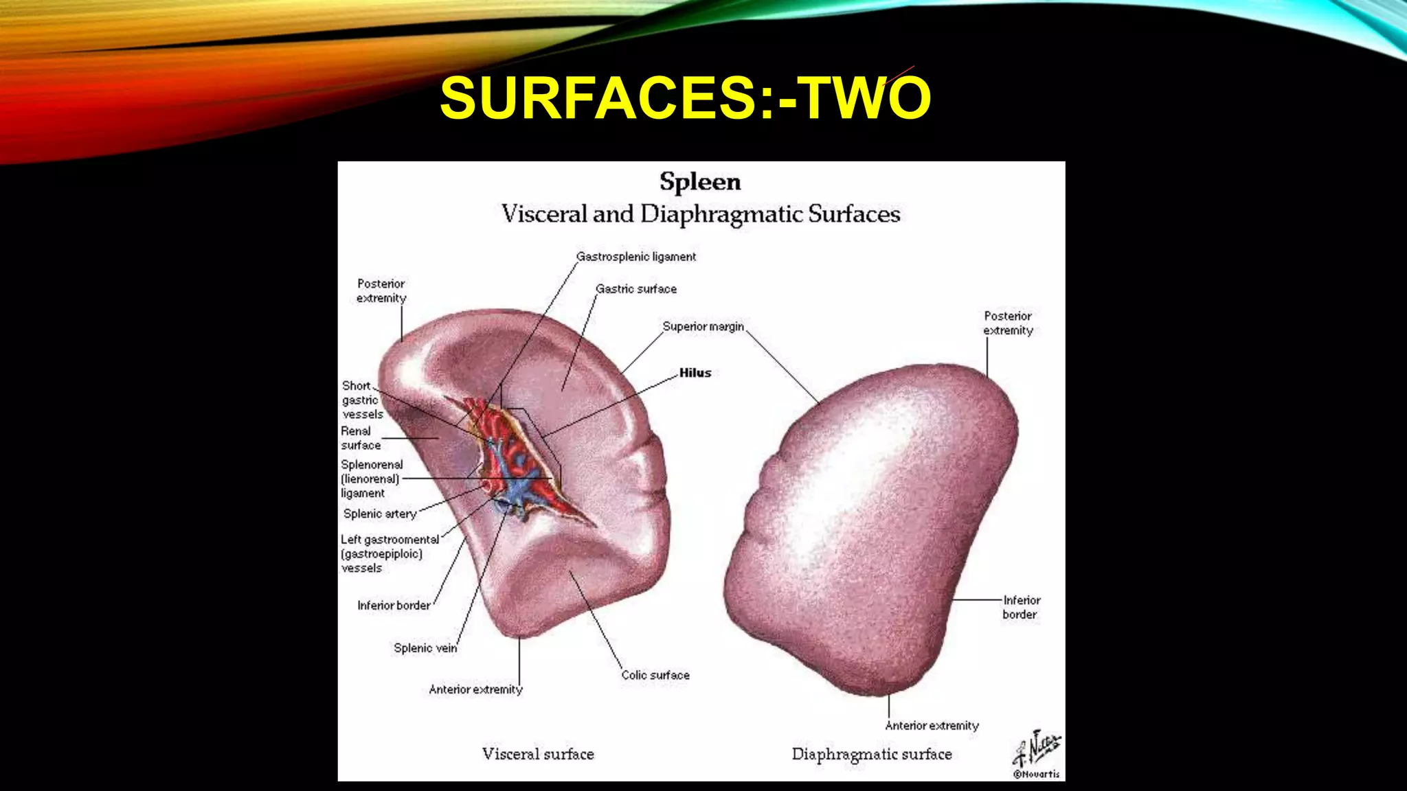


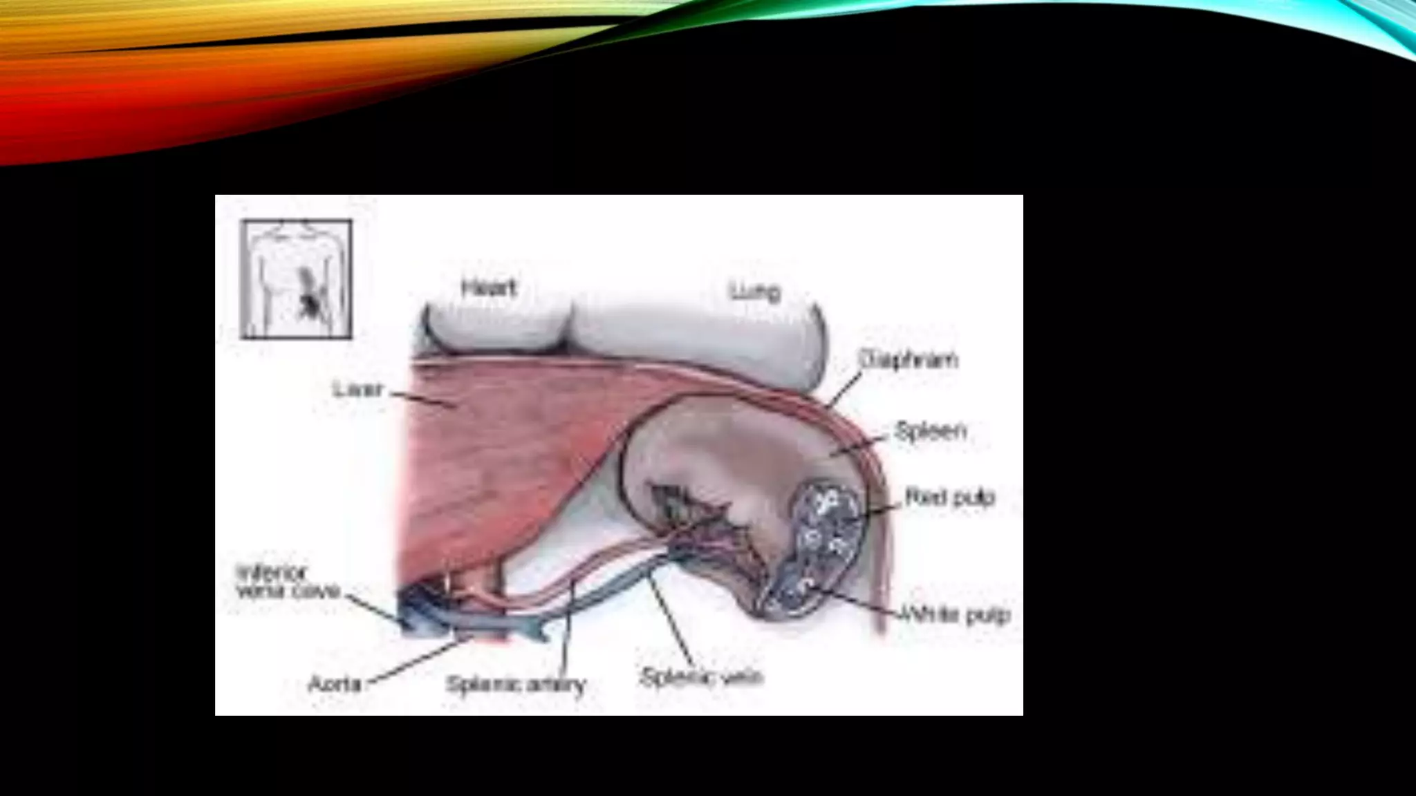


The spleen is a hematopoietic organ located in the left upper quadrant of the abdomen. It filters blood and fights infections. The spleen develops from mesenchymal cells in the dorsal mesogastrium and has dimensions of approximately 12x7x3 cm and a weight of 150 grams in adults. It receives a blood supply from the splenic artery and drains into the portal vein system. The spleen can be surgically removed (splenectomy) in cases of trauma or certain blood disorders.


