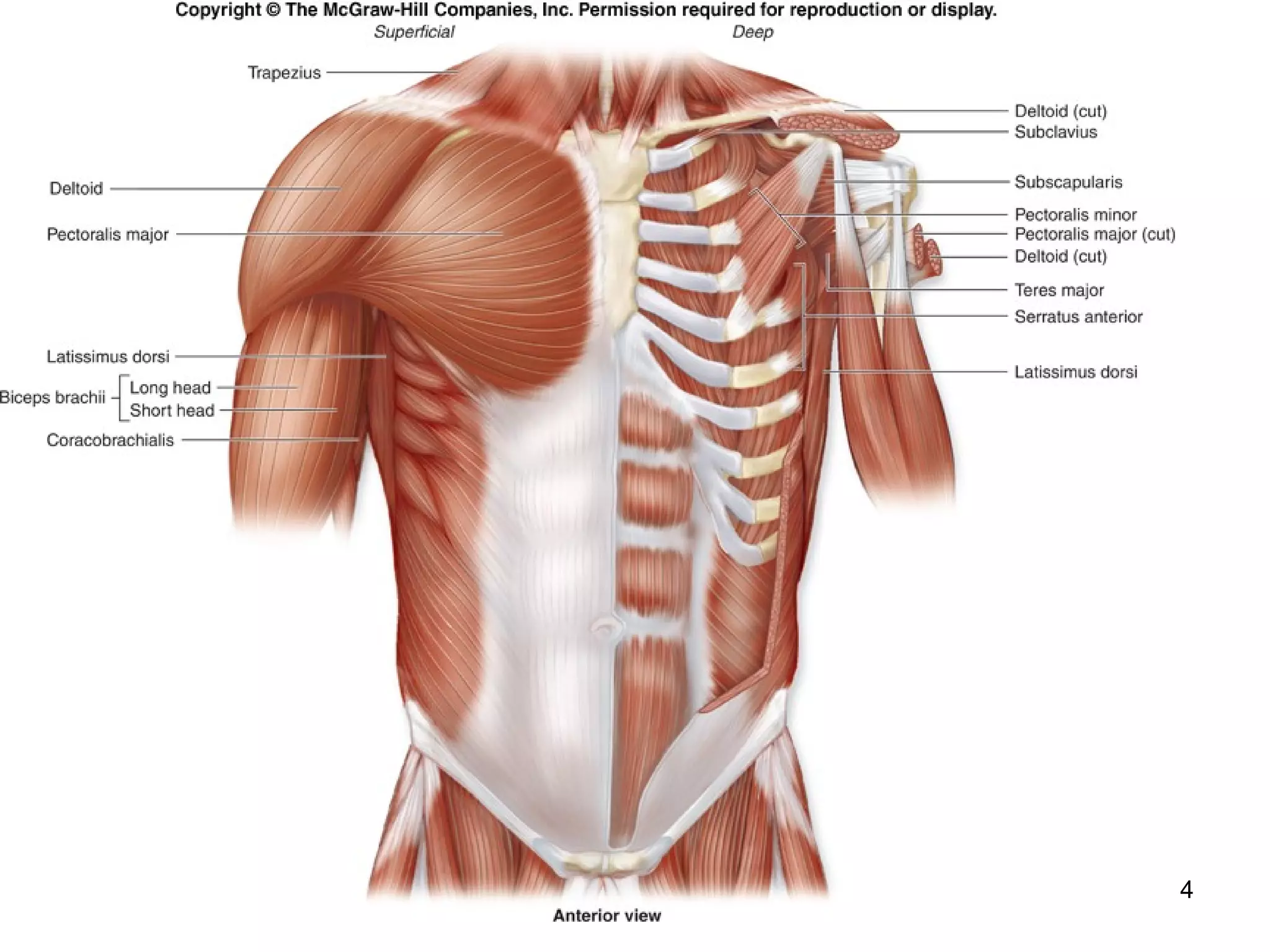
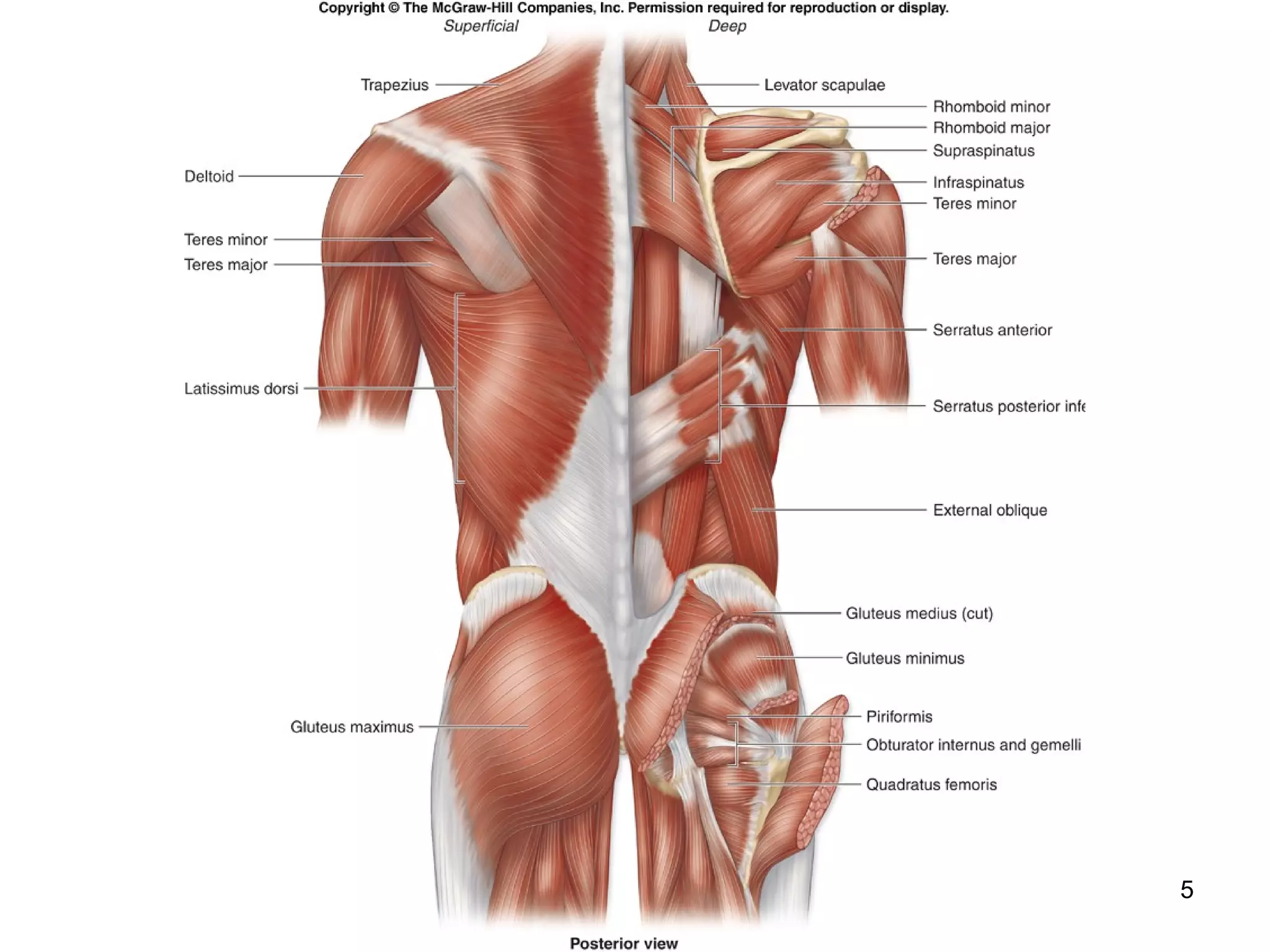
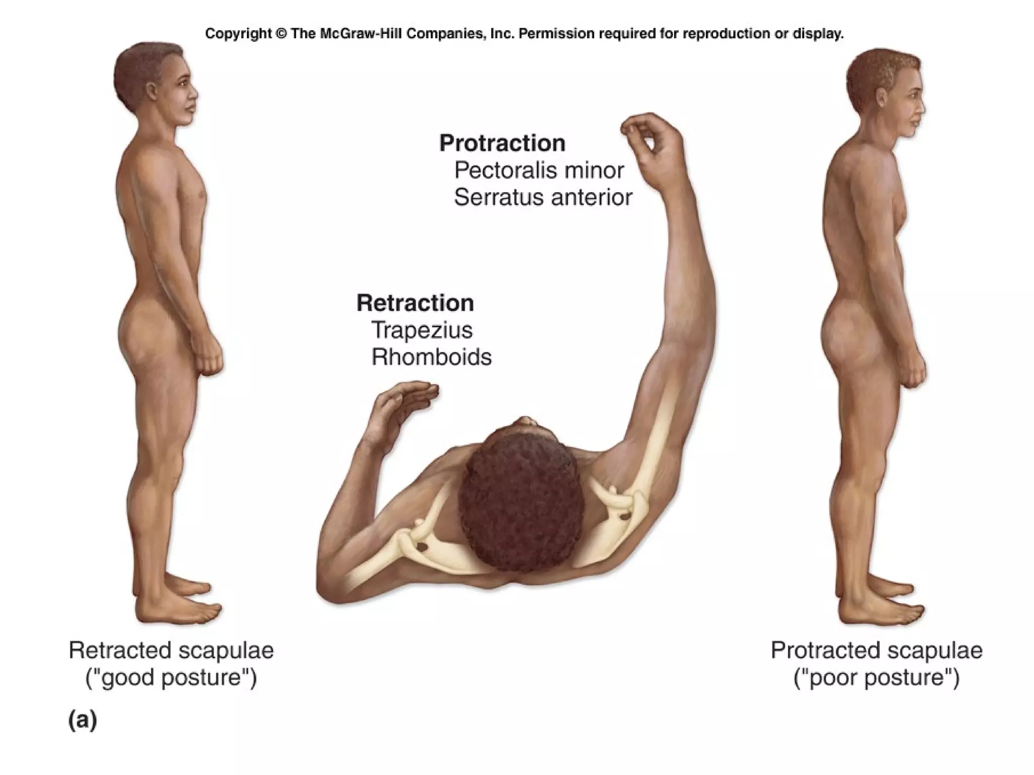
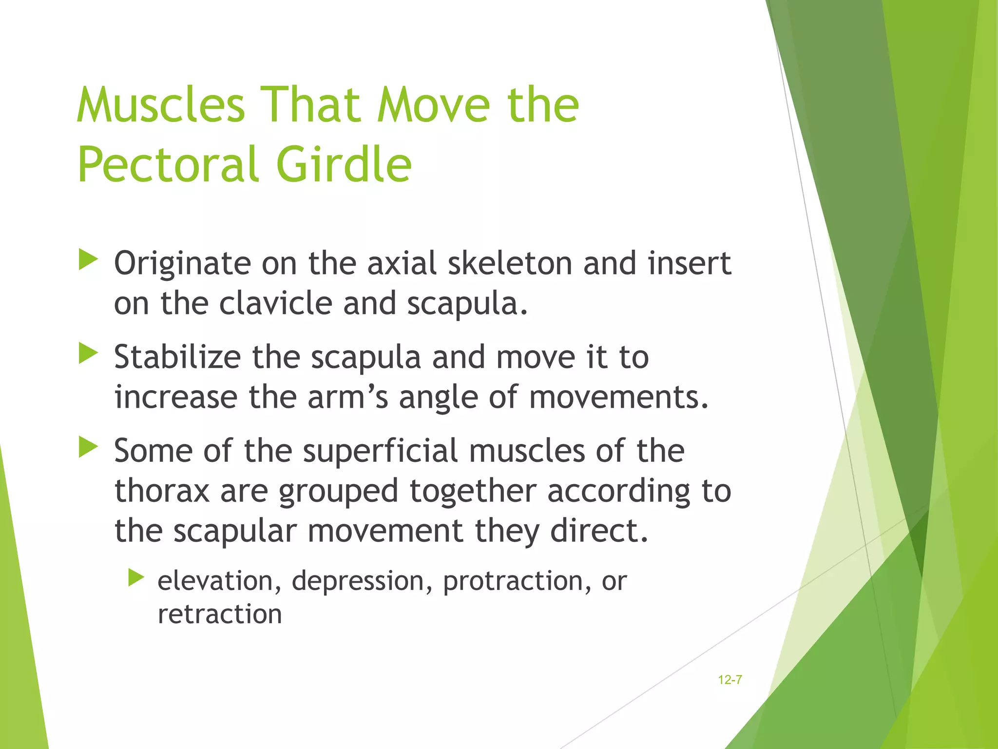
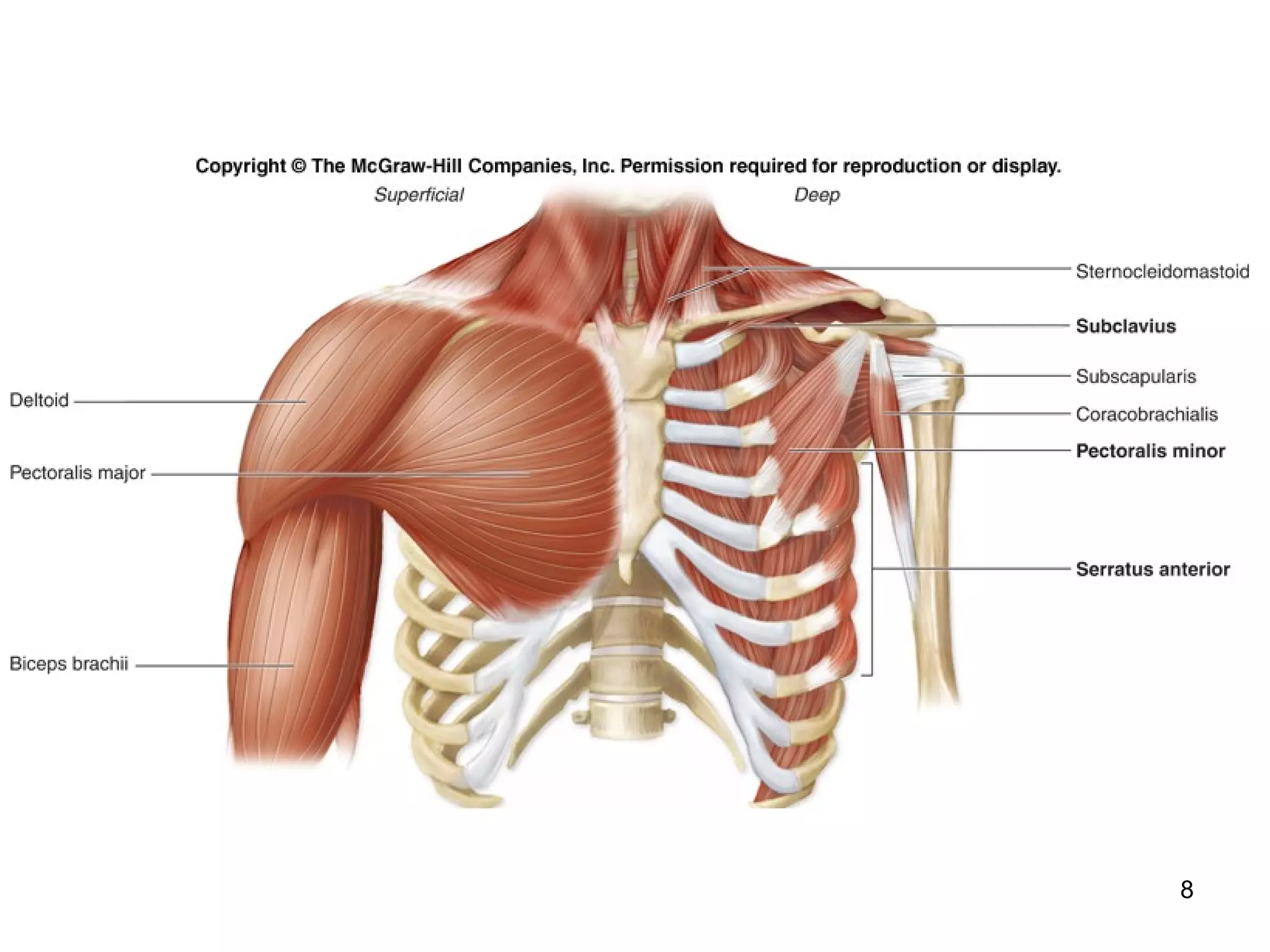
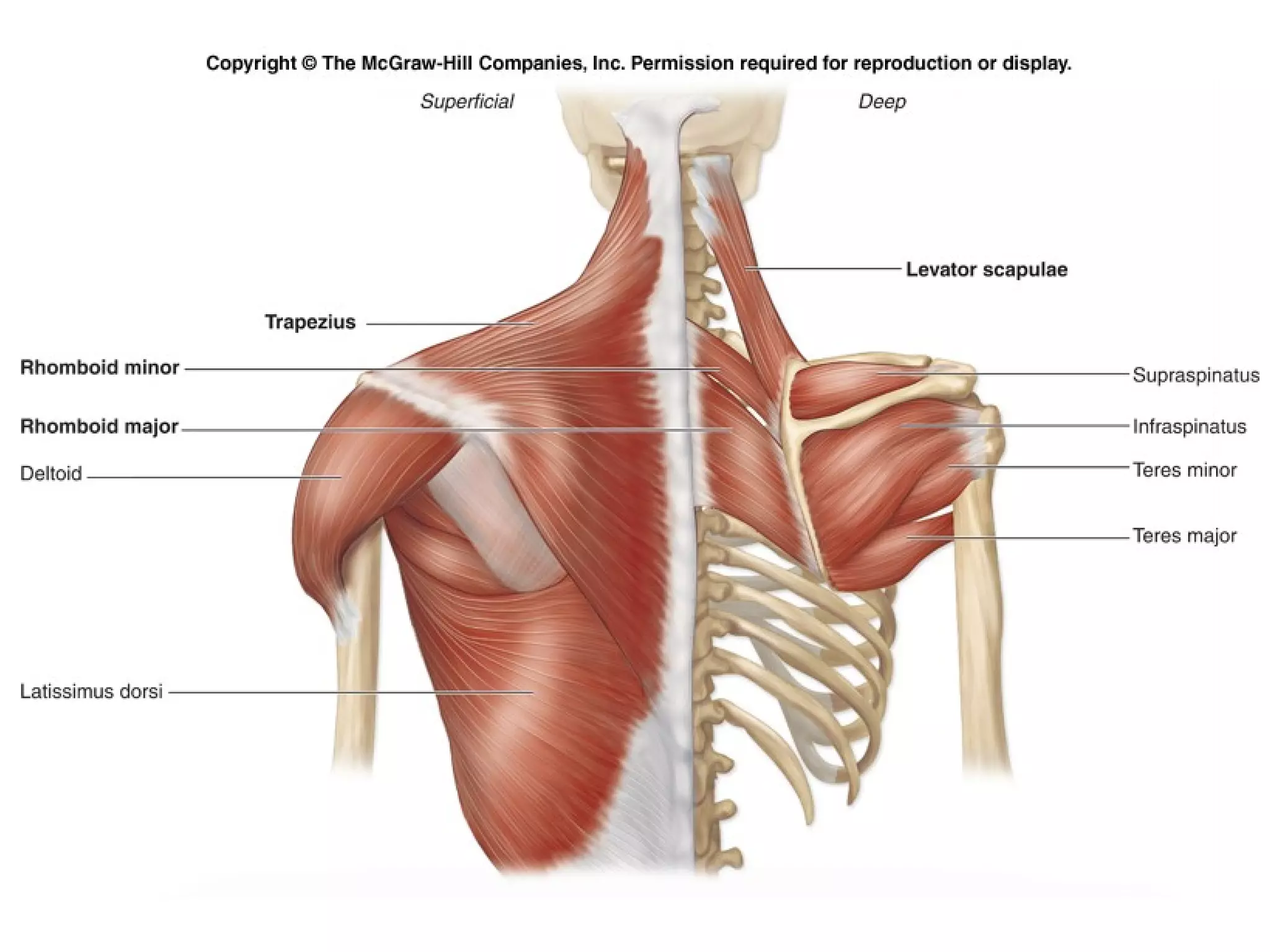
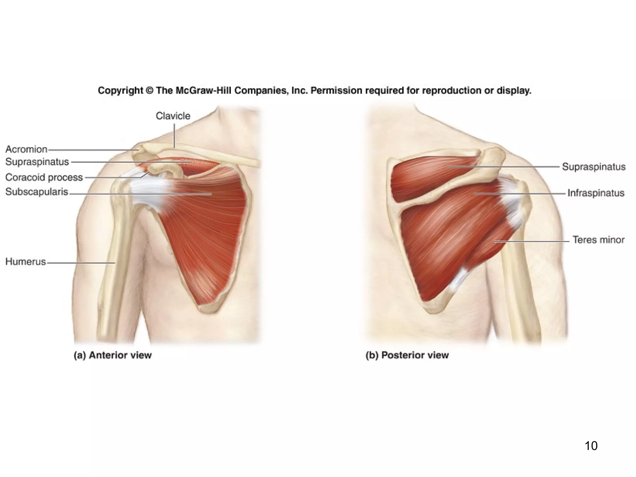
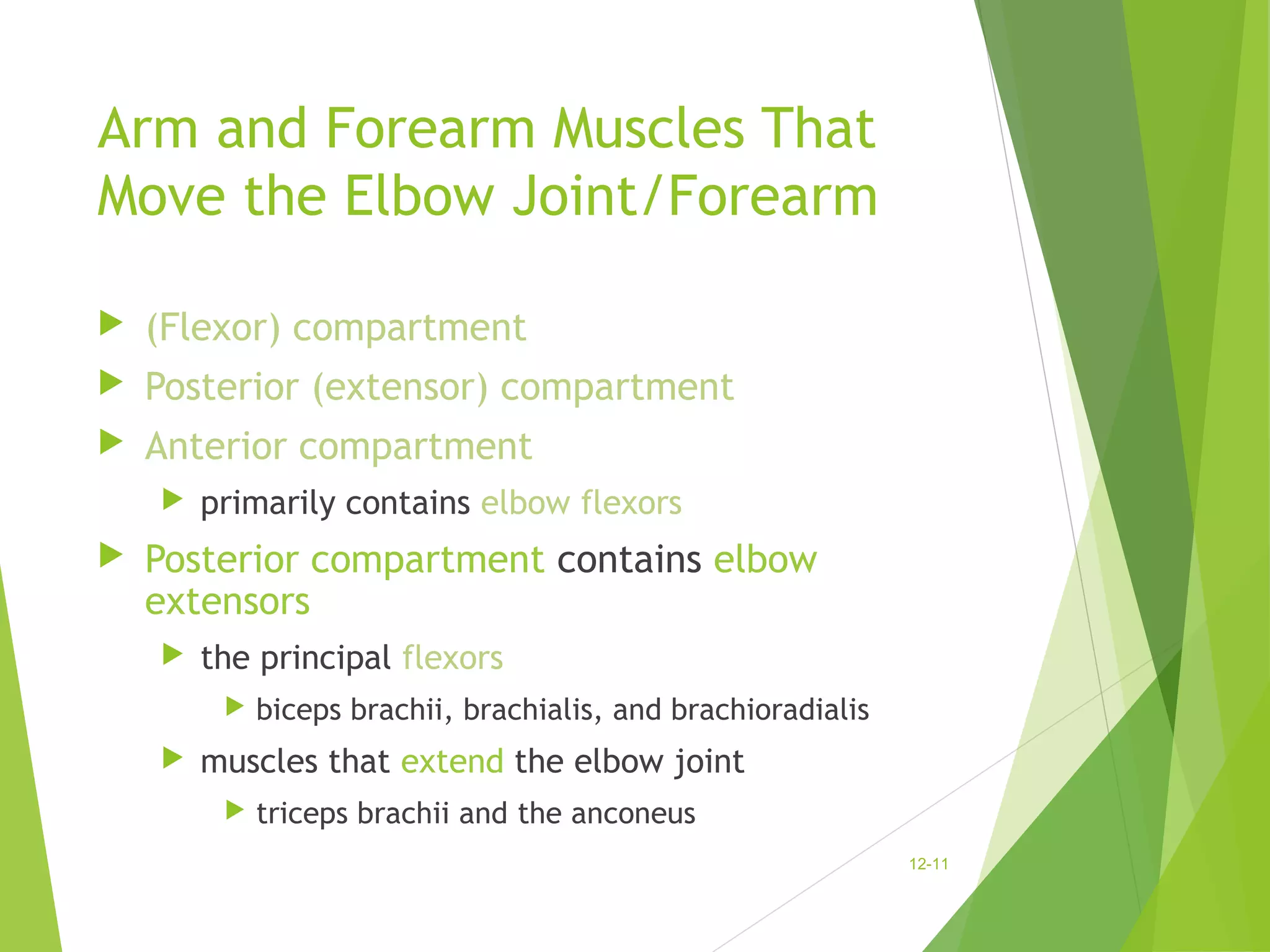
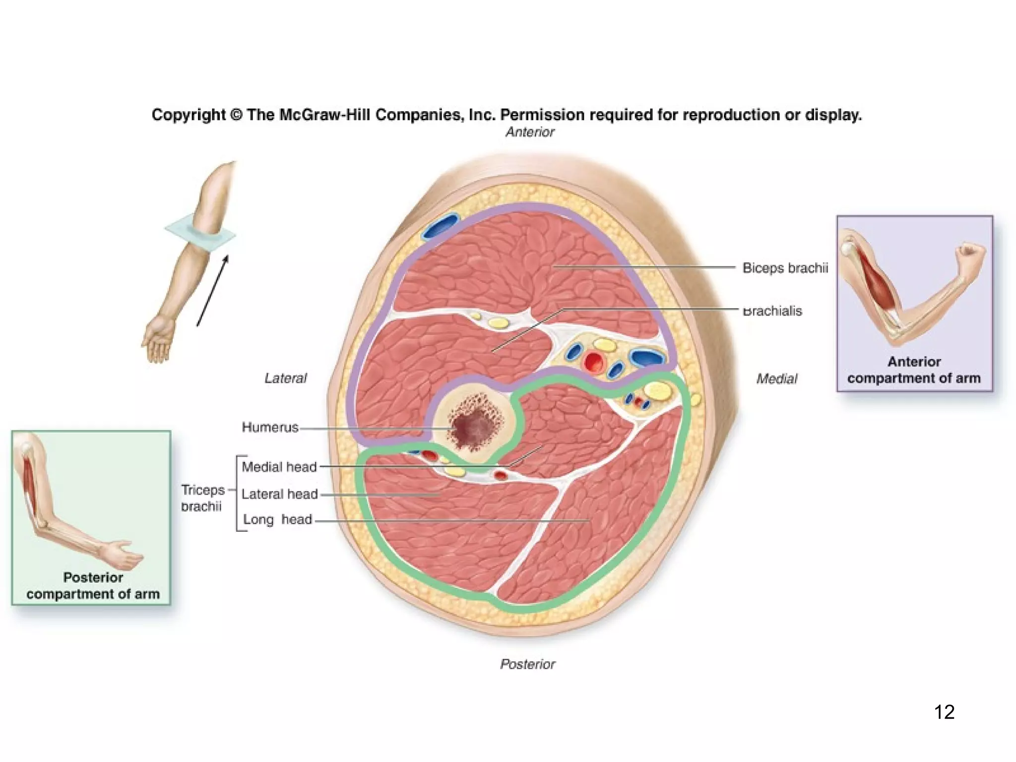
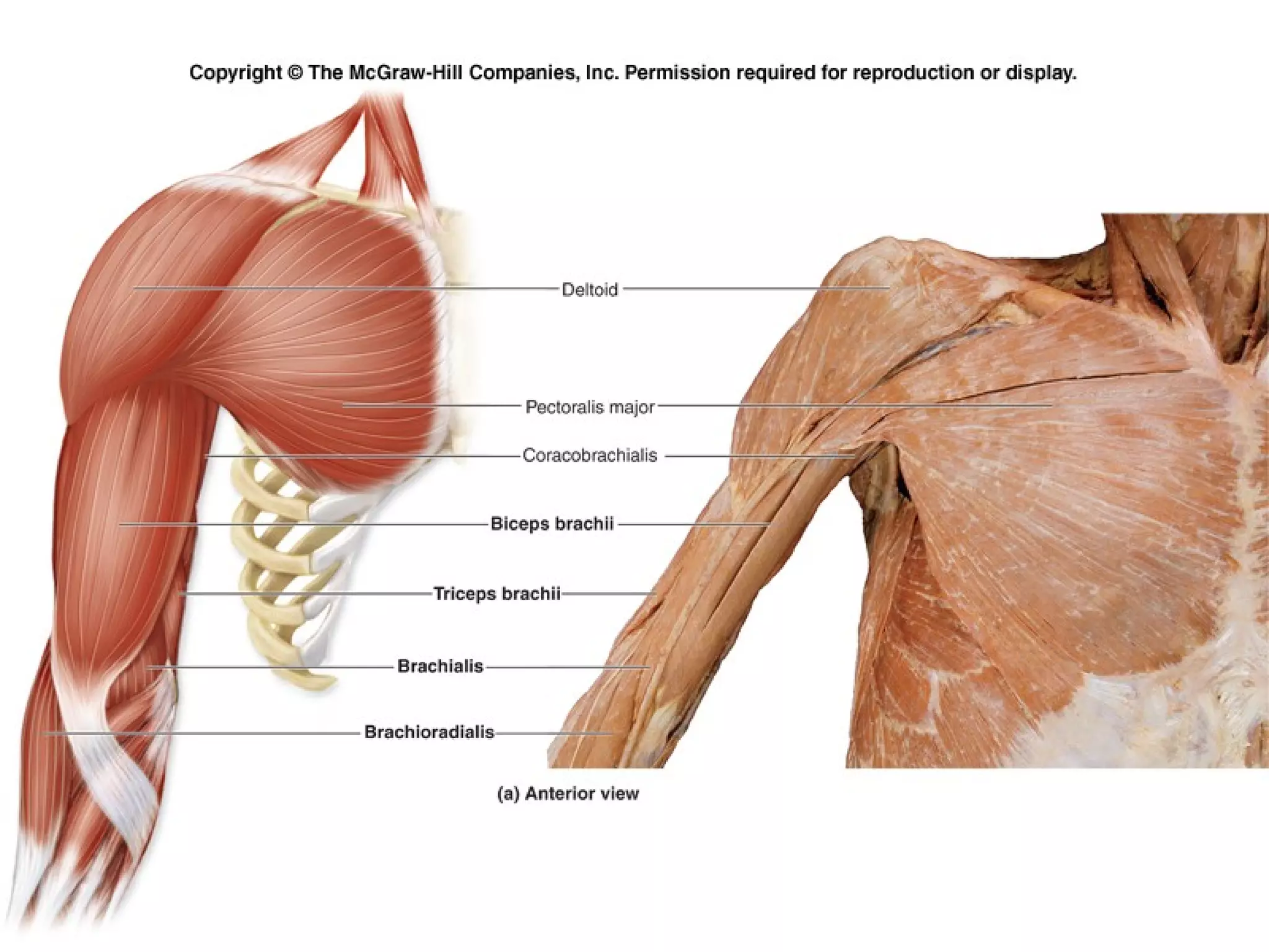
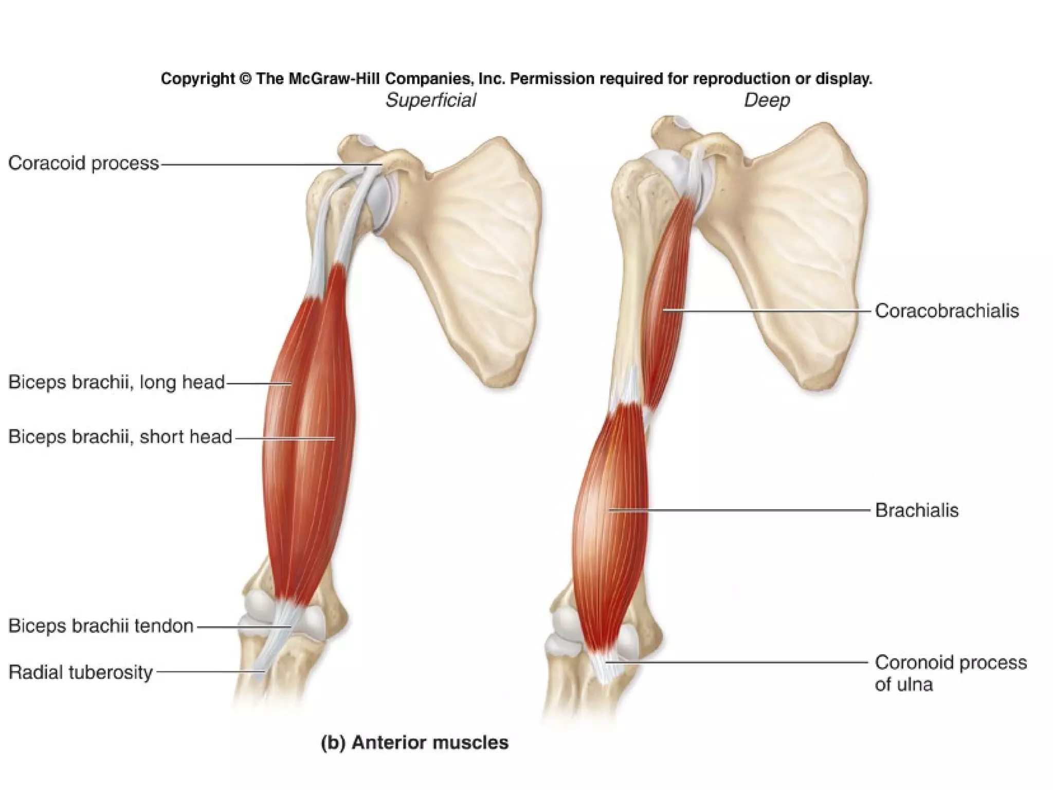
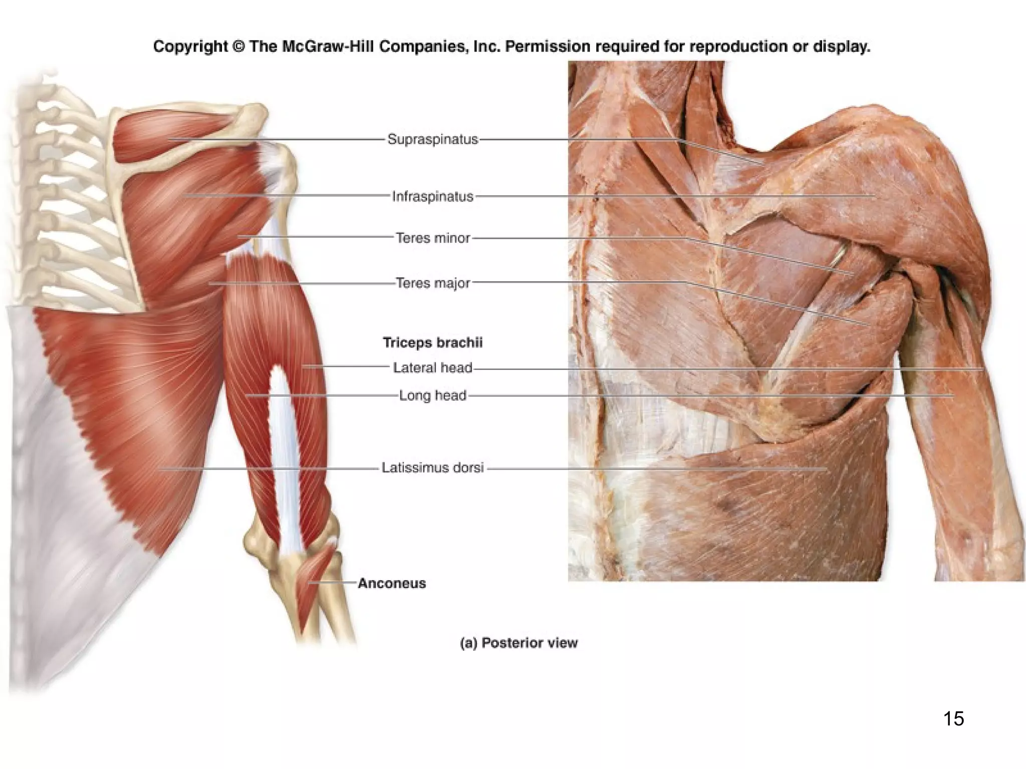
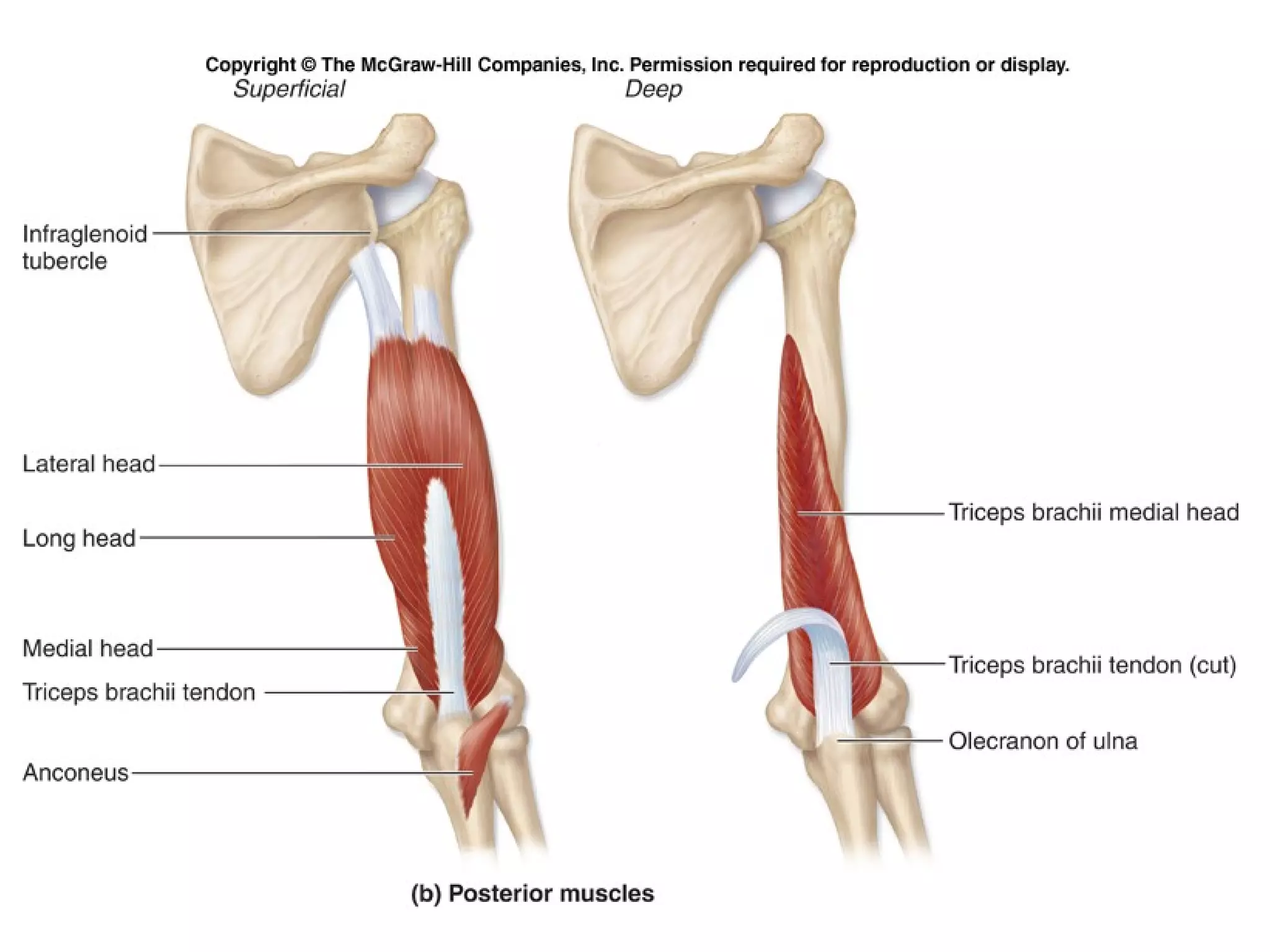
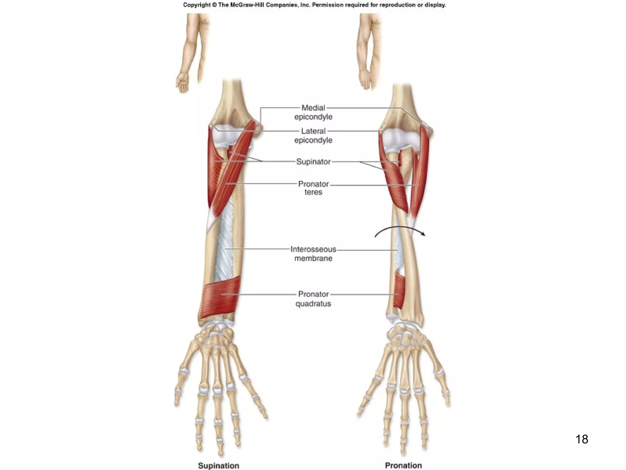
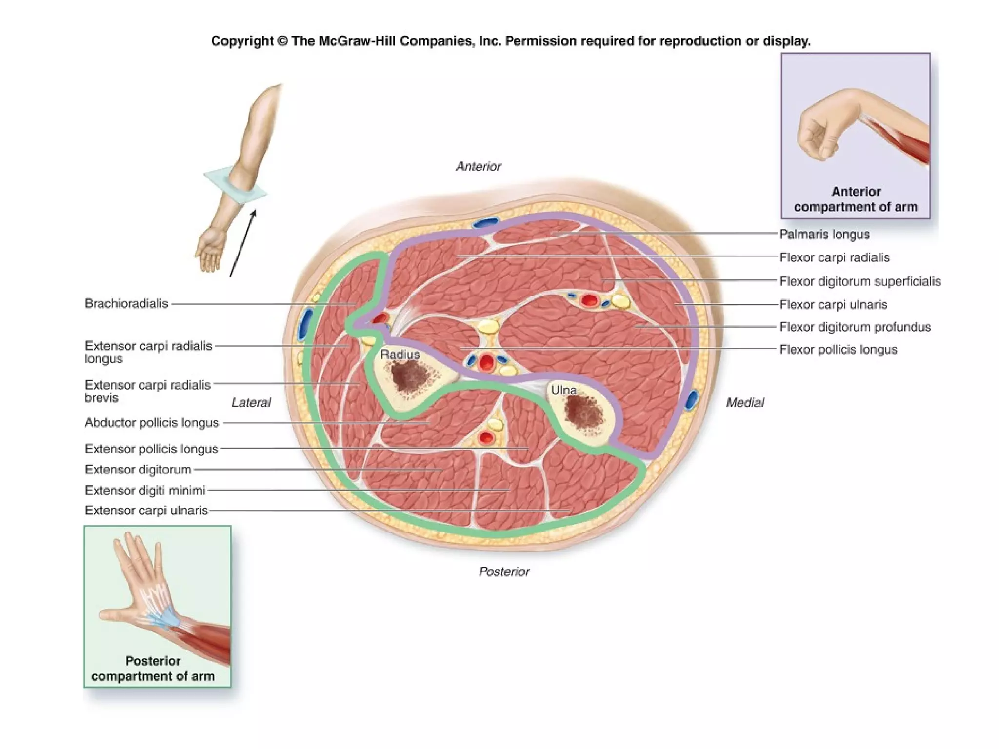
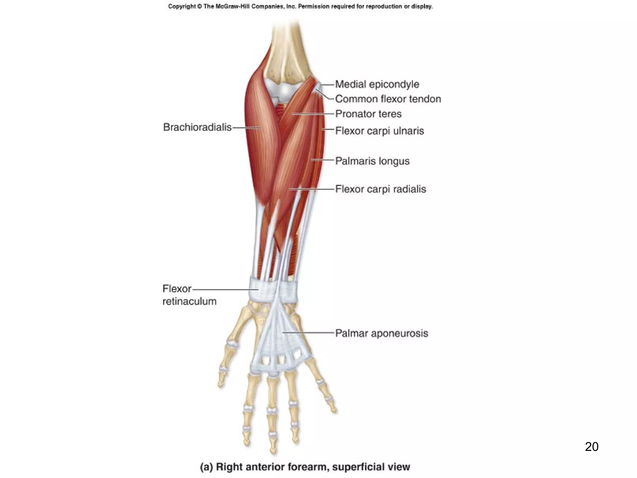
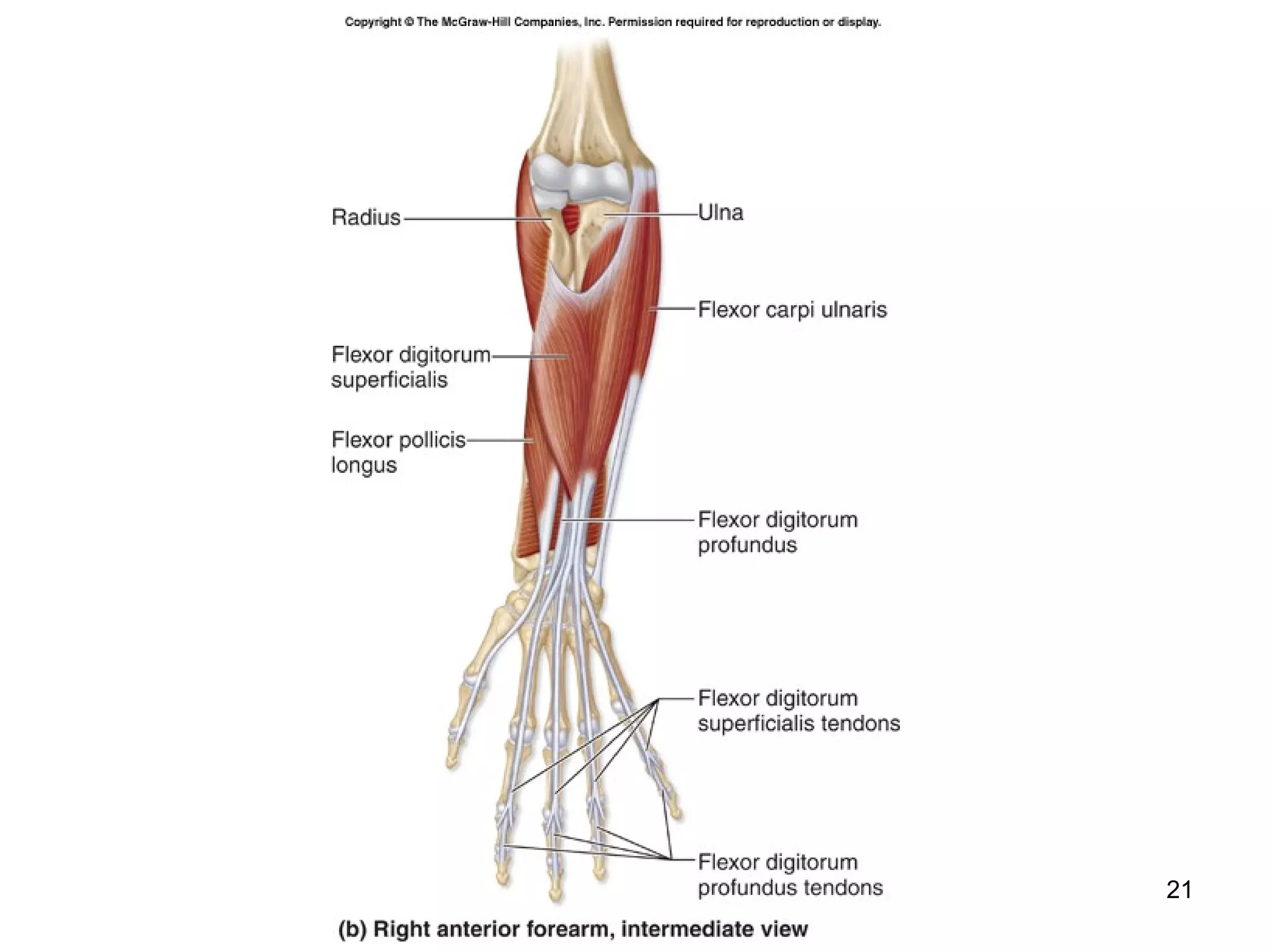
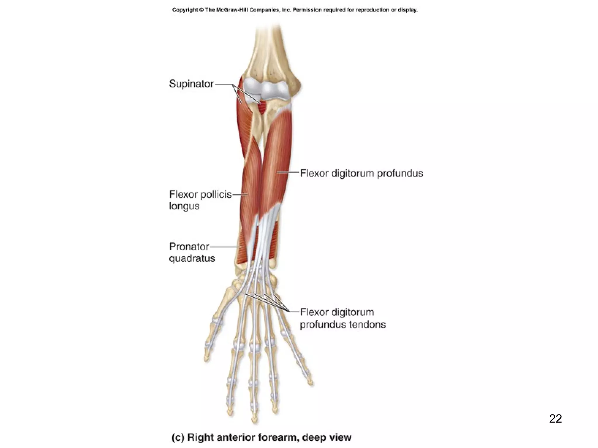
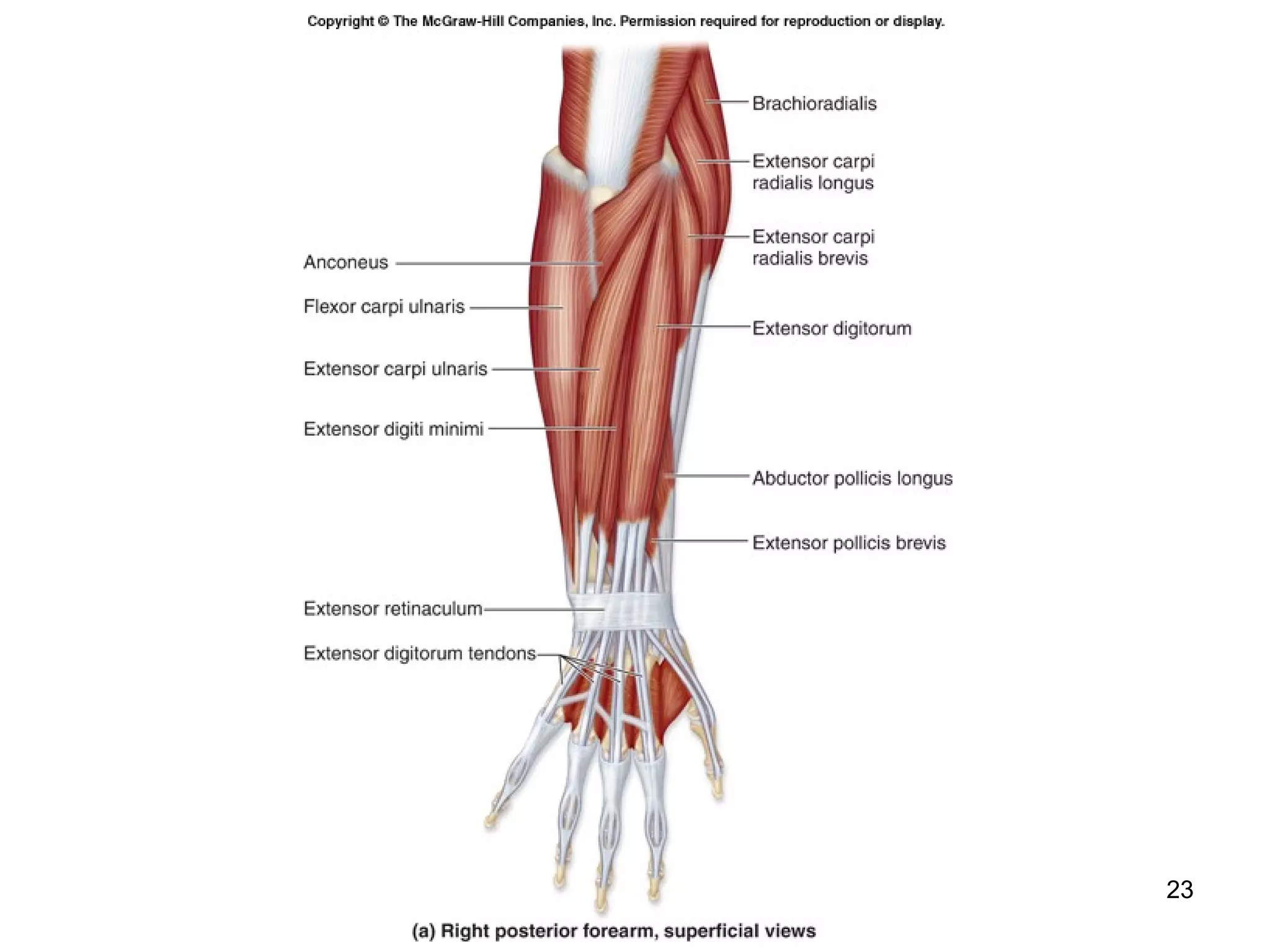
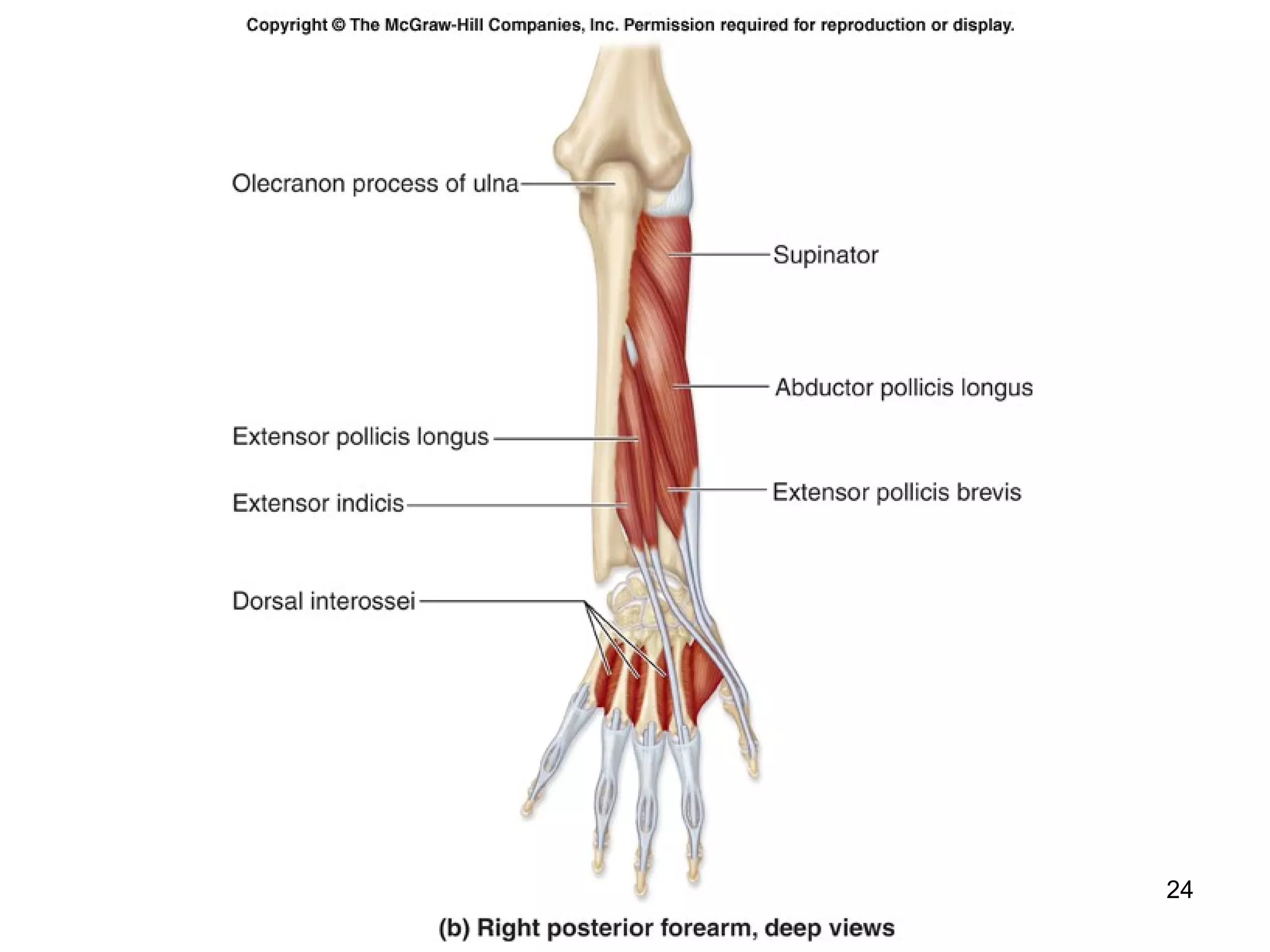
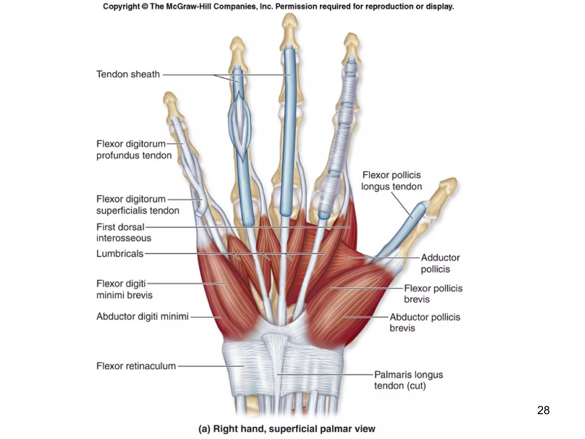
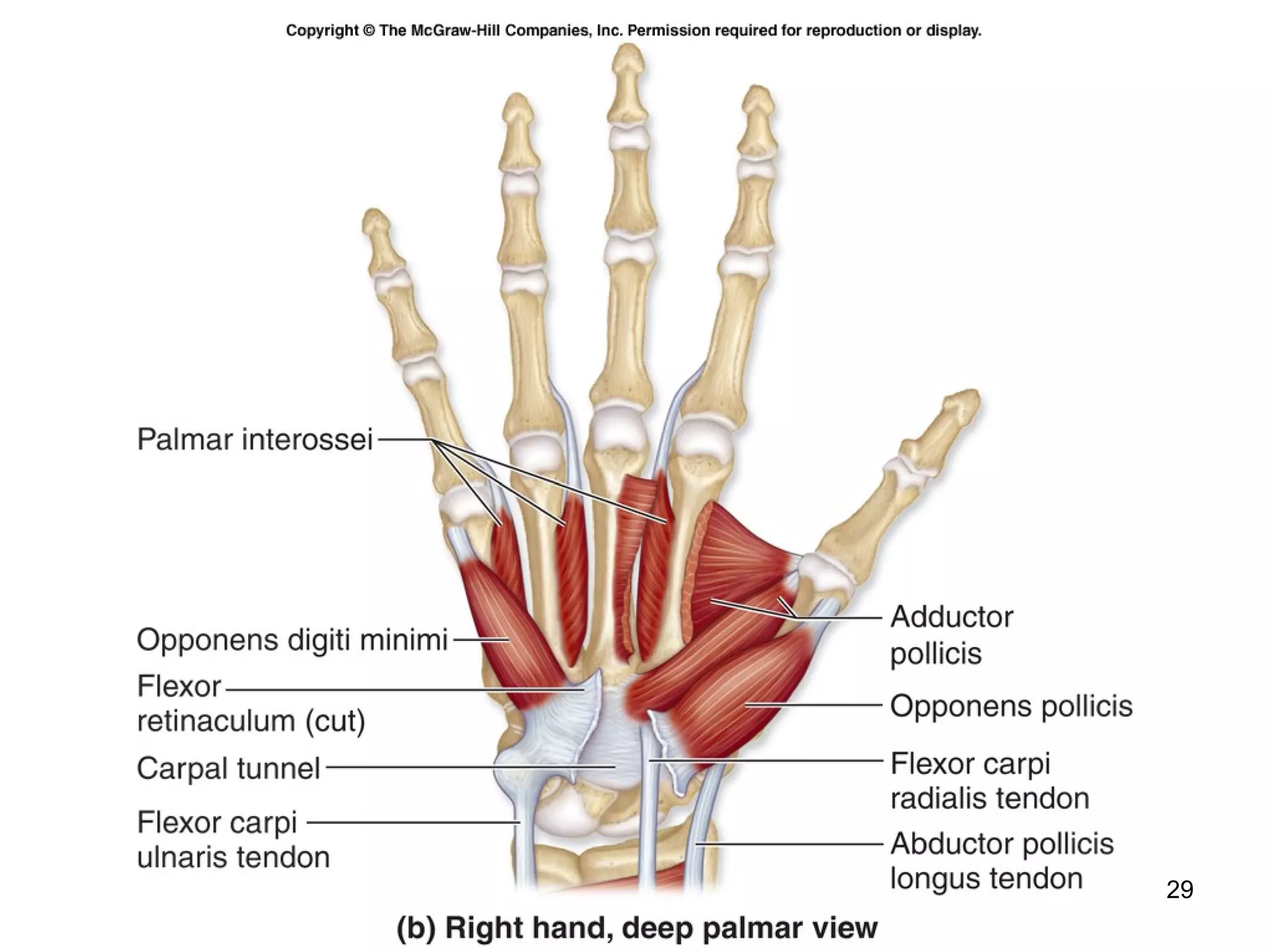
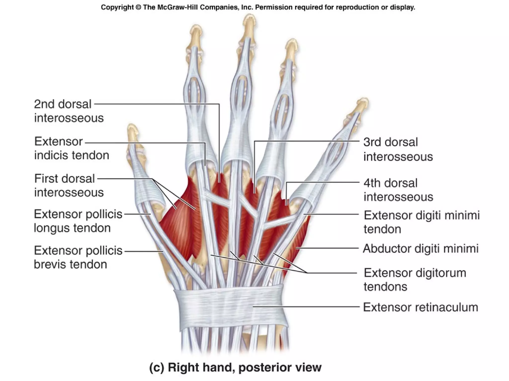
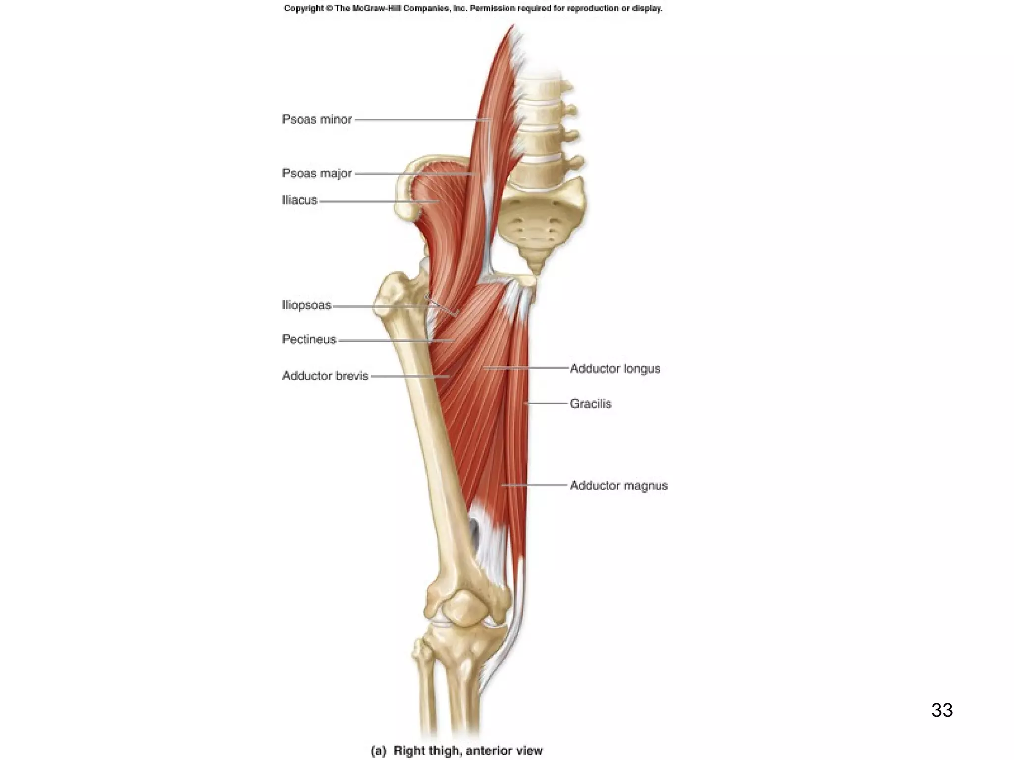
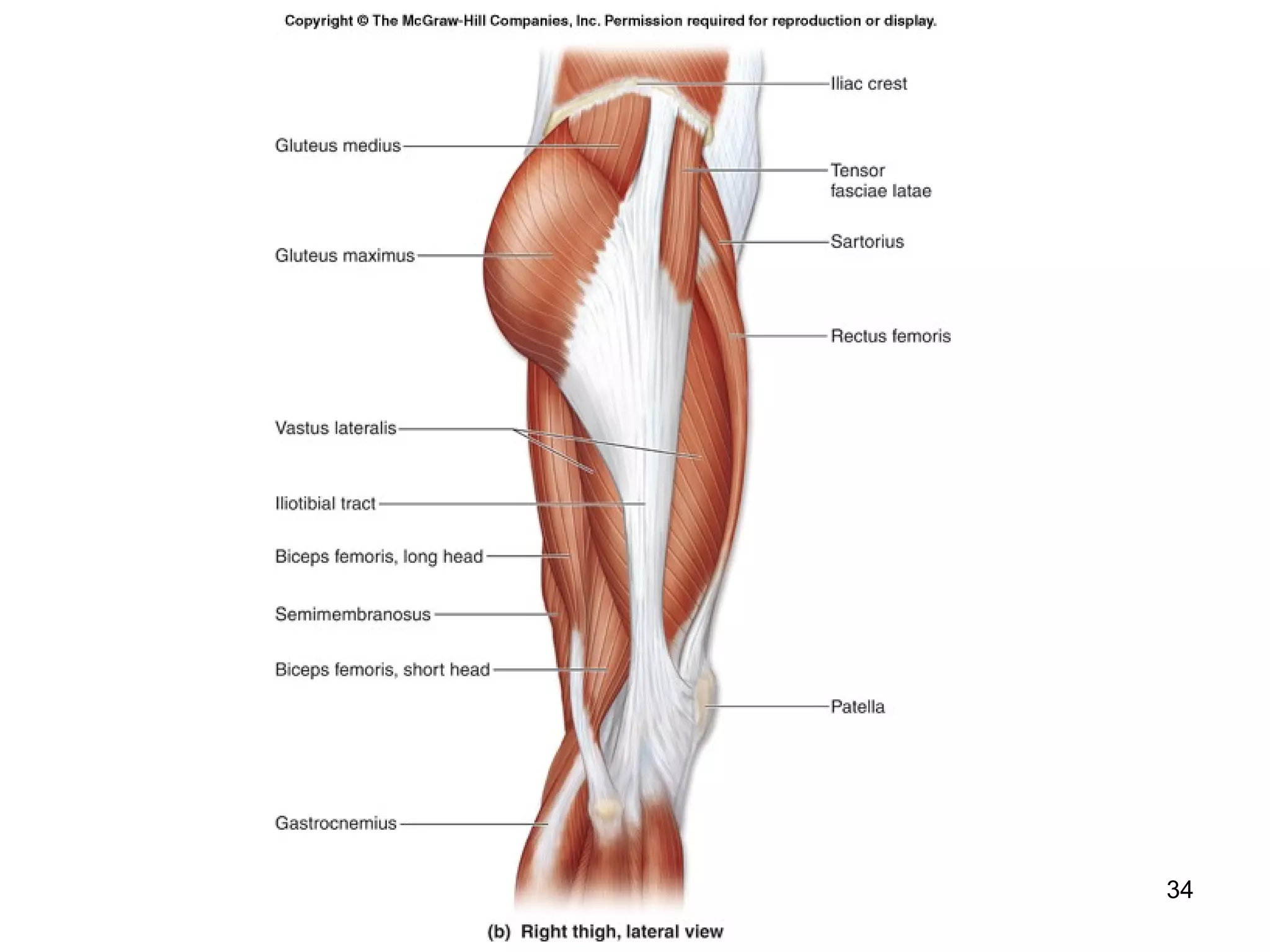
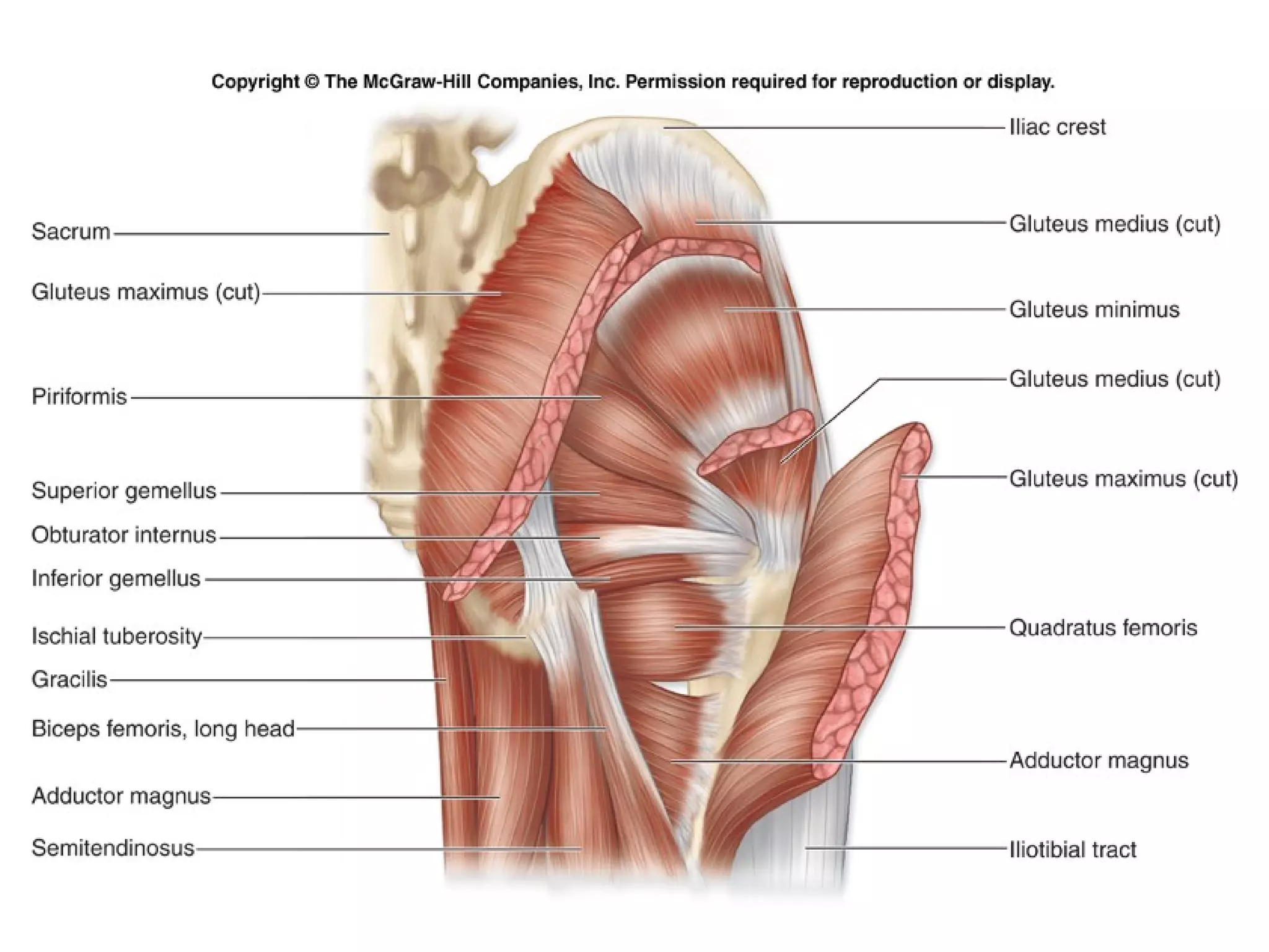
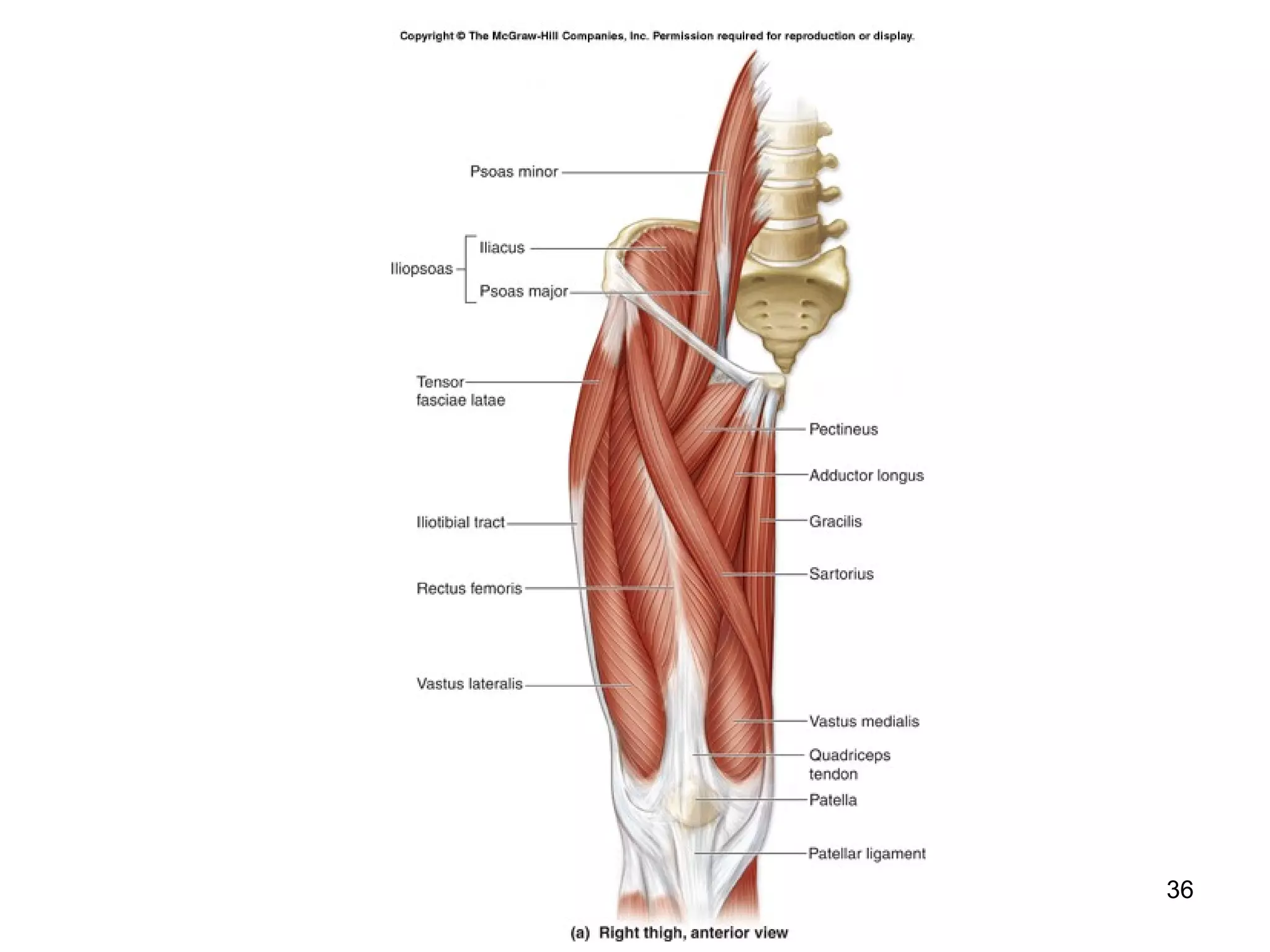
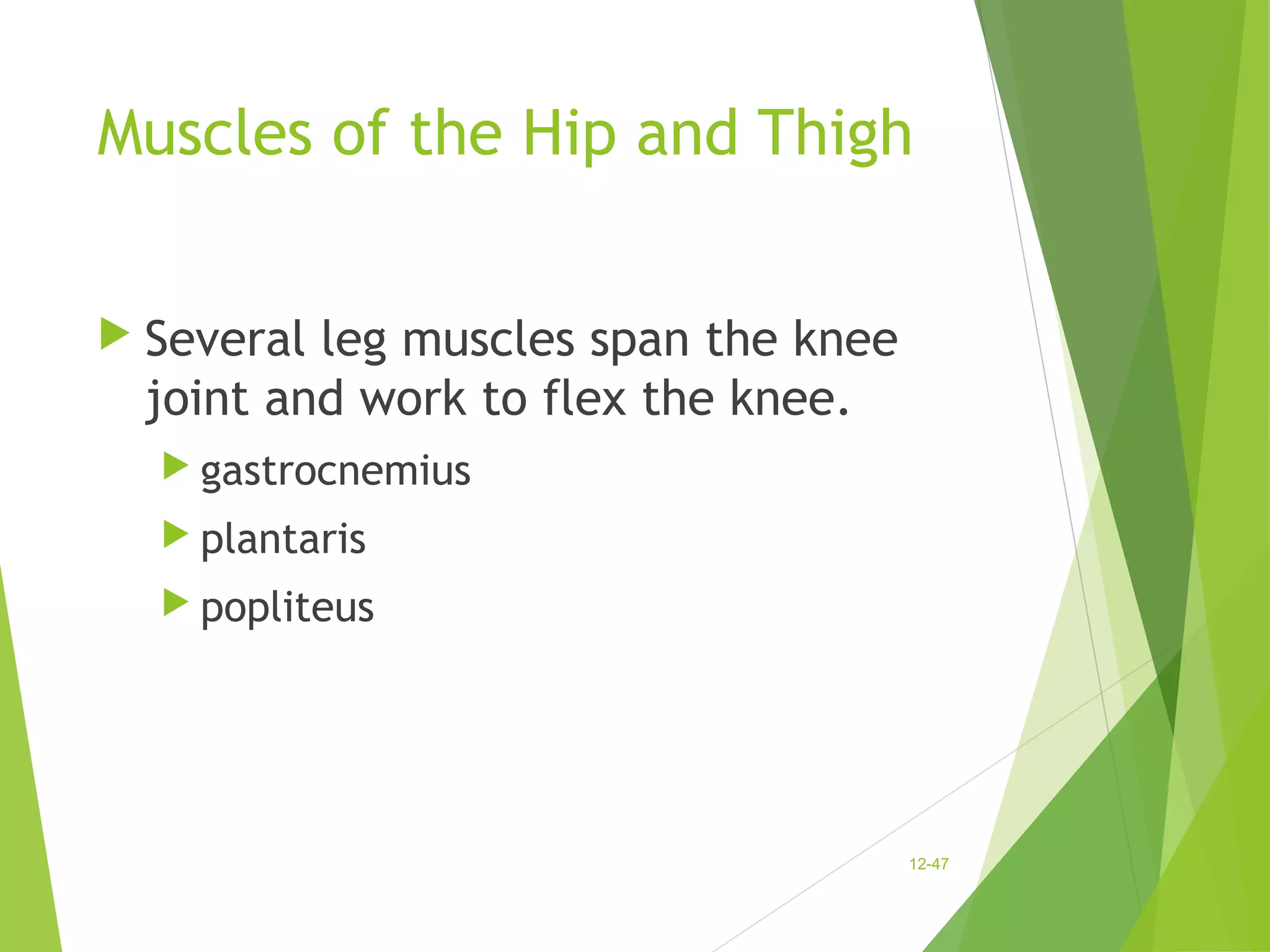
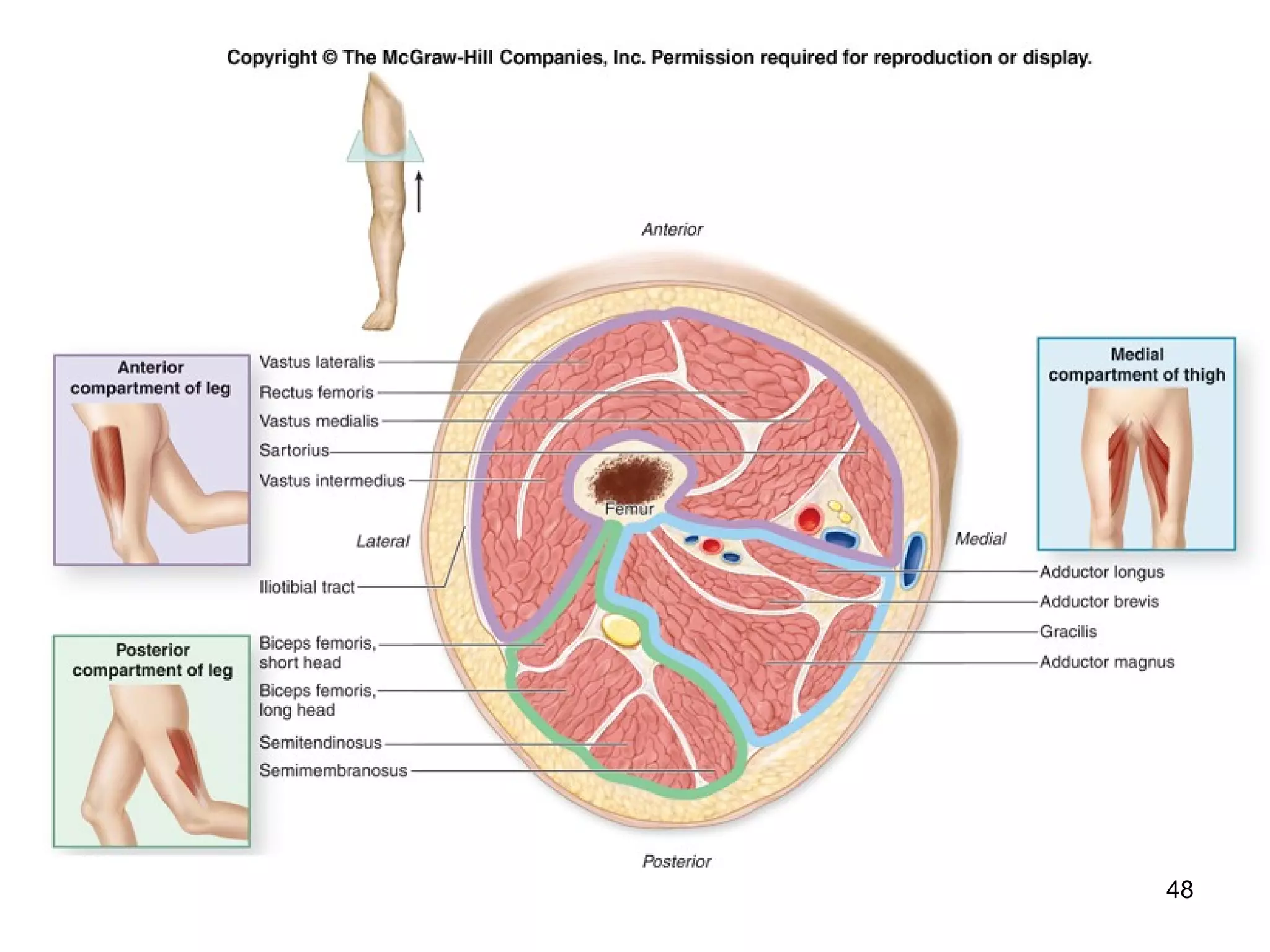
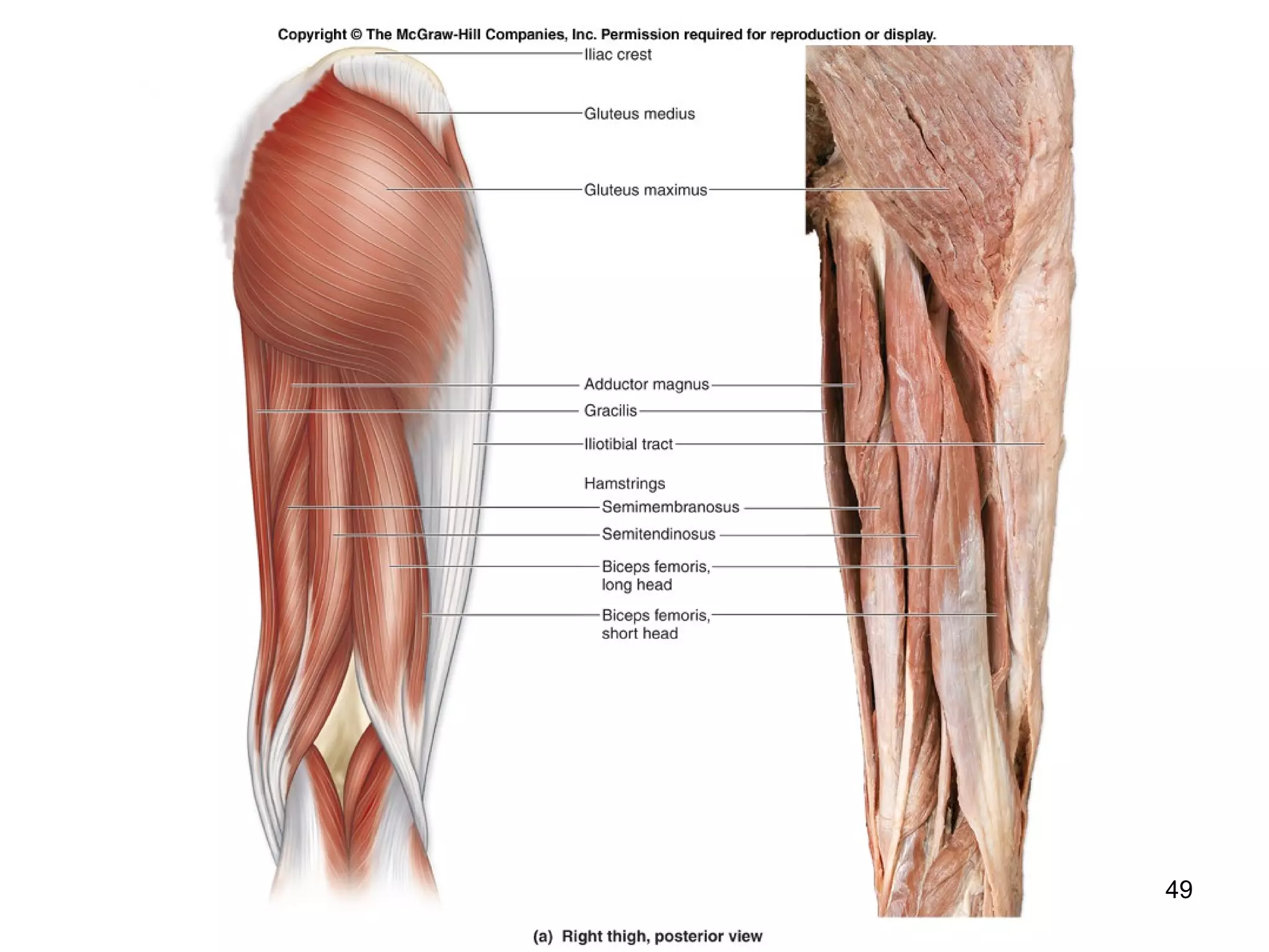
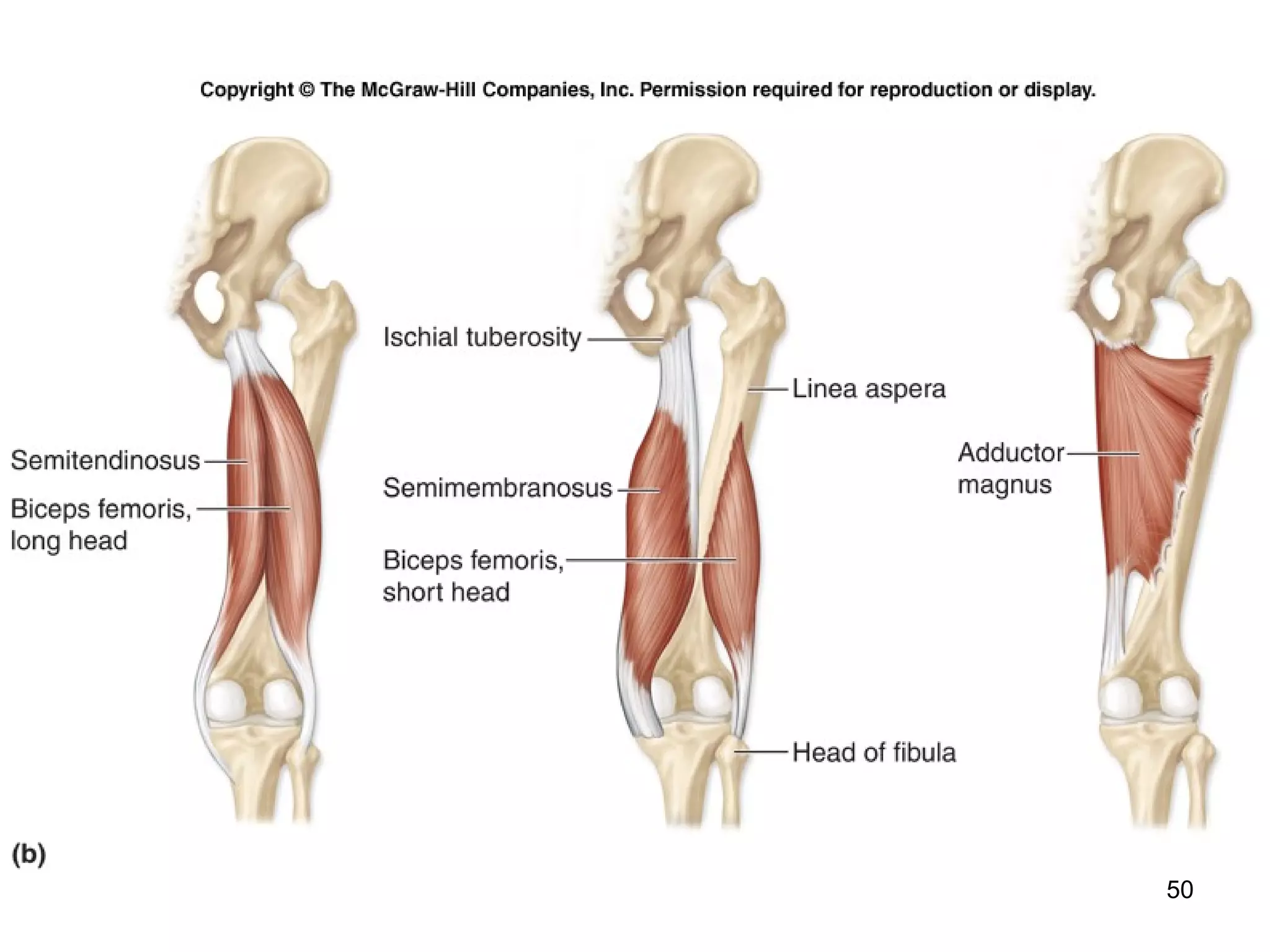
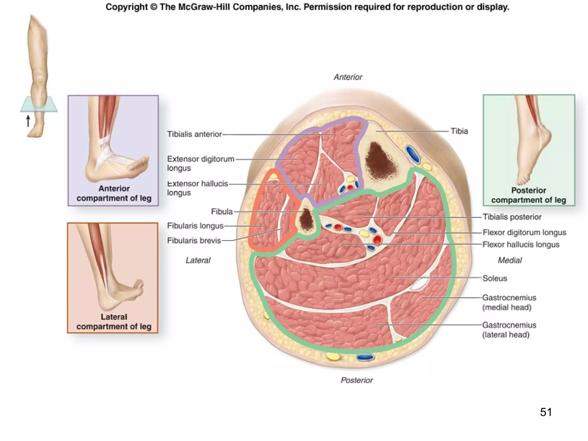
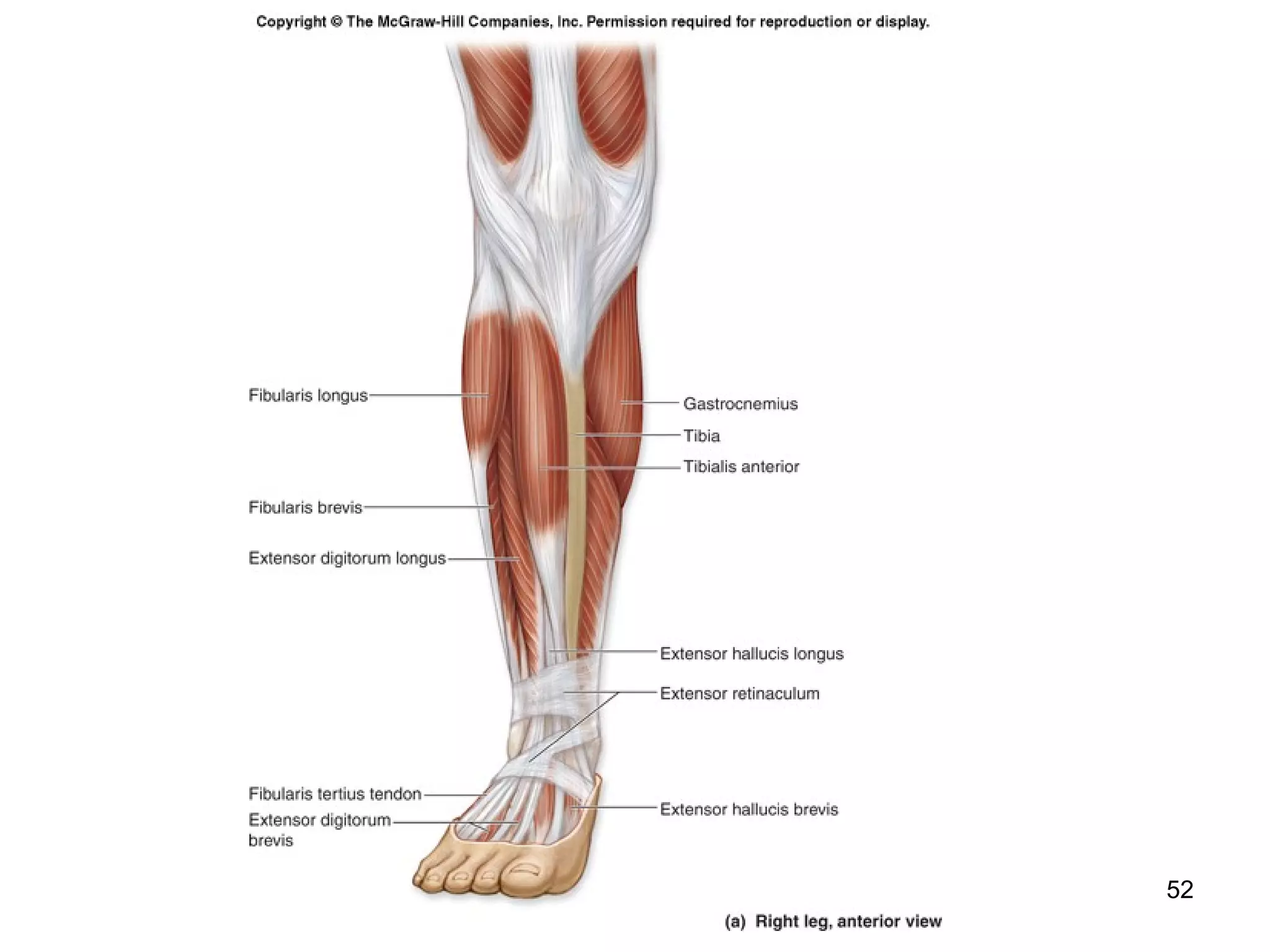
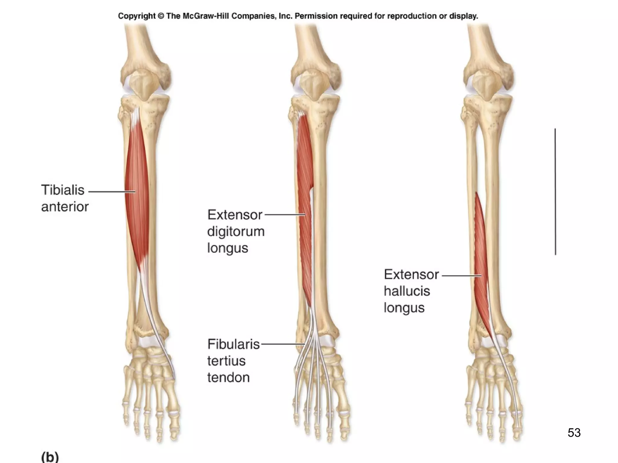
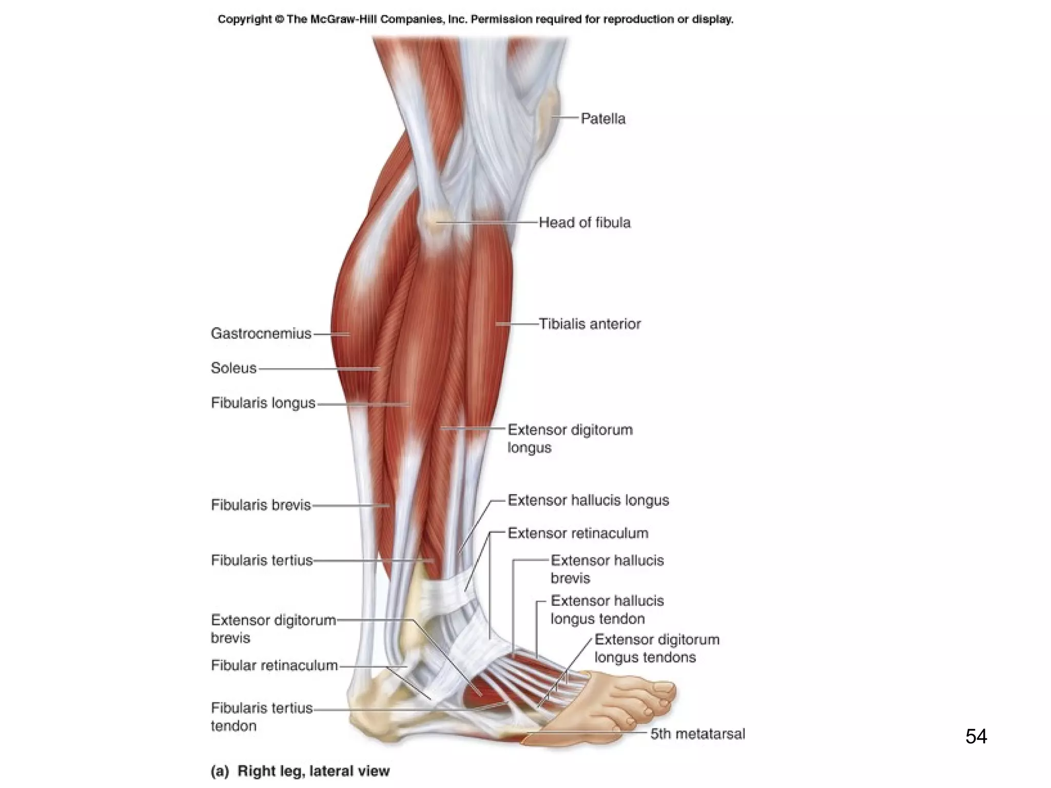
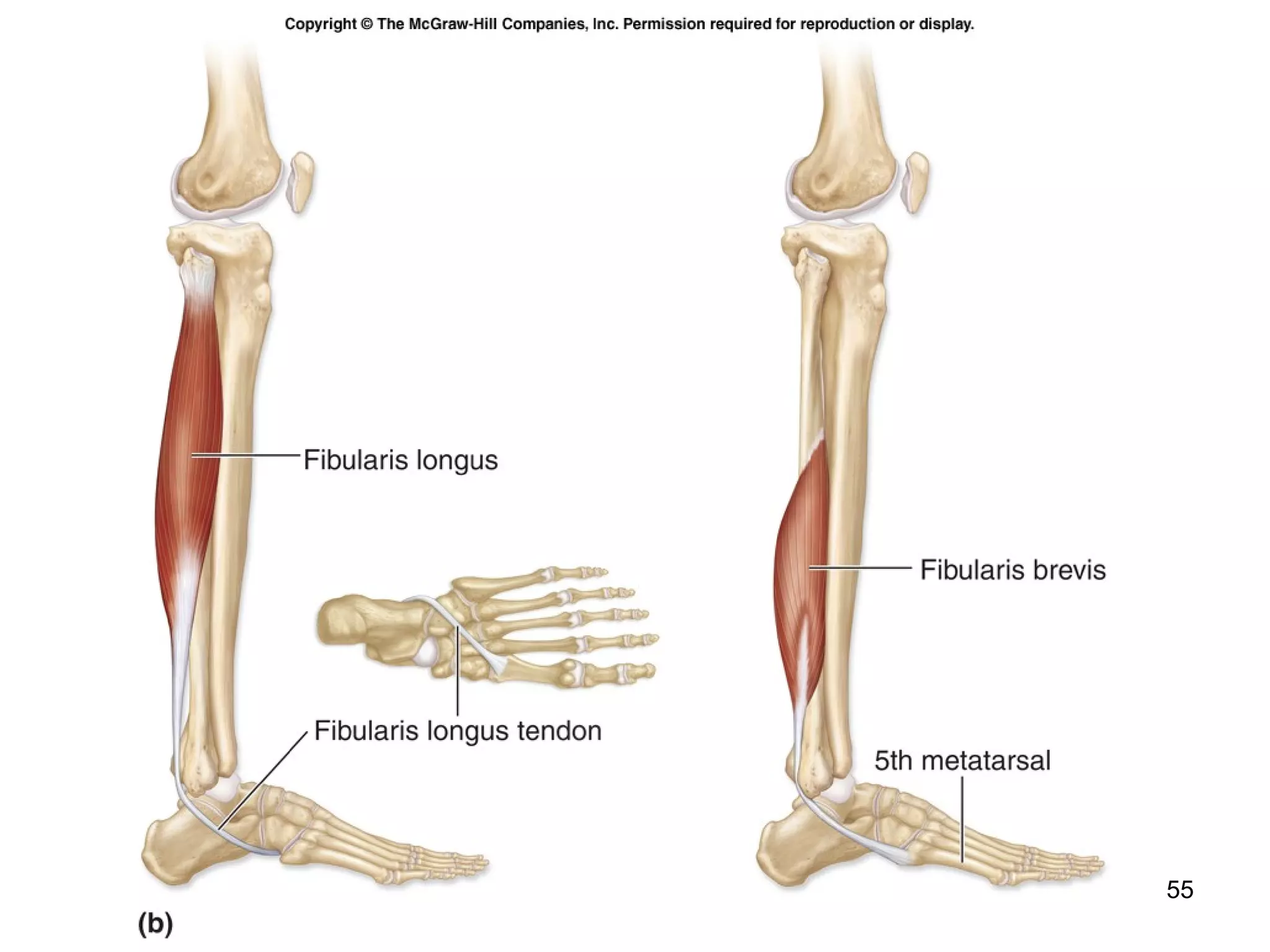
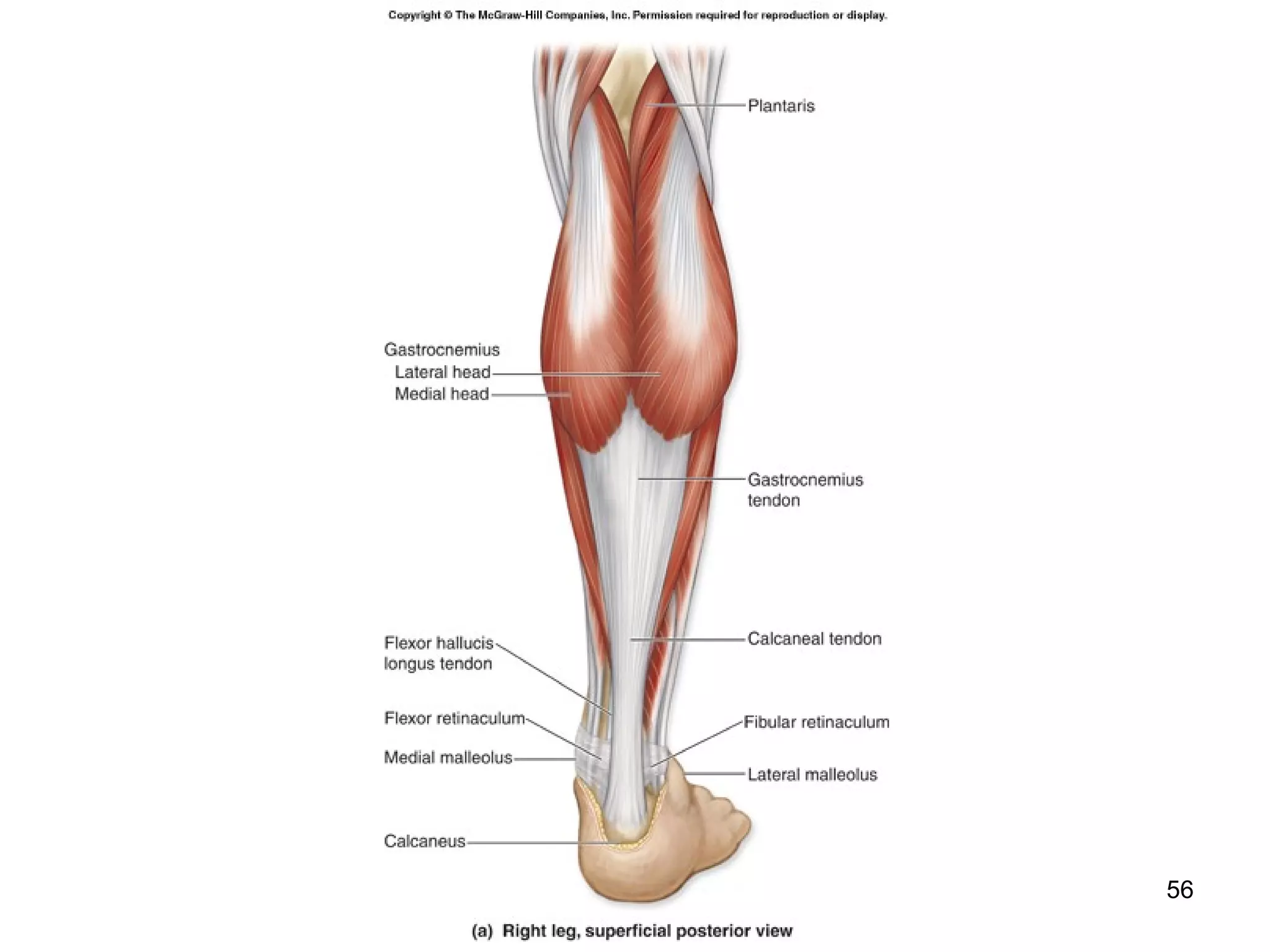
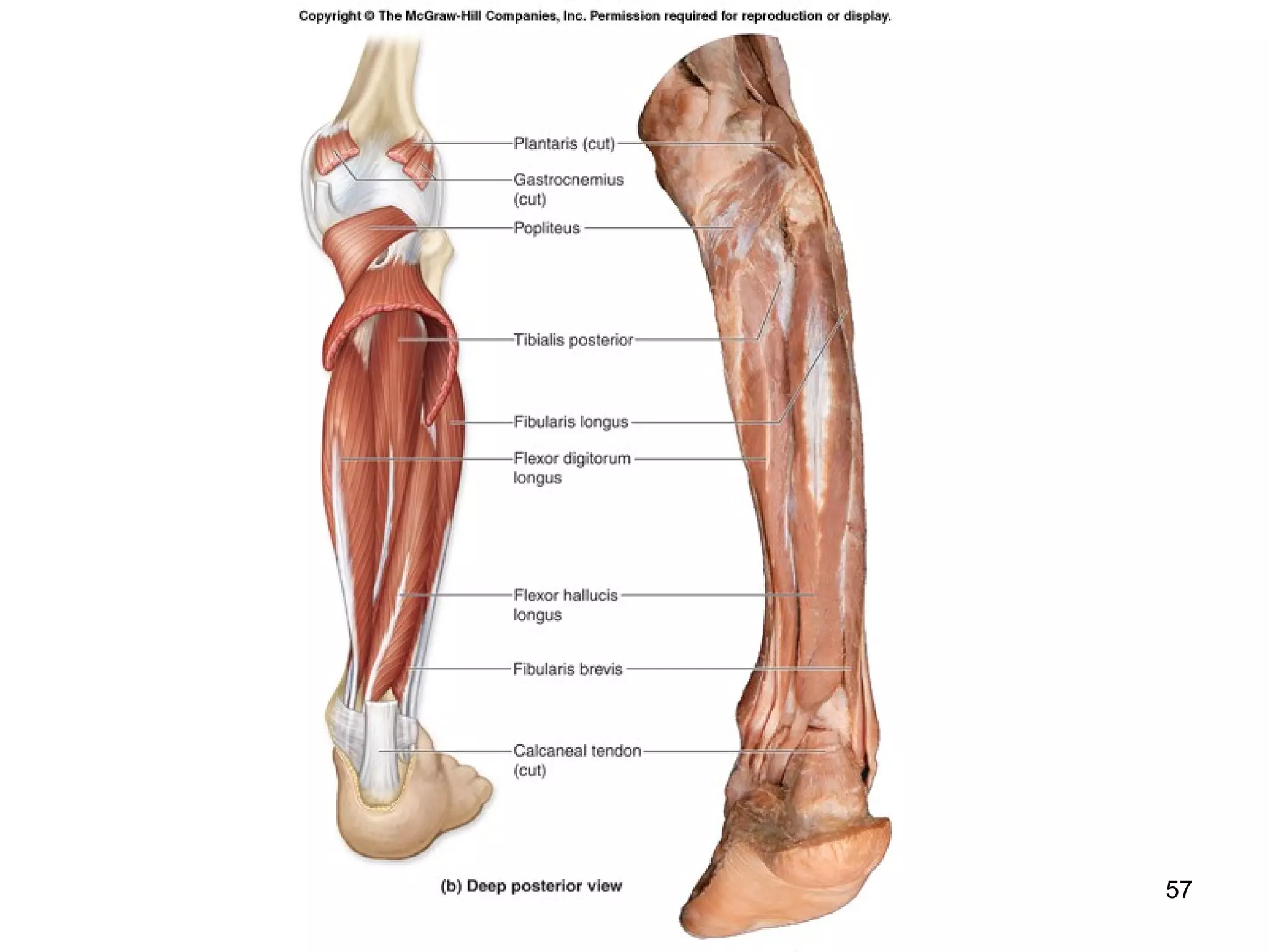
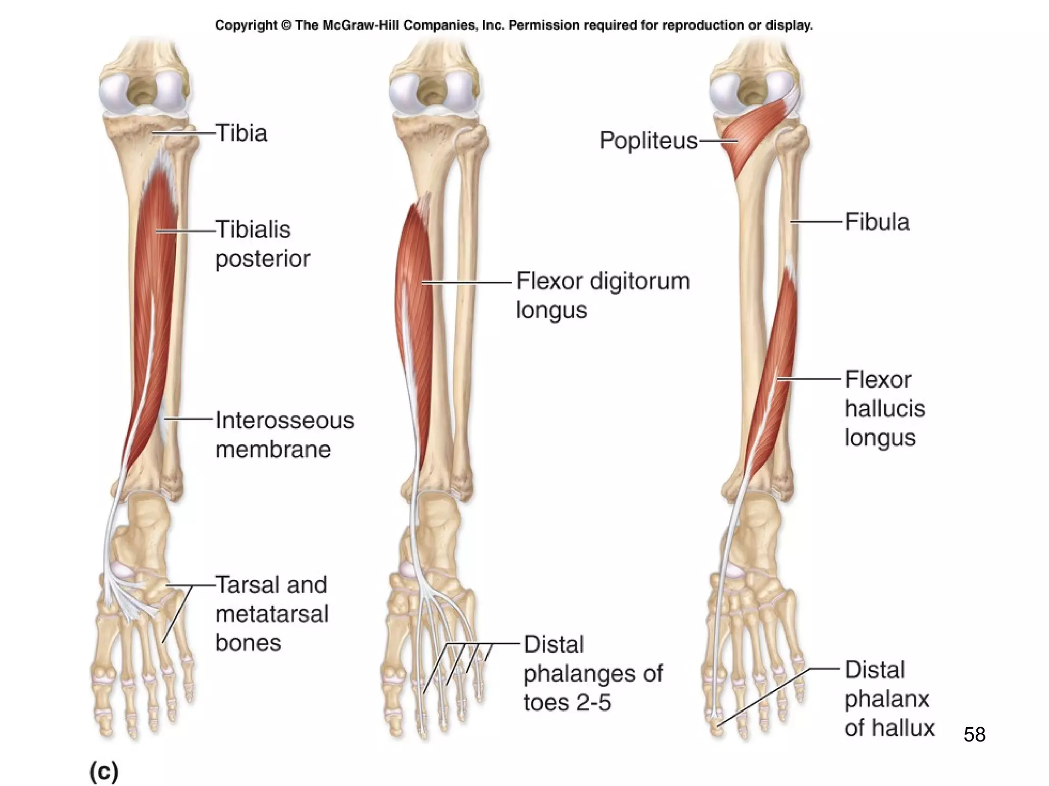
Human Anatomy 
Appendicular Muscles 
Pavemedicine.com 
12-1
Appendicular Muscles 
 Control the movements of the upper and 
lower limbs. 
 Stabilize and control the movements of 
the pectoral and pelvic girdles. 
 Organized into groups based on their 
location in the body or the part of the 
skeleton they move. 
 Work in groups that are either synergistic 
or antagonistic. 
12-2
Appendicular Muscles 
 Organized into specific groups. 
muscles that move the pectoral girdle 
muscles that move the glenohumeral 
joint/arm 
 arm and forearm muscles that move 
the elbow joint/forearm 
 forearm muscles that move the wrist 
joint, hand, and fingers 
 intrinsic muscles of the hand 
12-3
4
5
6
Muscles That Move the 
Pectoral Girdle 
 Originate on the axial skeleton and insert 
on the clavicle and scapula. 
 Stabilize the scapula and move it to 
increase the arm’s angle of movements. 
 Some of the superficial muscles of the 
thorax are grouped together according to 
the scapular movement they direct. 
 elevation, depression, protraction, or 
retraction 
12-7
8
9
10
Arm and Forearm Muscles That 
Move the Elbow Joint/Forearm 
 (Flexor) compartment 
 Posterior (extensor) compartment 
 Anterior compartment 
 primarily contains elbow flexors 
 Posterior compartment contains elbow 
extensors 
 the principal flexors 
 biceps brachii, brachialis, and brachioradialis 
 muscles that extend the elbow joint 
 triceps brachii and the anconeus 
12-11
12
13
14
15
16
Forearm Muscles That 
Supinate and Pronate 
 Supinator muscle supinates the forearm. 
 Contraction of the pronator teres and 
pronator quadratus pronates the forearm. 
 Biceps brachii helps supinate the forearm. 
12-17
18
19
20
21
22
23
24
Forearm Muscles That Move the 
Wrist Joint, Hand, and Fingers 
 Muscles in the forearm move the hand at 
the wrist and/or the fingers. 
 Extrinsic muscles of the wrist and hand 
originate on the forearm, not the wrist or 
hand. 
12-25
Forearm Muscles That Move the 
Wrist Joint, Hand, and Fingers 
 Both the pronator teres and the pronator quadratus 
are located in the anterior compartment of the 
forearm. 
 their primary function is pronation 
 The supinator muscle is in the posterior 
compartment of the forearm. 
 its primary function is supination 
 Tendons of forearm muscles typically are surrounded 
by tendon (synovial) sheaths and held adjacent to 
the skeletal elements by strong fascial structures. 
 At the wrist, the deep fascia of the forearm forms 
thickened, fibrous bands termed retinacula. 
12-26
Intrinsic Muscles of the Hand 
 Small muscles that both originate and insert 
on the hand. 
 They are housed entirely within the palm. 
 thenar group forms the thick, fleshy mass 
(thenar eminence) at the base of the 
thumb 
 hypothenar group forms a smaller fleshy 
mass (hypothenar eminence) at the base 
of the little finger 
 midpalmar group occupies the space 
between the first two groups 
12-27
28
29
30
Muscles That Move the Pelvic 
Girdle and Lower Limb 
 The most powerful and largest muscles in the body. 
 Several of these muscles cross and act upon two joints— 
the coxal joint (hip) and the knee joint. 
12-31
Muscles That Move the Coxal 
Joint/Thigh 
 Most muscles that act on the coxal 
joint/thigh originate on the os coxae. 
 Stabilize the highly movable coxal joint 
and support the body during standing and 
walking. 
 Majority of the muscles that move the 
thigh at the coxal joint originate on the 
pelvic girdle and insert on the femur. 
12-32
33
34
35
36
Muscles of the Hip and Thigh 
 Multiple muscles insert on the anterior 
thigh and flex the coxal joint. 
 the psoas major and the iliacus have 
different origins, but they share the 
common insertion at the lesser trochanter 
of the femur 
 they merge and insert on the femur as the 
iliopsoas 
 work synergistically to flex and laterally 
rotate the thigh 
 the sartorius crosses over the anterior 
thigh and helps flex the thigh 
12-37
Muscles of the Hip and Thigh 
 Five muscles are located in the medial 
compartment of the thigh. 
 Adduct the thigh and perform additional 
functions. 
 Adductor longus, adductor brevis, gracilis, 
and pectineus also flex the thigh. 
 Adductor magnus extends and laterally 
rotates the thigh. 
 Tensor fasciae latae abducts and medially 
rotates the thigh. 
12-38
Muscles of the Hip and Thigh 
 The gluteus maximus. 
 the largest and heaviest of the three gluteal muscles 
 one of the largest muscles in the body 
 is the chief extensor of the thigh 
 laterally rotates the thigh 
 Deep to the gluteus maximus is the gluteus medius. 
 a powerful abductor of the thigh 
 medially rotates the thigh 
 intramuscular injections are often given here 
 The smallest of the gluteal muscles is the gluteus minimus. 
 lies deep to the gluteus medius 
 works with the gluteus medius to abduct and medially 
rotate the thigh 
12-39
Muscles of the Hip and Thigh 
 Deep to the gluteal muscles are a group 
of muscles that collectively laterally 
rotate the thigh/coxal joint. 
 piriformis 
 superior gemellus 
 obturator externus 
 inferior gemellus 
 obturator internus 
 quadratus femoris 
12-40
41
Muscles of the Hip and Thigh 
 The posterior thigh contains a group of 
muscles that are collectively referred to as 
the hamstrings. 
 biceps femoris 
 semimembranosus 
 semitendinosus 
 Share a common origin on the ischial 
tuberosity of the os coxae. 
 Insert on the leg. 
 Move both the thigh and the knee. 
 Primary thigh movement is extension. 
12-42
Muscles of the Hip and Thigh 
 Vastus lateralis. 
 forms the anterolateral surface of the thigh 
 Vastus medialis. 
 forms the anteromedial surface of the thigh 
 Vastus intermedius. 
 positioned deep to the rectus femoris, and 
sandwiched between the other two vastus 
muscles 
 All four converge on a single quadriceps tendon, 
which extends to the patella and then continues 
inferiorly as the patellar ligament and inserts on the 
tibial tuberosity. 
12-43
Muscles of the Hip and Thigh 
 Patella becomes encased in this tendon and ligament. 
 Quadriceps femoris is the great extensor muscle of the leg 
 extends the knee 
 acts with the iliopsoas to flex the hip while the leg is 
off the ground 
 Sartorius projects obliquely across the anterior surface of 
the thigh from the lateral to the medial side. 
 acts on both the coxal and knee joints, flexing and 
laterally rotating the coxal joint while flexing and 
medially rotating the knee joint 
 the longest in the body 
12-44
Muscles of the Hip and Thigh 
 The medial (adductor) compartment of the thigh. 
 muscles that adduct the coxal joint 
 adduct the thigh 
 gracilis also flexes the knee joint/leg 
 The posterior (flexor) compartment of the thigh contains 
the three hamstring muscles discussed previously. 
 These muscles also flex the knee. The biceps femoris is a 
two-headed muscle that inserts on the lateral side of the 
leg. 
 The long head of the biceps femoris originates on the 
ischial tuberosity with the semimembranosus and 
semitendinosus. 
12-45
Muscles of the Hip and Thigh 
 The short head of the biceps femoris originates on 
the linea aspera of the femur. 
 The short head cannot move the hip joint, but it does 
help the other hamstring muscles in flexing the knee. 
 Semimembranosus is deep to the semitendinosus. 
 originates from the ischial tuberosity and attaches to the 
medial side of the leg 
 Semitendinosus is superficial to the 
semimembranosus and is attached to the medial leg. 
12-46
Muscles of the Hip and Thigh 
 Several leg muscles span the knee 
joint and work to flex the knee. 
 gastrocnemius 
 plantaris 
 popliteus 
12-47
48
49
50
51
52
53
54
55
56
57
58
Leg Muscles 
 Muscles that move the ankle, foot, and 
toes are housed within the leg. 
 called the crural muscles 
 help flex the knee joint/leg 
 three compartments (anterior, lateral, and 
posterior) each with its own nerve and blood 
supply 
12-59
Leg Muscles 
 Anterior compartment leg muscles 
 dorsiflex the foot and/or extend the toes 
 Extensor digitorum longus 
 sends four long tendons to attach to the dorsal surface 
of toes 2–5 
 dorsiflexes the foot and extends toes 2–5 
 Extensor hallucis longus 
 sends a tendon to the dorsum of the great toe (hallux) 
 dorsiflexes the foot and extends the great toe 
 Fibularis (peroneus) tertius 
 extends from the extensor digitorum longus muscle 
 dorsiflexes and weakly everts the foot 
12-60
Leg Muscles 
 Tibialis anterior 
 primary dorsiflexor of the foot at the 
ankle 
 attaches to the medial plantar side of the 
foot 
 also inverts the foot 
 analogous to the wrist 
 tendons are held tightly against the ankle 
by multiple deep fascia thickenings 
(extensor retinaculum) 
12-61
Leg Muscles 
 The lateral compartment leg muscles 
 contains two synergistic muscles that evert and plantar 
flex the foot 
 very powerful evertors of the foot 
 plantar flexion is a secondary function for them 
 Fibularis (peroneus) longus 
 superficial lateral muscle that covers the fibula 
 its tendon attaches to the plantar side of the foot 
 the fibularis (peroneus) brevis lies deep to the fibularis 
longus 
 its tendon inserts onto the base of the fifth 
metatarsal 
12-62
Leg Muscles 
 The deep layer of the posterior compartment 
contains four muscles. 
 The flexor digitorum longus. 
 attaches to the distal phalanges of toes 2–5 
 plantar flexes the foot 
 flexes the MP, PIP, and DIP joints of toes 2–5 
 Flexor hallucis longus. 
 originates on the fibula, and yet 
 its tendon travels medially and runs along the 
plantar side of the foot to attach to the distal 
phalanx of the great toe 
 plantar flexes the foot and flexes the great toe 
12-63
Leg Muscles 
 Tibialis posterior 
 plantar flexes and inverts the foot 
 Popliteus 
 forms the floor of the popliteal fossa, and acts 
to flex the leg 
 medially rotates the tibia slightly to “unlock” 
the fully extended knee joint 
 originates and inserts in the popliteal region 
 only moves the knee, not the foot 
12-64
65
66
67
Leg Muscles 
 The superficial muscles and most of 
the deep muscles plantar flex the foot 
at the ankle. 
 The superficial layer of the posterior 
compartment contains three muscles. 
 gastrocnemius 
 soleus 
 plantaris 
12-68
Leg Muscles 
Gastrocnemius is the most superficial 
muscle 
 referred to as the “calf” 
 spans both the knee and the ankle joints 
 flexes the knee joint and plantar flexes the foot 
Soleus 
 broad, flat muscle deep to the gastrocnemius 
 plantar flexes the foot 
Plantaris 
 small muscle that is absent in some individuals 
 projects obliquely between the gastrocnemius and soleus muscles 
 weak knee flexor and plantar flexor of the foot 
12-69
Intrinsic Muscles of the Foot 
 Originate and insert within the foot. 
 Support the arches and move the toes to aid locomotion. 
 Most are comparable to the intrinsic muscles of the hand. 
 Rarely perform all the precise movements their names 
suggest. 
 The dorsal group contains only two muscles. 
 extensor hallucis brevis 
 extends the MP joint of the great toe 
 extensor digitorum brevis 
 extends the MP and PIP joints of toes 2–4 
12-70

Appendicular muscles

  • 1.
    Human Anatomy AppendicularMuscles Pavemedicine.com 12-1
  • 2.
    Appendicular Muscles Control the movements of the upper and lower limbs.  Stabilize and control the movements of the pectoral and pelvic girdles.  Organized into groups based on their location in the body or the part of the skeleton they move.  Work in groups that are either synergistic or antagonistic. 12-2
  • 3.
    Appendicular Muscles Organized into specific groups. muscles that move the pectoral girdle muscles that move the glenohumeral joint/arm  arm and forearm muscles that move the elbow joint/forearm  forearm muscles that move the wrist joint, hand, and fingers  intrinsic muscles of the hand 12-3
  • 4.
  • 5.
  • 6.
  • 7.
    Muscles That Movethe Pectoral Girdle  Originate on the axial skeleton and insert on the clavicle and scapula.  Stabilize the scapula and move it to increase the arm’s angle of movements.  Some of the superficial muscles of the thorax are grouped together according to the scapular movement they direct.  elevation, depression, protraction, or retraction 12-7
  • 8.
  • 9.
  • 10.
  • 11.
    Arm and ForearmMuscles That Move the Elbow Joint/Forearm  (Flexor) compartment  Posterior (extensor) compartment  Anterior compartment  primarily contains elbow flexors  Posterior compartment contains elbow extensors  the principal flexors  biceps brachii, brachialis, and brachioradialis  muscles that extend the elbow joint  triceps brachii and the anconeus 12-11
  • 12.
  • 13.
  • 14.
  • 15.
  • 16.
  • 17.
    Forearm Muscles That Supinate and Pronate  Supinator muscle supinates the forearm.  Contraction of the pronator teres and pronator quadratus pronates the forearm.  Biceps brachii helps supinate the forearm. 12-17
  • 18.
  • 19.
  • 20.
  • 21.
  • 22.
  • 23.
  • 24.
  • 25.
    Forearm Muscles ThatMove the Wrist Joint, Hand, and Fingers  Muscles in the forearm move the hand at the wrist and/or the fingers.  Extrinsic muscles of the wrist and hand originate on the forearm, not the wrist or hand. 12-25
  • 26.
    Forearm Muscles ThatMove the Wrist Joint, Hand, and Fingers  Both the pronator teres and the pronator quadratus are located in the anterior compartment of the forearm.  their primary function is pronation  The supinator muscle is in the posterior compartment of the forearm.  its primary function is supination  Tendons of forearm muscles typically are surrounded by tendon (synovial) sheaths and held adjacent to the skeletal elements by strong fascial structures.  At the wrist, the deep fascia of the forearm forms thickened, fibrous bands termed retinacula. 12-26
  • 27.
    Intrinsic Muscles ofthe Hand  Small muscles that both originate and insert on the hand.  They are housed entirely within the palm.  thenar group forms the thick, fleshy mass (thenar eminence) at the base of the thumb  hypothenar group forms a smaller fleshy mass (hypothenar eminence) at the base of the little finger  midpalmar group occupies the space between the first two groups 12-27
  • 28.
  • 29.
  • 30.
  • 31.
    Muscles That Movethe Pelvic Girdle and Lower Limb  The most powerful and largest muscles in the body.  Several of these muscles cross and act upon two joints— the coxal joint (hip) and the knee joint. 12-31
  • 32.
    Muscles That Movethe Coxal Joint/Thigh  Most muscles that act on the coxal joint/thigh originate on the os coxae.  Stabilize the highly movable coxal joint and support the body during standing and walking.  Majority of the muscles that move the thigh at the coxal joint originate on the pelvic girdle and insert on the femur. 12-32
  • 33.
  • 34.
  • 35.
  • 36.
  • 37.
    Muscles of theHip and Thigh  Multiple muscles insert on the anterior thigh and flex the coxal joint.  the psoas major and the iliacus have different origins, but they share the common insertion at the lesser trochanter of the femur  they merge and insert on the femur as the iliopsoas  work synergistically to flex and laterally rotate the thigh  the sartorius crosses over the anterior thigh and helps flex the thigh 12-37
  • 38.
    Muscles of theHip and Thigh  Five muscles are located in the medial compartment of the thigh.  Adduct the thigh and perform additional functions.  Adductor longus, adductor brevis, gracilis, and pectineus also flex the thigh.  Adductor magnus extends and laterally rotates the thigh.  Tensor fasciae latae abducts and medially rotates the thigh. 12-38
  • 39.
    Muscles of theHip and Thigh  The gluteus maximus.  the largest and heaviest of the three gluteal muscles  one of the largest muscles in the body  is the chief extensor of the thigh  laterally rotates the thigh  Deep to the gluteus maximus is the gluteus medius.  a powerful abductor of the thigh  medially rotates the thigh  intramuscular injections are often given here  The smallest of the gluteal muscles is the gluteus minimus.  lies deep to the gluteus medius  works with the gluteus medius to abduct and medially rotate the thigh 12-39
  • 40.
    Muscles of theHip and Thigh  Deep to the gluteal muscles are a group of muscles that collectively laterally rotate the thigh/coxal joint.  piriformis  superior gemellus  obturator externus  inferior gemellus  obturator internus  quadratus femoris 12-40
  • 41.
  • 42.
    Muscles of theHip and Thigh  The posterior thigh contains a group of muscles that are collectively referred to as the hamstrings.  biceps femoris  semimembranosus  semitendinosus  Share a common origin on the ischial tuberosity of the os coxae.  Insert on the leg.  Move both the thigh and the knee.  Primary thigh movement is extension. 12-42
  • 43.
    Muscles of theHip and Thigh  Vastus lateralis.  forms the anterolateral surface of the thigh  Vastus medialis.  forms the anteromedial surface of the thigh  Vastus intermedius.  positioned deep to the rectus femoris, and sandwiched between the other two vastus muscles  All four converge on a single quadriceps tendon, which extends to the patella and then continues inferiorly as the patellar ligament and inserts on the tibial tuberosity. 12-43
  • 44.
    Muscles of theHip and Thigh  Patella becomes encased in this tendon and ligament.  Quadriceps femoris is the great extensor muscle of the leg  extends the knee  acts with the iliopsoas to flex the hip while the leg is off the ground  Sartorius projects obliquely across the anterior surface of the thigh from the lateral to the medial side.  acts on both the coxal and knee joints, flexing and laterally rotating the coxal joint while flexing and medially rotating the knee joint  the longest in the body 12-44
  • 45.
    Muscles of theHip and Thigh  The medial (adductor) compartment of the thigh.  muscles that adduct the coxal joint  adduct the thigh  gracilis also flexes the knee joint/leg  The posterior (flexor) compartment of the thigh contains the three hamstring muscles discussed previously.  These muscles also flex the knee. The biceps femoris is a two-headed muscle that inserts on the lateral side of the leg.  The long head of the biceps femoris originates on the ischial tuberosity with the semimembranosus and semitendinosus. 12-45
  • 46.
    Muscles of theHip and Thigh  The short head of the biceps femoris originates on the linea aspera of the femur.  The short head cannot move the hip joint, but it does help the other hamstring muscles in flexing the knee.  Semimembranosus is deep to the semitendinosus.  originates from the ischial tuberosity and attaches to the medial side of the leg  Semitendinosus is superficial to the semimembranosus and is attached to the medial leg. 12-46
  • 47.
    Muscles of theHip and Thigh  Several leg muscles span the knee joint and work to flex the knee.  gastrocnemius  plantaris  popliteus 12-47
  • 48.
  • 49.
  • 50.
  • 51.
  • 52.
  • 53.
  • 54.
  • 55.
  • 56.
  • 57.
  • 58.
  • 59.
    Leg Muscles Muscles that move the ankle, foot, and toes are housed within the leg.  called the crural muscles  help flex the knee joint/leg  three compartments (anterior, lateral, and posterior) each with its own nerve and blood supply 12-59
  • 60.
    Leg Muscles Anterior compartment leg muscles  dorsiflex the foot and/or extend the toes  Extensor digitorum longus  sends four long tendons to attach to the dorsal surface of toes 2–5  dorsiflexes the foot and extends toes 2–5  Extensor hallucis longus  sends a tendon to the dorsum of the great toe (hallux)  dorsiflexes the foot and extends the great toe  Fibularis (peroneus) tertius  extends from the extensor digitorum longus muscle  dorsiflexes and weakly everts the foot 12-60
  • 61.
    Leg Muscles Tibialis anterior  primary dorsiflexor of the foot at the ankle  attaches to the medial plantar side of the foot  also inverts the foot  analogous to the wrist  tendons are held tightly against the ankle by multiple deep fascia thickenings (extensor retinaculum) 12-61
  • 62.
    Leg Muscles The lateral compartment leg muscles  contains two synergistic muscles that evert and plantar flex the foot  very powerful evertors of the foot  plantar flexion is a secondary function for them  Fibularis (peroneus) longus  superficial lateral muscle that covers the fibula  its tendon attaches to the plantar side of the foot  the fibularis (peroneus) brevis lies deep to the fibularis longus  its tendon inserts onto the base of the fifth metatarsal 12-62
  • 63.
    Leg Muscles The deep layer of the posterior compartment contains four muscles.  The flexor digitorum longus.  attaches to the distal phalanges of toes 2–5  plantar flexes the foot  flexes the MP, PIP, and DIP joints of toes 2–5  Flexor hallucis longus.  originates on the fibula, and yet  its tendon travels medially and runs along the plantar side of the foot to attach to the distal phalanx of the great toe  plantar flexes the foot and flexes the great toe 12-63
  • 64.
    Leg Muscles Tibialis posterior  plantar flexes and inverts the foot  Popliteus  forms the floor of the popliteal fossa, and acts to flex the leg  medially rotates the tibia slightly to “unlock” the fully extended knee joint  originates and inserts in the popliteal region  only moves the knee, not the foot 12-64
  • 65.
  • 66.
  • 67.
  • 68.
    Leg Muscles The superficial muscles and most of the deep muscles plantar flex the foot at the ankle.  The superficial layer of the posterior compartment contains three muscles.  gastrocnemius  soleus  plantaris 12-68
  • 69.
    Leg Muscles Gastrocnemiusis the most superficial muscle  referred to as the “calf”  spans both the knee and the ankle joints  flexes the knee joint and plantar flexes the foot Soleus  broad, flat muscle deep to the gastrocnemius  plantar flexes the foot Plantaris  small muscle that is absent in some individuals  projects obliquely between the gastrocnemius and soleus muscles  weak knee flexor and plantar flexor of the foot 12-69
  • 70.
    Intrinsic Muscles ofthe Foot  Originate and insert within the foot.  Support the arches and move the toes to aid locomotion.  Most are comparable to the intrinsic muscles of the hand.  Rarely perform all the precise movements their names suggest.  The dorsal group contains only two muscles.  extensor hallucis brevis  extends the MP joint of the great toe  extensor digitorum brevis  extends the MP and PIP joints of toes 2–4 12-70