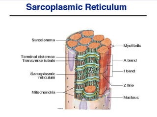
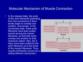
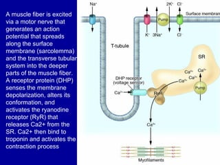
2
Structure of Skeletal
Muscle:
Skeletal Muscle
• Skeletal muscles are usually attached to bone by tendons composed
of connective tissue. This connective tissue also ensheathe the entire
muscle & is called epimysium. Skeletal muscles consist of numerous
subunits or bundles called fasicles
• Fascicles are also surrounded by connective tissue (called the
perimysium) and each fascicle is composed of numerous muscle
fibers (or muscle cells).
• Muscle cells, ensheathed by endomysium, consist of many fibrils (or
myofibrils), and these myofibrils are made up of long protein
molecules called myofilaments.
Types of Myofilaments
• Myofibrils; There are two
types of myofilaments
Actin and Myosin
Filaments.
• Each muscle fiber contains
several hundred to several
thousand myofibrils,
Each myofibril is
composed of about 1500
myosin filaments and
3000 actin filaments,
The thick filaments
are myosin, and the
thin filaments are
actin.
• myosin and actin filaments
partially interdigitate and thus
cause the myofibrils to have
alternate light and dark
bands, The light bands
contain only actin filaments
and are called I bands
because they are isotropic to
polarized light.
• The dark bands contain
myosin filaments, as well as
the ends of the actin
filaments where they overlap
the myosin, and are called A
bands because they are
anisotropic to polarized light.
• The portion of the myofibril
(or of the whole muscle fiber)
that lies between two
successive Z discs is called
a sarcomere.
Electron micrograph of muscle myofibrils showing the detailed organization of actin and
myosin filaments. Note the mitochondria lying between the myofibrils.
Titin Filamentous Molecules.
• The side-by-side relationship between the
myosin and actin filaments is achieved by
a large number of filamentous molecules
of a protein called titin. Also, because it is
filamentous, it is very springy. These
springy titin molecules act as a framework
that holds the myosin and actin filaments
in place
Sarcoplasm.
• Myofibrils of each muscle fiber are
suspended side by side in the muscle
fiber. The spaces between the myofibrils
are filled with intracellular fluid called
sarcoplasm, containing large quantities of
potassium, magnesium, and phosphate,
plus multiple protein enzymes.
• mitochondria that lie parallel to the
myofibrils, supply large amounts of energy
in the form of adenosine triphosphate
(ATP.
• Sarcoplasmic Reticulum. in the sarcoplasm
surrounding the myofibrils of each muscle fiber
is an extensive reticulum the sarcoplasmic
reticulum. This reticulum has a special
organization that is extremely important in
controlling muscle contraction.
• The very rapidly contracting types of muscle
fibers have extensive sarcoplasmic reticula.
General Mechanism of Muscle Contraction
• The initiation and execution of muscle
contraction occur in the following sequential
steps.
• An action potential travels along a motor nerve
to its endings on muscle fibers.
• At each ending, the nerve secretes a small
amount of the neurotransmitter substance
acetylcholine.
• The acetylcholine acts on a local area of the
muscle fiber membrane to open multiple
"acetylcholine-gated" channels through protein
molecules floating in the membrane.
• Opening of the acetylcholine-gated channels
allows large quantities of sodium ions to diffuse
to the interior of the muscle fiber membrane.
This initiates an action potential at the
membrane.
• The action potential travels along the muscle fiber
membrane in the same way that action potentials travel
along nerve fiber membranes.
• The action potential depolarizes the muscle membrane,
and much of the action potential electricity flows through
the center of the muscle fiber. Here it causes the
sarcoplasmic reticulum to release large quantities of
calcium ions that have been stored within this reticulum.
• The calcium ions initiate attractive forces between the
actin and myosin filaments, causing them to slide
alongside each other, which is the contractile process.
• After a fraction of a second, the calcium ions are
pumped back into the sarcoplasmic reticulum by a Ca++
membrane pump, and they remain stored in the
reticulum until a new muscle action potential comes
along; this removal of calcium ions from the myofibrils
causes the muscle contraction to cease.
Molecular Mechanism of Muscle Contraction
• In the relaxed state, the ends
of the actin filaments extending
from two successive Z discs
barely begin to overlap one
another. Conversely, in the
contracted state, these actin
filaments have been pulled
inward among the myosin
filaments, so that their ends
overlap one another to their
maximum extent. Also, the Z
discs have been pulled by the
actin filaments up to the ends
of the myosin filaments. Thus,
muscle contraction occurs by a
sliding filament mechanism.
skeletal muscle contraction.flv
A muscle fiber is excited
via a motor nerve that
generates an action
potential that spreads
along the surface
membrane (sarcolemma)
and the transverse tubular
system into the deeper
parts of the muscle fiber.
A receptor protein (DHP)
senses the membrane
depolarization, alters its
conformation, and
activates the ryanodine
receptor (RyR) that
releases Ca2+ from the
SR. Ca2+ then bind to
troponin and activates the
contraction process
Sarcoplasmic reticulum (SR) membranes in close proximity to a T-tubule. 'RyR'
are proteins the aid in the release of calcium from the SR, 'SERCA2' are proteins
that aid in the transport of calcium into the SR

Structure of skeletal muscle

  • 2.
  • 3.
  • 4.
    Skeletal Muscle • Skeletalmuscles are usually attached to bone by tendons composed of connective tissue. This connective tissue also ensheathe the entire muscle & is called epimysium. Skeletal muscles consist of numerous subunits or bundles called fasicles • Fascicles are also surrounded by connective tissue (called the perimysium) and each fascicle is composed of numerous muscle fibers (or muscle cells). • Muscle cells, ensheathed by endomysium, consist of many fibrils (or myofibrils), and these myofibrils are made up of long protein molecules called myofilaments.
  • 5.
    Types of Myofilaments •Myofibrils; There are two types of myofilaments Actin and Myosin Filaments. • Each muscle fiber contains several hundred to several thousand myofibrils, Each myofibril is composed of about 1500 myosin filaments and 3000 actin filaments, The thick filaments are myosin, and the thin filaments are actin.
  • 6.
    • myosin andactin filaments partially interdigitate and thus cause the myofibrils to have alternate light and dark bands, The light bands contain only actin filaments and are called I bands because they are isotropic to polarized light. • The dark bands contain myosin filaments, as well as the ends of the actin filaments where they overlap the myosin, and are called A bands because they are anisotropic to polarized light. • The portion of the myofibril (or of the whole muscle fiber) that lies between two successive Z discs is called a sarcomere.
  • 7.
    Electron micrograph ofmuscle myofibrils showing the detailed organization of actin and myosin filaments. Note the mitochondria lying between the myofibrils.
  • 8.
    Titin Filamentous Molecules. •The side-by-side relationship between the myosin and actin filaments is achieved by a large number of filamentous molecules of a protein called titin. Also, because it is filamentous, it is very springy. These springy titin molecules act as a framework that holds the myosin and actin filaments in place
  • 9.
    Sarcoplasm. • Myofibrils ofeach muscle fiber are suspended side by side in the muscle fiber. The spaces between the myofibrils are filled with intracellular fluid called sarcoplasm, containing large quantities of potassium, magnesium, and phosphate, plus multiple protein enzymes. • mitochondria that lie parallel to the myofibrils, supply large amounts of energy in the form of adenosine triphosphate (ATP.
  • 11.
    • Sarcoplasmic Reticulum.in the sarcoplasm surrounding the myofibrils of each muscle fiber is an extensive reticulum the sarcoplasmic reticulum. This reticulum has a special organization that is extremely important in controlling muscle contraction. • The very rapidly contracting types of muscle fibers have extensive sarcoplasmic reticula.
  • 14.
    General Mechanism ofMuscle Contraction • The initiation and execution of muscle contraction occur in the following sequential steps. • An action potential travels along a motor nerve to its endings on muscle fibers. • At each ending, the nerve secretes a small amount of the neurotransmitter substance acetylcholine. • The acetylcholine acts on a local area of the muscle fiber membrane to open multiple "acetylcholine-gated" channels through protein molecules floating in the membrane. • Opening of the acetylcholine-gated channels allows large quantities of sodium ions to diffuse to the interior of the muscle fiber membrane. This initiates an action potential at the membrane.
  • 15.
    • The actionpotential travels along the muscle fiber membrane in the same way that action potentials travel along nerve fiber membranes. • The action potential depolarizes the muscle membrane, and much of the action potential electricity flows through the center of the muscle fiber. Here it causes the sarcoplasmic reticulum to release large quantities of calcium ions that have been stored within this reticulum. • The calcium ions initiate attractive forces between the actin and myosin filaments, causing them to slide alongside each other, which is the contractile process. • After a fraction of a second, the calcium ions are pumped back into the sarcoplasmic reticulum by a Ca++ membrane pump, and they remain stored in the reticulum until a new muscle action potential comes along; this removal of calcium ions from the myofibrils causes the muscle contraction to cease.
  • 16.
    Molecular Mechanism ofMuscle Contraction • In the relaxed state, the ends of the actin filaments extending from two successive Z discs barely begin to overlap one another. Conversely, in the contracted state, these actin filaments have been pulled inward among the myosin filaments, so that their ends overlap one another to their maximum extent. Also, the Z discs have been pulled by the actin filaments up to the ends of the myosin filaments. Thus, muscle contraction occurs by a sliding filament mechanism.
  • 17.
  • 18.
    A muscle fiberis excited via a motor nerve that generates an action potential that spreads along the surface membrane (sarcolemma) and the transverse tubular system into the deeper parts of the muscle fiber. A receptor protein (DHP) senses the membrane depolarization, alters its conformation, and activates the ryanodine receptor (RyR) that releases Ca2+ from the SR. Ca2+ then bind to troponin and activates the contraction process
  • 19.
    Sarcoplasmic reticulum (SR)membranes in close proximity to a T-tubule. 'RyR' are proteins the aid in the release of calcium from the SR, 'SERCA2' are proteins that aid in the transport of calcium into the SR