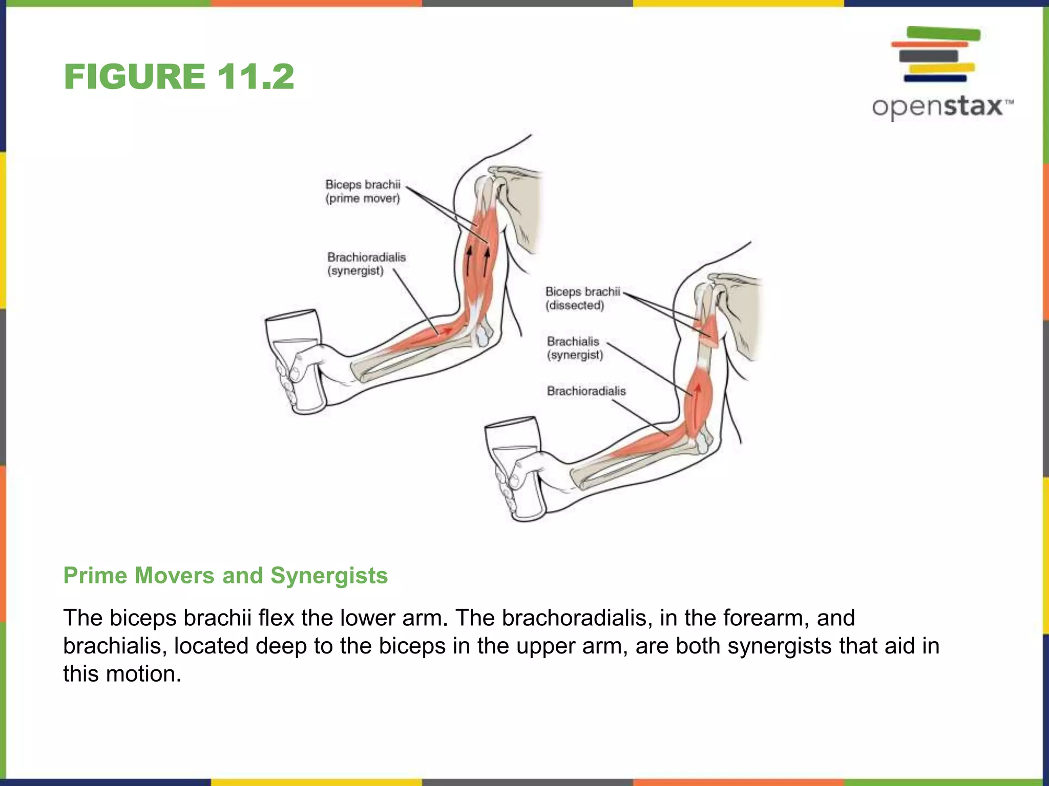
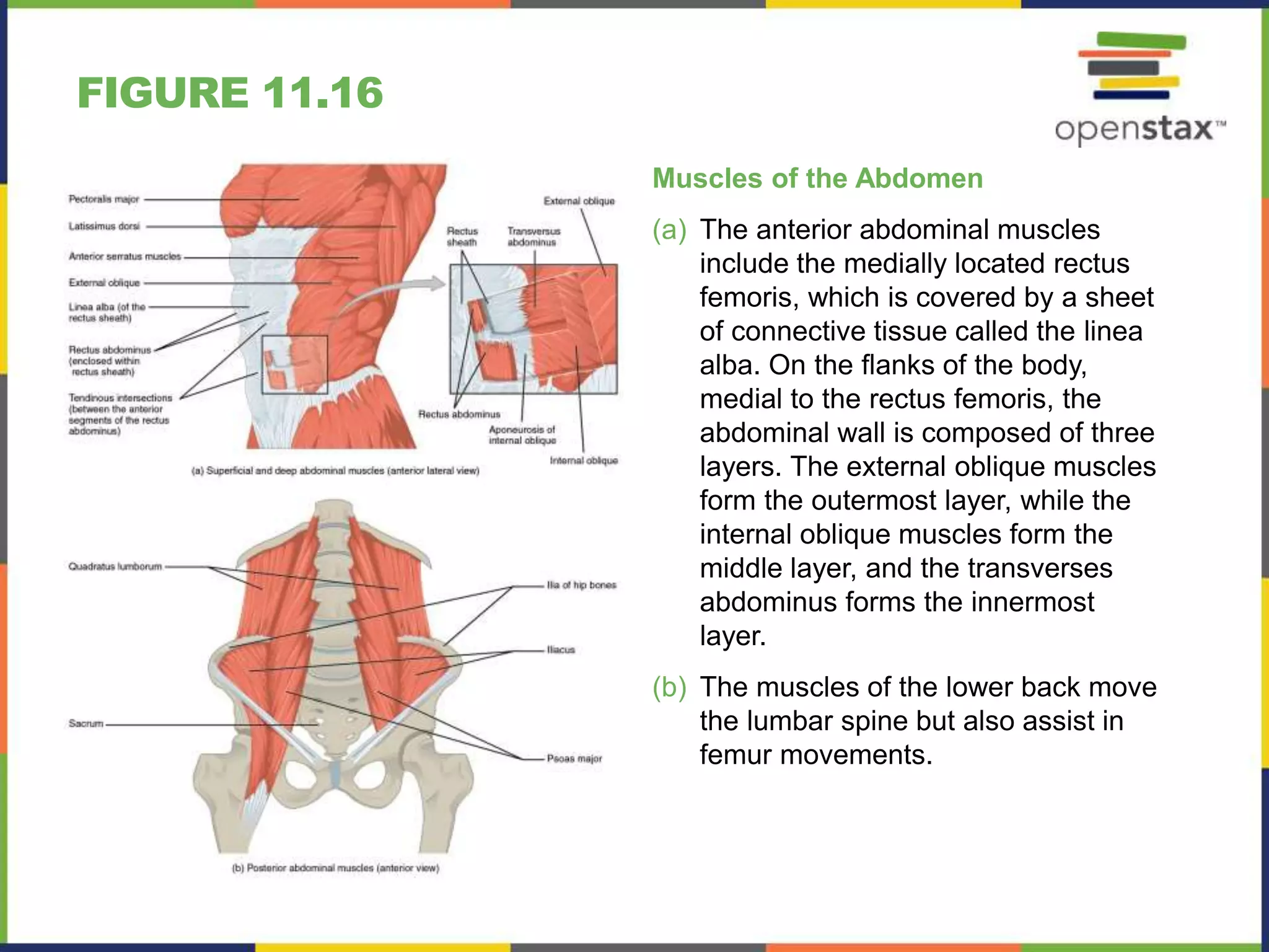
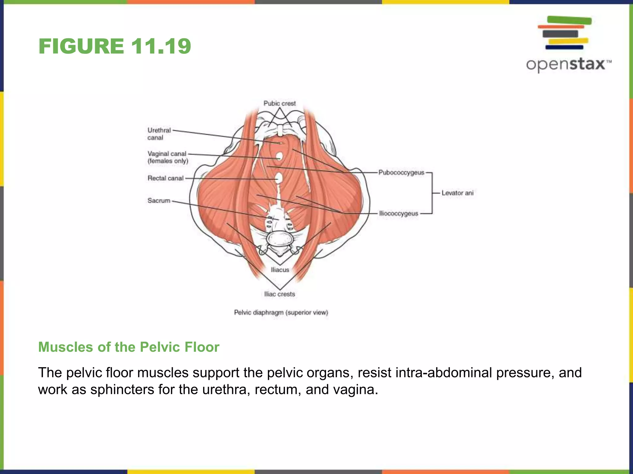
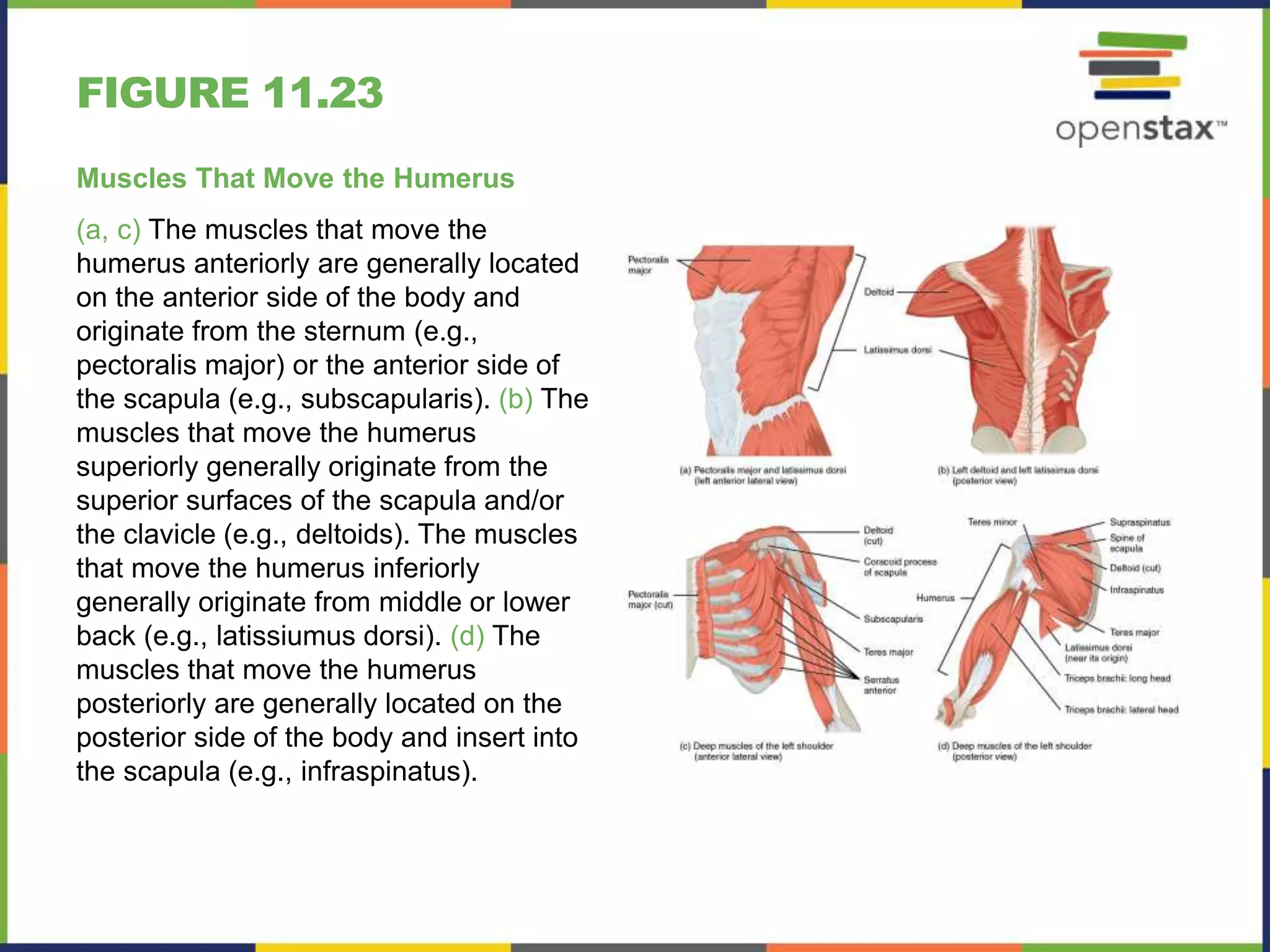
ANATOMY & PHYSIOLOGY
Chapter 11 THE MUSCULAR SYSTEM
PowerPoint Image Slideshow
FIGURE 11.1
A Body in Motion
The muscular system allows us to move, flex and contort our bodies. Practicing yoga,
as pictured here, is a good example of the voluntary use of the muscular system.
(credit: Dmitry Yanchylenko)
FIGURE 11.2
Prime Movers and Synergists
The biceps brachii flex the lower arm. The brachoradialis, in the forearm, and
brachialis, located deep to the biceps in the upper arm, are both synergists that aid in
this motion.
FIGURE 11.3
Muscle Shapes and Fiber Alignment
The skeletal muscles of the body typically come in seven different general shapes.
FIGURE 11.4
Biceps Brachii Muscle Contraction
The large mass at the center of a muscle is called the belly. Tendons emerge from both ends
of the belly and connect the muscle to the bones, allowing the skeleton to move. The tendons
of the bicep connect to the upper arm and the forearm. (credit: Victoria Garcia)
FIGURE 11.5
Overview of the Muscular System
On the anterior and posterior views of
the muscular system above, superficial
muscles (those at the surface) are shown
on the right side of the body while deep
muscles (those underneath the
superficial muscles) are shown on the left
half of the body. For the legs, superficial
muscles are shown in the anterior view
while the posterior view shows both
superficial and deep muscles.
FIGURE 11.6
Understanding a Muscle Name from the Latin
FIGURE 11.7
Muscles of Facial Expression
Many of the muscles of facial expression insert into the skin surrounding the eyelids,
nose and mouth, producing facial expressions by moving the skin rather than bones.
FIGURE 11.8
Muscles in Facial Expression
FIGURE 11.9
Muscles of the Eyes
(a) The extrinsic eye muscles originate outside of the eye on the skull.
(b) Each muscle inserts onto the eyeball.
FIGURE 11.10
Muscles That Move the Lower Jaw
The muscles that move the lower jaw are typically located within the cheek and
originate from processes in the skull. This provides the jaw muscles with the large
amount of leverage needed for chewing.
FIGURE 11.11
Muscles that Move the Tongue
FIGURE 11.12
Muscles for Tongue Movement,
Swallowing, and Speech
FIGURE 11.13
Muscles of the Anterior Neck
The anterior muscles of the neck facilitate swallowing and speech. The suprahyoid
muscles originate from above the hyoid bone in the chin region. The infrahyoid muscles
originate below the hyoid bone in the lower neck.
FIGURE 11.14
Posterior and Lateral Views of the Neck
The superficial and deep muscles of the neck are responsible for moving the head,
cervical vertebrae, and scapulas.
FIGURE 11.15
Muscles of the Neck and Back
The large, complex muscles of the neck
and back move the head, shoulders, and
vertebral column.
FIGURE 11.16
Muscles of the Abdomen
(a) The anterior abdominal muscles
include the medially located rectus
femoris, which is covered by a sheet
of connective tissue called the linea
alba. On the flanks of the body,
medial to the rectus femoris, the
abdominal wall is composed of three
layers. The external oblique muscles
form the outermost layer, while the
internal oblique muscles form the
middle layer, and the transverses
abdominus forms the innermost
layer.
(b) The muscles of the lower back move
the lumbar spine but also assist in
femur movements.
FIGURE 11.17
Muscles of the Diaphragm
The diaphragm separates the thoracic and abdominal cavities.
FIGURE 11.18
Intercostal Muscles
The external intercostals are located laterally on the sides of the body. The internal
intercostals are located medially near the sternum. The innermost intercostals are
located deep to both the internal and external intercostals.
FIGURE 11.19
Muscles of the Pelvic Floor
The pelvic floor muscles support the pelvic organs, resist intra-abdominal pressure, and
work as sphincters for the urethra, rectum, and vagina.
FIGURE 11.20
Muscles of the Perineum
The perineum muscles play roles in urination in both sexes, ejaculation in men, and
vaginal contraction in women.
FIGURE 11.21
Muscles of the Perineum Common to Men and Women
FIGURE 11.22
Muscles That Position the Pectoral Girdle
The muscles that stabilize the pectoral girdle make it a steady base on which other
muscles can move the arm. Note that the pectoralis major and deltoid, which move the
humerus, are cut here to show the deeper positioning muscles.
FIGURE 11.23
Muscles That Move the Humerus
(a, c) The muscles that move the
humerus anteriorly are generally located
on the anterior side of the body and
originate from the sternum (e.g.,
pectoralis major) or the anterior side of
the scapula (e.g., subscapularis). (b) The
muscles that move the humerus
superiorly generally originate from the
superior surfaces of the scapula and/or
the clavicle (e.g., deltoids). The muscles
that move the humerus inferiorly
generally originate from middle or lower
back (e.g., latissiumus dorsi). (d) The
muscles that move the humerus
posteriorly are generally located on the
posterior side of the body and insert into
the scapula (e.g., infraspinatus).
FIGURE 11.24
Muscles That Move the Humerus
FIGURE 11.25
Muscles That Move the Forearm
The muscles originating in the upper arm
flex, extend, pronate, and supinate the
forearm. The muscles originating in the
forearm move the wrists, hands, and
fingers.
FIGURE 11.26
Muscles That Move the Forearm
FIGURE 11.27
Muscles That Move the Wrist, Hands,
and Forearm
FIGURE 11.28
Intrinsic Muscles of the Hand
The intrinsic muscles of the hand both originate and insert within the hand. These
muscles provide the fine motor control of the fingers by flexing, extending, abducting,
and adducting the more distal finger and thumb segments.
FIGURE 11.29
Hip and Thigh Muscles
The large and powerful muscles of the
hip that move the femur generally
originate on the pelvic girdle and insert
into the femur. The muscles that move
the lower leg typically originate on the
femur and insert into the bones of the
knee joint. The anterior muscles of the
femur extend the lower leg but also aid in
flexing the thigh. The posterior muscles
of the femur flex the lower leg but also
aid in extending the thigh. A combination
of gluteal and thigh muscles also adduct,
abduct, and rotate the thigh and lower
leg.
FIGURE 11.30
Gluteal Region Muscles That Move the
Femur
FIGURE 11.31
Thigh Muscles That Move the Femur,
Tibia, and Fibula
FIGURE 11.32
Muscles of the Lower Leg
The muscles of the anterior compartment of the lower leg are generally responsible for
dorsiflexion, and the muscles of the posterior compartment of the lower leg are generally
responsible for plantar flexion. The lateral and medial muscles in both compartments invert,
evert, and rotate the foot.
FIGURE 11.33
Muscles That Move the Feet and Toes
FIGURE 11.34
Intrinsic Muscles of the Foot
The muscles along the dorsal side of the foot (a) generally extend the toes while the muscles of the plantar side of
the foot (b, c, d) generally flex the toes. The plantar muscles exist in three layers, providing the foot the strength to
counterbalance the weight of the body. In this diagram, these three layers are shown from a plantar view beginning
with the bottom-most layer just under the plantar skin of the foot (b) and ending with the top-most layer (d) located
just inferior to the foot and toe bones.
FIGURE 11.35
Intrinsic Muscles in the Foot
This OpenStax ancillary resource is © Rice University under a CC-BY 4.0
International license; it may be reproduced or modified but must be
attributed to OpenStax, Rice University and any changes must be noted.

Open stax anatomy& physiology ch11

  • 1.
    ANATOMY & PHYSIOLOGY Chapter11 THE MUSCULAR SYSTEM PowerPoint Image Slideshow
  • 2.
    FIGURE 11.1 A Bodyin Motion The muscular system allows us to move, flex and contort our bodies. Practicing yoga, as pictured here, is a good example of the voluntary use of the muscular system. (credit: Dmitry Yanchylenko)
  • 3.
    FIGURE 11.2 Prime Moversand Synergists The biceps brachii flex the lower arm. The brachoradialis, in the forearm, and brachialis, located deep to the biceps in the upper arm, are both synergists that aid in this motion.
  • 4.
    FIGURE 11.3 Muscle Shapesand Fiber Alignment The skeletal muscles of the body typically come in seven different general shapes.
  • 5.
    FIGURE 11.4 Biceps BrachiiMuscle Contraction The large mass at the center of a muscle is called the belly. Tendons emerge from both ends of the belly and connect the muscle to the bones, allowing the skeleton to move. The tendons of the bicep connect to the upper arm and the forearm. (credit: Victoria Garcia)
  • 6.
    FIGURE 11.5 Overview ofthe Muscular System On the anterior and posterior views of the muscular system above, superficial muscles (those at the surface) are shown on the right side of the body while deep muscles (those underneath the superficial muscles) are shown on the left half of the body. For the legs, superficial muscles are shown in the anterior view while the posterior view shows both superficial and deep muscles.
  • 7.
    FIGURE 11.6 Understanding aMuscle Name from the Latin
  • 8.
    FIGURE 11.7 Muscles ofFacial Expression Many of the muscles of facial expression insert into the skin surrounding the eyelids, nose and mouth, producing facial expressions by moving the skin rather than bones.
  • 9.
    FIGURE 11.8 Muscles inFacial Expression
  • 10.
    FIGURE 11.9 Muscles ofthe Eyes (a) The extrinsic eye muscles originate outside of the eye on the skull. (b) Each muscle inserts onto the eyeball.
  • 11.
    FIGURE 11.10 Muscles ThatMove the Lower Jaw The muscles that move the lower jaw are typically located within the cheek and originate from processes in the skull. This provides the jaw muscles with the large amount of leverage needed for chewing.
  • 12.
  • 13.
    FIGURE 11.12 Muscles forTongue Movement, Swallowing, and Speech
  • 14.
    FIGURE 11.13 Muscles ofthe Anterior Neck The anterior muscles of the neck facilitate swallowing and speech. The suprahyoid muscles originate from above the hyoid bone in the chin region. The infrahyoid muscles originate below the hyoid bone in the lower neck.
  • 15.
    FIGURE 11.14 Posterior andLateral Views of the Neck The superficial and deep muscles of the neck are responsible for moving the head, cervical vertebrae, and scapulas.
  • 16.
    FIGURE 11.15 Muscles ofthe Neck and Back The large, complex muscles of the neck and back move the head, shoulders, and vertebral column.
  • 17.
    FIGURE 11.16 Muscles ofthe Abdomen (a) The anterior abdominal muscles include the medially located rectus femoris, which is covered by a sheet of connective tissue called the linea alba. On the flanks of the body, medial to the rectus femoris, the abdominal wall is composed of three layers. The external oblique muscles form the outermost layer, while the internal oblique muscles form the middle layer, and the transverses abdominus forms the innermost layer. (b) The muscles of the lower back move the lumbar spine but also assist in femur movements.
  • 18.
    FIGURE 11.17 Muscles ofthe Diaphragm The diaphragm separates the thoracic and abdominal cavities.
  • 19.
    FIGURE 11.18 Intercostal Muscles Theexternal intercostals are located laterally on the sides of the body. The internal intercostals are located medially near the sternum. The innermost intercostals are located deep to both the internal and external intercostals.
  • 20.
    FIGURE 11.19 Muscles ofthe Pelvic Floor The pelvic floor muscles support the pelvic organs, resist intra-abdominal pressure, and work as sphincters for the urethra, rectum, and vagina.
  • 21.
    FIGURE 11.20 Muscles ofthe Perineum The perineum muscles play roles in urination in both sexes, ejaculation in men, and vaginal contraction in women.
  • 22.
    FIGURE 11.21 Muscles ofthe Perineum Common to Men and Women
  • 23.
    FIGURE 11.22 Muscles ThatPosition the Pectoral Girdle The muscles that stabilize the pectoral girdle make it a steady base on which other muscles can move the arm. Note that the pectoralis major and deltoid, which move the humerus, are cut here to show the deeper positioning muscles.
  • 24.
    FIGURE 11.23 Muscles ThatMove the Humerus (a, c) The muscles that move the humerus anteriorly are generally located on the anterior side of the body and originate from the sternum (e.g., pectoralis major) or the anterior side of the scapula (e.g., subscapularis). (b) The muscles that move the humerus superiorly generally originate from the superior surfaces of the scapula and/or the clavicle (e.g., deltoids). The muscles that move the humerus inferiorly generally originate from middle or lower back (e.g., latissiumus dorsi). (d) The muscles that move the humerus posteriorly are generally located on the posterior side of the body and insert into the scapula (e.g., infraspinatus).
  • 25.
    FIGURE 11.24 Muscles ThatMove the Humerus
  • 26.
    FIGURE 11.25 Muscles ThatMove the Forearm The muscles originating in the upper arm flex, extend, pronate, and supinate the forearm. The muscles originating in the forearm move the wrists, hands, and fingers.
  • 27.
    FIGURE 11.26 Muscles ThatMove the Forearm
  • 28.
    FIGURE 11.27 Muscles ThatMove the Wrist, Hands, and Forearm
  • 29.
    FIGURE 11.28 Intrinsic Musclesof the Hand The intrinsic muscles of the hand both originate and insert within the hand. These muscles provide the fine motor control of the fingers by flexing, extending, abducting, and adducting the more distal finger and thumb segments.
  • 30.
    FIGURE 11.29 Hip andThigh Muscles The large and powerful muscles of the hip that move the femur generally originate on the pelvic girdle and insert into the femur. The muscles that move the lower leg typically originate on the femur and insert into the bones of the knee joint. The anterior muscles of the femur extend the lower leg but also aid in flexing the thigh. The posterior muscles of the femur flex the lower leg but also aid in extending the thigh. A combination of gluteal and thigh muscles also adduct, abduct, and rotate the thigh and lower leg.
  • 31.
    FIGURE 11.30 Gluteal RegionMuscles That Move the Femur
  • 32.
    FIGURE 11.31 Thigh MusclesThat Move the Femur, Tibia, and Fibula
  • 33.
    FIGURE 11.32 Muscles ofthe Lower Leg The muscles of the anterior compartment of the lower leg are generally responsible for dorsiflexion, and the muscles of the posterior compartment of the lower leg are generally responsible for plantar flexion. The lateral and medial muscles in both compartments invert, evert, and rotate the foot.
  • 34.
    FIGURE 11.33 Muscles ThatMove the Feet and Toes
  • 35.
    FIGURE 11.34 Intrinsic Musclesof the Foot The muscles along the dorsal side of the foot (a) generally extend the toes while the muscles of the plantar side of the foot (b, c, d) generally flex the toes. The plantar muscles exist in three layers, providing the foot the strength to counterbalance the weight of the body. In this diagram, these three layers are shown from a plantar view beginning with the bottom-most layer just under the plantar skin of the foot (b) and ending with the top-most layer (d) located just inferior to the foot and toe bones.
  • 36.
  • 37.
    This OpenStax ancillaryresource is © Rice University under a CC-BY 4.0 International license; it may be reproduced or modified but must be attributed to OpenStax, Rice University and any changes must be noted.