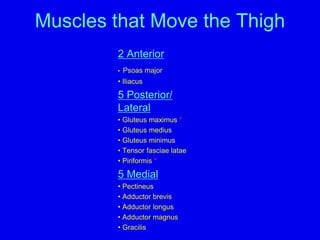
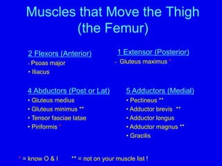
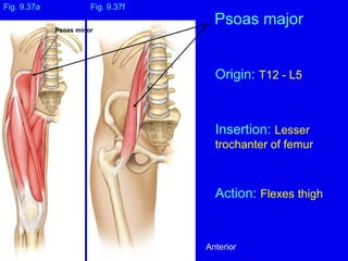
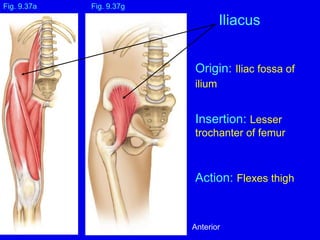
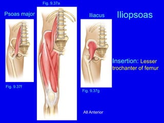
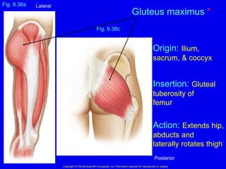
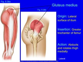
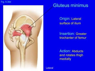
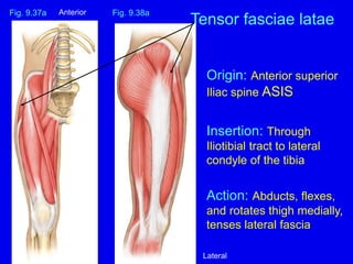
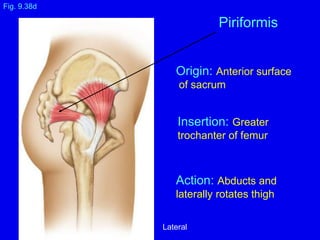
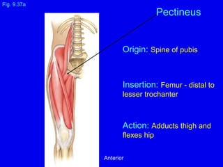
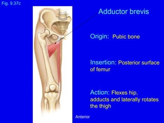
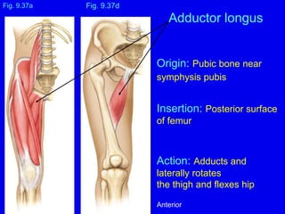
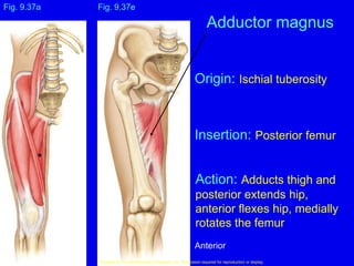
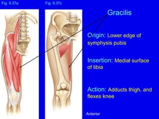
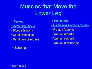
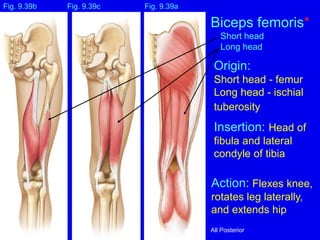
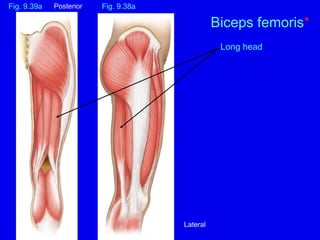
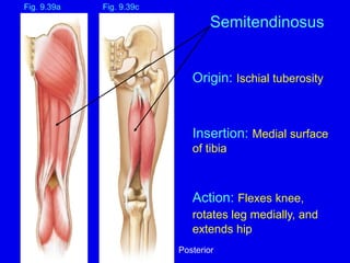
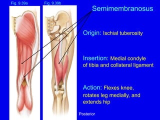
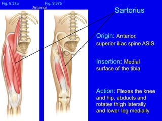
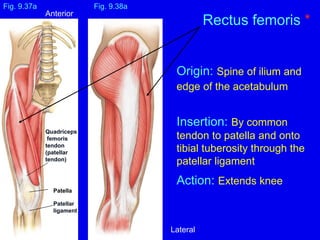
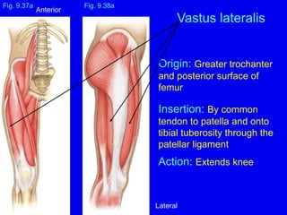
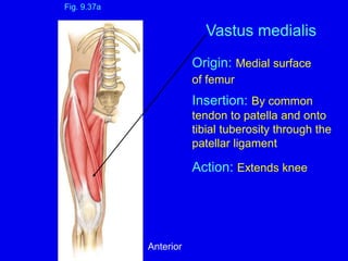
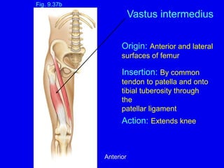
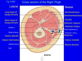
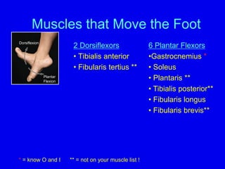
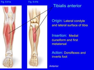
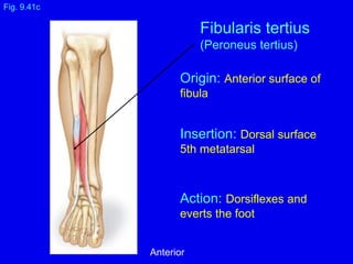
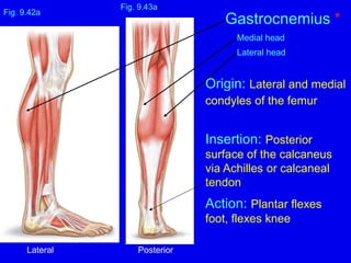
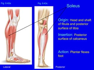
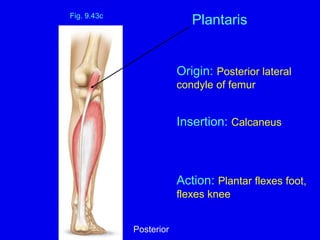
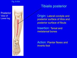
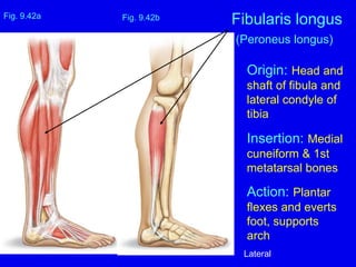
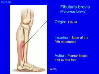
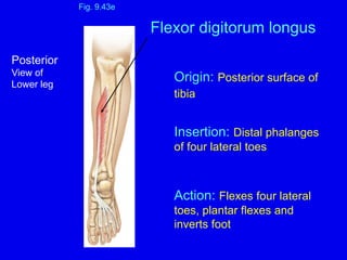
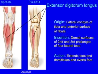
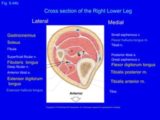
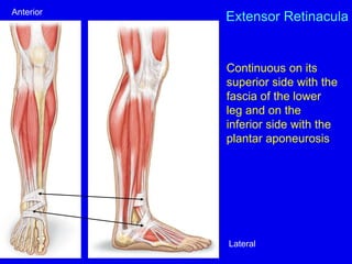
7. Fig. 9.38c
Copyright ยฉ The McGraw-Hill Companies, Inc. Permission required for reproduction or display.
Gluteus maximus *
Fig. 9.38a
Origin: Ilium,
sacrum, & coccyx
Insertion: Gluteal
tuberosity of
femur
Action: Extends hip,
abducts and
laterally rotates thigh
Lateral
Posterior
8. Fig. 9.38b
Gluteus medius
Copyright ยฉ The McGraw-Hill Companies, Inc. Permission required for reproduction or display.
Fig. 9.38a
Origin: Lateral
surface of ilium
Insertion: Greater
trochanter of femur
Action: Abducts
and rotates thigh
medially
Lateral
9. Fig. 9.38d
Gluteus minimus
Origin: Lateral
surface of ilium
Insertion: Greater
trochanter of femur
Action: Abducts
and rotates thigh
medially
Lateral
10. Origin: Anterior superior
Iliac spine ASIS
Insertion: Through
Iliotibial tract to lateral
condyle of the tibia
Action: Abducts, flexes,
and rotates thigh medially,
tenses lateral fascia
Fig. 9.37a
Tensor fasciae latae
Fig. 9.38a
Anterior
Lateral
11. Origin: Anterior surface
of sacrum
Insertion: Greater
trochanter of femur
Action: Abducts and
laterally rotates thigh
Fig. 9.38d
Piriformis
Lateral
12. Origin: Spine of pubis
Insertion: Femur - distal to
lesser trochanter
Action: Adducts thigh and
flexes hip
Fig. 9.37a
Pectineus
Anterior
13. Origin: Pubic bone
Insertion: Posterior surface
of femur
Action: Flexes hip,
adducts and laterally rotates
the thigh
Fig. 9.37c
Adductor brevis
Anterior
14. Origin: Pubic bone near
symphysis pubis
Insertion: Posterior surface
of femur
Action: Adducts and
laterally rotates
the thigh and flexes hip
Fig. 9.37a
Adductor longus
Fig. 9.37d
Anterior
15. Fig. 9.37e
Copyright ยฉ The McGraw-Hill Companies, Inc. Permission required for reproduction or display.
Adductor magnus
Fig. 9.37a
Origin: Ischial tuberosity
Insertion: Posterior femur
Action: Adducts thigh and
posterior extends hip,
anterior flexes hip, medially
rotates the femur
Anterior
16. Origin: Lower edge of
symphysis pubis
Insertion: Medial surface
of tibia
Action: Adducts thigh, and
flexes knee
Fig. 9.37a
Gracilis
Fig. 9.37c
Anterior
17. Muscles that Move the
Lower Leg
4 Flexors
Hamstring Group
โข Biceps femoris *
โข Semitendinosus
โข Semimembranosus
4 Extensors
Quadriceps Femoris Group
โข Rectus femoris *
โข Vastus lateralis
โข Vastus medialis
โข Vastus intermedius
โข Sartorius
* = know O and I
18. Origin:
Short head - femur
Long head - ischial
tuberosity
Insertion: Head of
fibula and lateral
condyle of tibia
Action: Flexes knee,
rotates leg laterally,
and extends hip
Fig. 9.39c
Biceps femoris*
Short head
Long head
Fig. 9.39a
Fig. 9.39b
All Posterior
20. Origin: Ischial tuberosity
Insertion: Medial surface
of tibia
Action: Flexes knee,
rotates leg medially, and
extends hip
Fig. 9.39c
Fig. 9.39a
Semitendinosus
Posterior
21. Origin: Ischial tuberosity
Insertion: Medial condyle
of tibia and collateral ligament
Action: Flexes knee,
rotates leg medially, and
extends hip
Fig. 9.39b
Semimembranosus
Fig. 9.39a
Posterior
22. Origin: Anterior,
superior iliac spine ASIS
Insertion: Medial
surface of the tibia
Action: Flexes the knee
and hip, abducts and
rotates thigh laterally
and lower leg medially
Fig. 9.37a
Sartorius
Fig. 9.37b
Anterior
23. Origin: Spine of ilium and
edge of the acetabulum
Insertion: By common
tendon to patella and onto
tibial tuberosity through the
patellar ligament
Action: Extends knee
Fig. 9.37a
Rectus femoris *
Fig. 9.38a
Patella
Patellar
ligament
Quadriceps
femoris
tendon
(patellar
tendon)
Anterior
Lateral
24. Origin: Greater trochanter
and posterior surface of
femur
Insertion: By common
tendon to patella and onto
tibial tuberosity through the
patellar ligament
Action: Extends knee
Fig. 9.37a Fig. 9.38a
Vastus lateralis
Anterior
Lateral
25. Origin: Medial surface
of femur
Insertion: By common
tendon to patella and onto
tibial tuberosity through the
patellar ligament
Action: Extends knee
Fig. 9.37a
Vastus medialis
Anterior
26. Fig. 9.37b
Vastus intermedius
Origin: Anterior and lateral
surfaces of femur
Insertion: By common
tendon to patella and onto
tibial tuberosity through
the
patellar ligament
Action: Extends knee
Anterior
27. Fig. 9.40b
Gracilis
Semimembranosus
Semitendinosus
Sartorius
Vastus medialis
Rectus femoris
Adipose tissue
Skin
Adductor magnus
Adductor longus
Great saphenous v.
Femoral v. and a.
Vastus intermedius
Shaft of femur
Sciatic n.
Vastus lateralis
Lateral Medial
Anterior
Long head of
biceps femoris
Short head of
biceps femoris
Cross section of the Right Thigh
28. Muscles that Move the Foot
2 Dorsiflexors
โข Tibialis anterior
โข Fibularis tertius **
Dorsiflexion
Plantar
Flexion
6 Plantar Flexors
โขGastrocnemius *
โข Soleus
โข Plantaris **
โข Tibialis posterior**
โข Fibularis longus
โข Fibularis brevis**
* = know O and I ** = not on your muscle list !
29. Origin: Lateral condyle
and lateral surface of tibia
Insertion: Medial
cuneiform and first
metatarsal
Action: Dorsiflexes and
inverts foot
Fig. 9.41a
Tibialis anterior
Fig. 9.41b
Anterior
30. Origin: Anterior surface of
fibula
Insertion: Dorsal surface
5th metatarsal
Action: Dorsiflexes and
everts the foot
Fig. 9.41c
Fibularis tertius
(Peroneus tertius)
Anterior
31. Origin: Lateral and medial
condyles of the femur
Insertion: Posterior
surface of the calcaneus
via Achilles or calcaneal
tendon
Action: Plantar flexes
foot, flexes knee
Fig. 9.43a
Gastrocnemius *
Medial head
Lateral head
Fig. 9.42a
Lateral Posterior
32. Origin: Head and shaft
of fibula and posterior
surface of tibia
Insertion: Posterior
surface of calcaneus
Action: Plantar flexes
foot
Fig. 9.43c
Soleus
Fig. 9.42a
Lateral Posterior
34. Origin: Lateral condyle and
posterior surface of tibia and
posterior surface of fibula
Insertion: Tarsal and
metatarsal bones
Action: Plantar flexes and
inverts foot
Fig. 9.43d
Tibialis posterior
Posterior
View of
Lower leg
35. Origin: Head and
shaft of fibula and
lateral condyle of
tibia
Insertion: Medial
cuneiform & 1st
metatarsal bones
Action: Plantar
flexes and everts
foot, supports
arch
Fig. 9.42a
Fibularis longus
(Peroneus longus)
Fig. 9.42b
Lateral
36. Origin: Fibula
Insertion: Base of the
fifth metatarsal
Action: Plantar flexes
and everts foot
Fig. 9.42c
Fibularis brevis
(Peroneus brevis)
Lateral
37. Muscles that Move the Toes
โข Flexor digitorum longus
โข Extensor digitorum longus
38. Origin: Posterior surface of
tibia
Insertion: Distal phalanges
of four lateral toes
Action: Flexes four lateral
toes, plantar flexes and
inverts foot
Fig. 9.43e
Flexor digitorum longus
Posterior
View of
Lower leg
39. Origin: Lateral condyle of
tibia and anterior surface
of fibula
Insertion: Dorsal surfaces
of 2nd and 3rd phalanges
of four lateral toes
Action: Extends toes and
dorsiflexes and everts foot
Fig. 9.41a
Extensor digitorum longus
Fig. 9.41d
Anterior
40. Fig. 9.44b
Copyright ยฉ The McGraw-Hill Companies, Inc. Permission required for reproduction or display.
Gastrocnemius
Fibularis longus
Deep fibular n.
Superficial fibular n.
Anterior tibial a.
Extensor digitorum
longus
Extensor hallucis longus
Fibula
Soleus
Tibia
Great saphenous v.
Tibialis posterior m.
Flexor digitorum longus
Small saphenous v.
Posterior tibial a.
Flexor hallucis longus m.
Tibial n.
Tibialis anterior m.
Anterior
Cross section of the Right Lower Leg
H
H
Lateral Medial
41. Extensor Retinacula
Continuous on its
superior side with the
fascia of the lower
leg and on the
inferior side with the
plantar aponeurosis
Anterior
Lateral