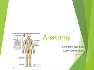
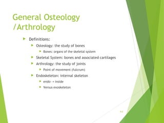
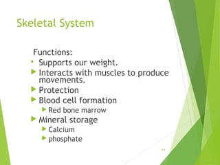
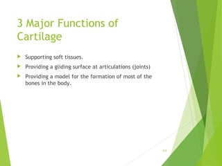
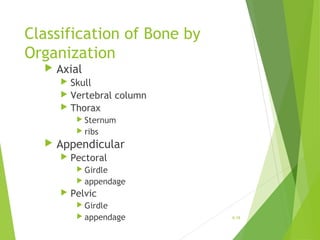
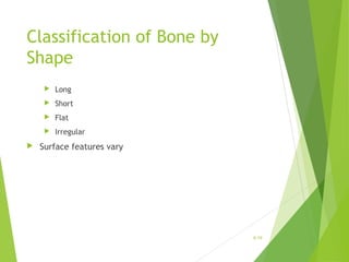
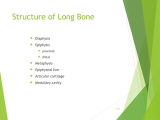
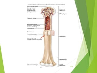
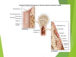
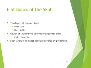
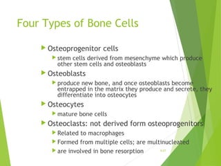
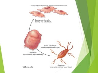
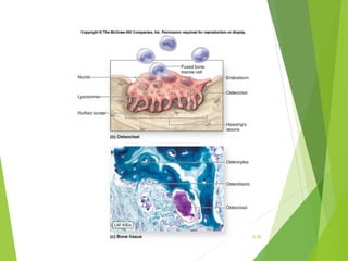
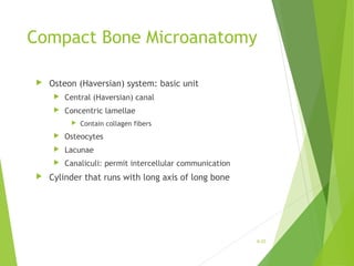
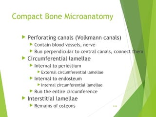
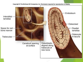
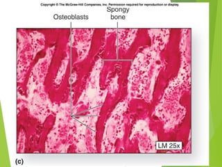
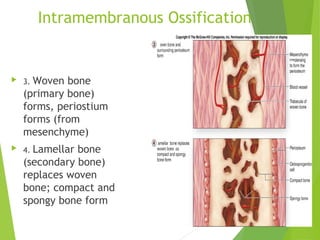
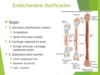
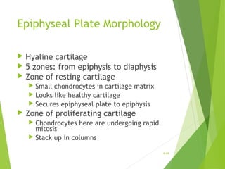
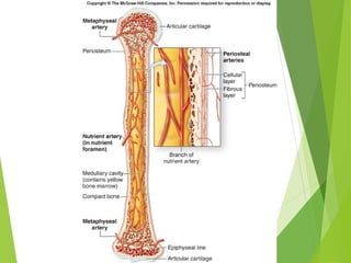
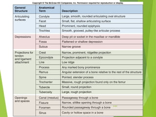
Human Anatomy
Cartilage and Bone
Connective Tissue
Pavemedicine.com
1
General Osteology
/Arthrology
 Definitions:
 Osteology: the study of bones
 Bones: organs of the skeletal system
 Skeletal System: bones and associated cartilages
 Arthrology: the study of joints
 Point of movement (fulcrum)
 Endoskeleton: internal skeleton
 endo- = inside
 Versus exoskeleton
6-2
Skeletal System
 Composed of dynamic living tissues
 Osseous tissue, cartilage, fibrous CT, blood, nervous
tissue.
 Continually rebuilds and remodels itself
 Changes over a lifetime
 Interacts with all of the other organ systems.
 Includes:
 bones of the skeleton
 Cartilage
 Ligaments
 other connective tissues that stabilize or connect the
bones. 6-3
Skeletal System
Functions:
 Supports our weight.
 Interacts with muscles to produce
movements.
 Protection
 Blood cell formation
 Red bone marrow
 Mineral storage
 Calcium
 phosphate
6-4
Cartilage Connective Tissue
Characteristics:
 Weaker than bone
 More flexible than bone
 Cells in an abundant matrix.
 Cell Types
 Chondroblasts
 Chondrocytes in lacunae
 Avascular
6-5
3 Major Functions of
Cartilage
 Supporting soft tissues.
 Providing a gliding surface at articulations (joints)
 Providing a model for the formation of most of the
bones in the body.
6-6
Types of Cartilage
 Three types of cartilage:
 Hyaline cartilage
 Most abundant kind
 Has a perichondrium (membrane)
 Associated with synovial joints
 Most bones first modeled in hyaline cartilage
 Fibrocartilage
 Has collagen fibers
 Intervertebral discs, pubic symphysis
 Elastic cartilage
 Has elastic fibers
 Ear, respiratory tubing
6-7
6-8
Growth Patterns of Cartilage
 Two main types:
 Interstitial Growth
 Appositional Growth.
 Interstitial Growth.
 Chondrocytes in lacuna undergoes mitosis.
 Two chondrocytes in one lacuna
 Will push apart, form separate lacuna
6-9
6-10
Growth Patterns of Cartilage
 Appositional Growth.
 Undifferentiated cells divide (mitosis)
 One daughter cell remains a stem cell, one
differentiates into a committed cell.
 Committed cell further differentiates into
chondroblast
 Located at edge of cartilage
 Both types common during growth
 Later, mostly appositional
 In adult, usually no growth unless for repair
6-11
Bone
 Bones are organs
 Bones are composed of all tissue types.
 Their primary component is osseous connective tissue.
 The matrix is sturdy and rigid due to calcification (also called
mineralization).
6-12
Functions of Bone
 Support.
 Protection.
 Movement
 Hemopoiesis
 Storage of minerals.
 Energy Reserves (marrow)
6-13
Support and Protection
 Bones provide structural support and serve as a
framework for the entire body.
 Bones protect many delicate tissues and organs from
injury and trauma.
6-14
Movement
 Muscles attach to the bones of the
skeleton
 contract and pull on bone
 functions as a series of levers.
6-15
Hemopoiesis
 Blood cell production in red bone marrow
 located in some spongy bone.
 Red bone marrow contains stem cells
 form all of the blood cell types.
6-16
Storage of Mineral and Energy
Reserves
 More than 90% of the body’s reserves
of the minerals calcium and phosphate
are stored and released by bone.
 Calcium: needed for
 muscle contraction
 blood clotting
 nerve impulse transmission.
 Phosphate: needed for
 ATP utilization
 structure of nucleic acids (DNA, RNA)
6-17
Classification of Bone by
Organization
 Axial
 Skull
 Vertebral column
 Thorax
 Sternum
 ribs
 Appendicular
 Pectoral
 Girdle
 appendage
 Pelvic
 Girdle
 appendage 6-18
Classification of Bone by
Shape
 Long
 Short
 Flat
 Irregular
 Surface features vary
6-19
6-20
Structure of Long Bone
 Diaphysis
 Epiphysis
 proximal
 distal
 Metaphysis
 Epiphyseal line
 Articular cartilage
 Medullary cavity
6-21
6-22
Structure of Long Bone
 Endostium: lines marrow cavity, incomplete
 Osteoprogenitor cells
 Osteoblasts
 Osteoclasts
 Periostium: covers bone everywhere but articular
surfaces
 Two layers
 Fibrous layer: outermost, dense irregular CT
 Site of tendon attachment
 Inner layer: next to compact bone
 Osteoblasts present in young bone
 Anchored to bone by perforating fibers (collagen)
6-23
6-24
Flat Bones of the Skull
 Two layers of compact bone
 Inner table
 Outer table
 Region of spongy bone sandwiched between them
 Called the diploe
 Both layers of compact bone are covered by periosteum
6-25
6-26
Four Types of Bone Cells
 Osteoprogenitor cells
 stem cells derived from mesenchyme which produce
other stem cells and osteoblasts
 Osteoblasts
 produce new bone, and once osteoblasts become
entrapped in the matrix they produce and secrete, they
differentiate into osteocytes
 Osteocytes
 mature bone cells
 Osteoclasts: not derived form osteoprogenitors
 Related to macrophages
 Formed from multiple cells; are multinucleated
 are involved in bone resorption 6-27
6-28
Osteoclasts
 Located in Howship’s lacuna
 Ruffled edge contacts bone
 Secrete hydrochloric acid
 Dissolves minerals
 osteolysis
 Lysosomes
 Secrete enzymes that dissolve matrix
6-29
6-30
Composition of Bone Matrix
 Organic components: one third
 Cells
 Collagen fibers
 Ground substance
 Inorganic components: two thirds
 Calcium phosphate
 Hydroxyapatite crystals: calcium phosphate and calcium
hydroxide
6-31
Types of Osseous Tissue
 Compact
 Dense, cortical
 Spongy
 Cancellous, trabecular
6-32
Compact Bone Microanatomy
 Osteon (Haversian) system: basic unit
 Central (Haversian) canal
 Concentric lamellae
 Contain collagen fibers
 Osteocytes
 Lacunae
 Canaliculi: permit intercellular communication
 Cylinder that runs with long axis of long bone
6-33
Compact Bone Microanatomy
 Perforating canals (Volkmann canals)
 Contain blood vessels, nerve
 Run perpendicular to central canals, connect them
 Circumferential lamellae
 Internal to periostium
 External circumferential lamellae
 Internal to endosteum
 Internal circumferential lamellae
 Run the entire circumference
 Interstitial lamellae
 Remains of osteons 6-34
6-35
6-36
6-37
Spongy Bone Microanatomy
 No osteons
 In trabeculae:
 Parallel lamellae
 Osteocytes in lacunae
 canaliculi
6-38
6-39
6-40
Ossification
 Osteogenesis: bone formation and
development
 Begins in the embryo: By the eighth through
twelfth weeks:
 the skeleton begins forming:
 from mesenchyme
 or from a hyaline cartilage model of bone.
 These models are replaced by hard bone
 Continues during childhood and adolescence.
 In the adult, ossification continues.6-41
Intramembranous Ossification
 Also called dermal ossification
 Produces:
 the flat bones of the skull (cranial vault)
 some of the facial bones (zygomatic bone,
maxilla), the mandible (lower jaw)
 the central part of the clavicle
(collarbone).
 It begins when mesenchyme becomes
thickened and condensed with a dense
supply of blood capillaries.
6-42
Intramembranous Ossification
 1. Ossification centers
form in thickened
mesenchyme
 Osteoprogenitors
develop, become
osteoblasts
 2. Osteoid (bone matrix)
calcifies
 Trapped osteoblasts
become osteocytes
6-43
Intramembranous Ossification
 3. Woven bone
(primary bone)
forms, periostium
forms (from
mesenchyme)
 4. Lamellar bone
(secondary bone)
replaces woven
bone; compact and
spongy bone form
6-44
Endochondral Ossification
 Begins with a hyaline cartilage model
 Produces most of the other bones of the skeleton
 Long bone will be used as an example.
6-45
Endochondral Ossification
 Steps:
 1. Cartilage model develops:
 Chondroblasts become
chondrocytes
 Perichondrium develops
 2. Cartilage calcification, bone collar
develops in shaft
 Chondrocytes hypertrophy, then
die
 Blood vessels grow toward cartilage
 Osteoblasts under perichondrium
form bone
 3. Primary Ossification center forms:
 Periosteal bud: osteoblasts and
blood vessels
 12th
week: most have formed 6-46
Endochondral Ossification
 Steps:
 3. Secondary Ossification centers:
 In epiphysis
 Some form post-natally
 4. Cartilage replaced by bone
 Except articular cartilage,
epiphyseal plate
 5. Epiphyseal plate ossifies:
 Forms epiphyseal line
 Between 10 and 25
 Last… clavicle 6-47
Epiphyseal Plate Morphology
 Hyaline cartilage
 5 zones: from epiphysis to diaphysis
 Zone of resting cartilage
 Small chondrocytes in cartilage matrix
 Looks like healthy cartilage
 Secures epiphyseal plate to epiphysis
 Zone of proliferating cartilage
 Chondrocytes here are undergoing rapid
mitosis
 Stack up in columns
6-48
Epiphyseal Plate Morphology
 Zone of hypertrophic cartilage
 Chondrocytes stop dividing
 Start hypertrophy
 Absorb matrix
 Zone of calcified cartilage
 Few cells thick
 Calcification of matrix
 Kills the chondrocytes
 Zone of ossification
 Invasion by capillaries and osteoprogenitor
cells
6-49
6-50
Bone Growth
 Interstitial growth occurs in the epiphyseal plate as
chondrocytes undergo mitosis
 Growth in length
 Appositional growth occurs within the periosteum.
 Growth in diameter, thickness
6-51
Bone Remodeling
 The continual deposition of new bone tissue and the
removal (resorption) of old bone tissue.
 helps maintain calcium and phosphate levels in body
fluids, and can be stimulated by stress on a bone
 occurs at both the periosteal and endosteal surfaces of a
bone
 Relative rates differ with age, bone
6-52
Blood Supply and Innervation
 Bone is highly vascularized, especially in regions containing red bone
marrow.
 Kinds of blood vessels
 Nutrient artery and the nutrient vein
 supply the diaphysis of a long bone
 Metaphyseal blood vessels
 Diaphyseal face of epiphyseal plate
 Periosteal blood vessels
 Supply superficial osteons on diaphysis.
6-53
6-54
6-55
Effects of Hormones
 Control and regulate growth patterns
in bone by altering the rates of both
osteoblast and osteoclast activity.
 Growth hormone (Pituitary gland):
affects bone growth by stimulating the
formation of another hormone,
somatomedin which is produced by the
liver.
 Somatomedin: directly stimulates
growth of cartilage in the epiphyseal
plate.
6-56
Effects of Hormones
 Thyroid hormone (Thyroid gland): stimulates
bone growth.
 Growth hormone and thyroid hormone
regulate and maintain normal activity at the
epiphyseal plates until puberty.
 Calcitonin (Thyroid gland): inhibits osteoclast
activity.
 Parathyroid Hormone (Parathyroid gland):
increases blood calcium levels, stimulates
osteoclast activity
 Sex Hormones: gonads
 Increase rate of bone formation
 Production associated with puberty
6-57
Effects of Vitamins
 Vitamin A: activates osteoblasts
 Vitamin C: normal synthesis of collagen
 Vitamin D: absorption and transport of calcium and
phosphate
6-58
6-59
6-60

Cartilage,bone,ct

  • 1.
    Human Anatomy Cartilage andBone Connective Tissue Pavemedicine.com 1
  • 2.
    General Osteology /Arthrology  Definitions: Osteology: the study of bones  Bones: organs of the skeletal system  Skeletal System: bones and associated cartilages  Arthrology: the study of joints  Point of movement (fulcrum)  Endoskeleton: internal skeleton  endo- = inside  Versus exoskeleton 6-2
  • 3.
    Skeletal System  Composedof dynamic living tissues  Osseous tissue, cartilage, fibrous CT, blood, nervous tissue.  Continually rebuilds and remodels itself  Changes over a lifetime  Interacts with all of the other organ systems.  Includes:  bones of the skeleton  Cartilage  Ligaments  other connective tissues that stabilize or connect the bones. 6-3
  • 4.
    Skeletal System Functions:  Supportsour weight.  Interacts with muscles to produce movements.  Protection  Blood cell formation  Red bone marrow  Mineral storage  Calcium  phosphate 6-4
  • 5.
    Cartilage Connective Tissue Characteristics: Weaker than bone  More flexible than bone  Cells in an abundant matrix.  Cell Types  Chondroblasts  Chondrocytes in lacunae  Avascular 6-5
  • 6.
    3 Major Functionsof Cartilage  Supporting soft tissues.  Providing a gliding surface at articulations (joints)  Providing a model for the formation of most of the bones in the body. 6-6
  • 7.
    Types of Cartilage Three types of cartilage:  Hyaline cartilage  Most abundant kind  Has a perichondrium (membrane)  Associated with synovial joints  Most bones first modeled in hyaline cartilage  Fibrocartilage  Has collagen fibers  Intervertebral discs, pubic symphysis  Elastic cartilage  Has elastic fibers  Ear, respiratory tubing 6-7
  • 8.
  • 9.
    Growth Patterns ofCartilage  Two main types:  Interstitial Growth  Appositional Growth.  Interstitial Growth.  Chondrocytes in lacuna undergoes mitosis.  Two chondrocytes in one lacuna  Will push apart, form separate lacuna 6-9
  • 10.
  • 11.
    Growth Patterns ofCartilage  Appositional Growth.  Undifferentiated cells divide (mitosis)  One daughter cell remains a stem cell, one differentiates into a committed cell.  Committed cell further differentiates into chondroblast  Located at edge of cartilage  Both types common during growth  Later, mostly appositional  In adult, usually no growth unless for repair 6-11
  • 12.
    Bone  Bones areorgans  Bones are composed of all tissue types.  Their primary component is osseous connective tissue.  The matrix is sturdy and rigid due to calcification (also called mineralization). 6-12
  • 13.
    Functions of Bone Support.  Protection.  Movement  Hemopoiesis  Storage of minerals.  Energy Reserves (marrow) 6-13
  • 14.
    Support and Protection Bones provide structural support and serve as a framework for the entire body.  Bones protect many delicate tissues and organs from injury and trauma. 6-14
  • 15.
    Movement  Muscles attachto the bones of the skeleton  contract and pull on bone  functions as a series of levers. 6-15
  • 16.
    Hemopoiesis  Blood cellproduction in red bone marrow  located in some spongy bone.  Red bone marrow contains stem cells  form all of the blood cell types. 6-16
  • 17.
    Storage of Mineraland Energy Reserves  More than 90% of the body’s reserves of the minerals calcium and phosphate are stored and released by bone.  Calcium: needed for  muscle contraction  blood clotting  nerve impulse transmission.  Phosphate: needed for  ATP utilization  structure of nucleic acids (DNA, RNA) 6-17
  • 18.
    Classification of Boneby Organization  Axial  Skull  Vertebral column  Thorax  Sternum  ribs  Appendicular  Pectoral  Girdle  appendage  Pelvic  Girdle  appendage 6-18
  • 19.
    Classification of Boneby Shape  Long  Short  Flat  Irregular  Surface features vary 6-19
  • 20.
  • 21.
    Structure of LongBone  Diaphysis  Epiphysis  proximal  distal  Metaphysis  Epiphyseal line  Articular cartilage  Medullary cavity 6-21
  • 22.
  • 23.
    Structure of LongBone  Endostium: lines marrow cavity, incomplete  Osteoprogenitor cells  Osteoblasts  Osteoclasts  Periostium: covers bone everywhere but articular surfaces  Two layers  Fibrous layer: outermost, dense irregular CT  Site of tendon attachment  Inner layer: next to compact bone  Osteoblasts present in young bone  Anchored to bone by perforating fibers (collagen) 6-23
  • 24.
  • 25.
    Flat Bones ofthe Skull  Two layers of compact bone  Inner table  Outer table  Region of spongy bone sandwiched between them  Called the diploe  Both layers of compact bone are covered by periosteum 6-25
  • 26.
  • 27.
    Four Types ofBone Cells  Osteoprogenitor cells  stem cells derived from mesenchyme which produce other stem cells and osteoblasts  Osteoblasts  produce new bone, and once osteoblasts become entrapped in the matrix they produce and secrete, they differentiate into osteocytes  Osteocytes  mature bone cells  Osteoclasts: not derived form osteoprogenitors  Related to macrophages  Formed from multiple cells; are multinucleated  are involved in bone resorption 6-27
  • 28.
  • 29.
    Osteoclasts  Located inHowship’s lacuna  Ruffled edge contacts bone  Secrete hydrochloric acid  Dissolves minerals  osteolysis  Lysosomes  Secrete enzymes that dissolve matrix 6-29
  • 30.
  • 31.
    Composition of BoneMatrix  Organic components: one third  Cells  Collagen fibers  Ground substance  Inorganic components: two thirds  Calcium phosphate  Hydroxyapatite crystals: calcium phosphate and calcium hydroxide 6-31
  • 32.
    Types of OsseousTissue  Compact  Dense, cortical  Spongy  Cancellous, trabecular 6-32
  • 33.
    Compact Bone Microanatomy Osteon (Haversian) system: basic unit  Central (Haversian) canal  Concentric lamellae  Contain collagen fibers  Osteocytes  Lacunae  Canaliculi: permit intercellular communication  Cylinder that runs with long axis of long bone 6-33
  • 34.
    Compact Bone Microanatomy Perforating canals (Volkmann canals)  Contain blood vessels, nerve  Run perpendicular to central canals, connect them  Circumferential lamellae  Internal to periostium  External circumferential lamellae  Internal to endosteum  Internal circumferential lamellae  Run the entire circumference  Interstitial lamellae  Remains of osteons 6-34
  • 35.
  • 36.
  • 37.
  • 38.
    Spongy Bone Microanatomy No osteons  In trabeculae:  Parallel lamellae  Osteocytes in lacunae  canaliculi 6-38
  • 39.
  • 40.
  • 41.
    Ossification  Osteogenesis: boneformation and development  Begins in the embryo: By the eighth through twelfth weeks:  the skeleton begins forming:  from mesenchyme  or from a hyaline cartilage model of bone.  These models are replaced by hard bone  Continues during childhood and adolescence.  In the adult, ossification continues.6-41
  • 42.
    Intramembranous Ossification  Alsocalled dermal ossification  Produces:  the flat bones of the skull (cranial vault)  some of the facial bones (zygomatic bone, maxilla), the mandible (lower jaw)  the central part of the clavicle (collarbone).  It begins when mesenchyme becomes thickened and condensed with a dense supply of blood capillaries. 6-42
  • 43.
    Intramembranous Ossification  1.Ossification centers form in thickened mesenchyme  Osteoprogenitors develop, become osteoblasts  2. Osteoid (bone matrix) calcifies  Trapped osteoblasts become osteocytes 6-43
  • 44.
    Intramembranous Ossification  3.Woven bone (primary bone) forms, periostium forms (from mesenchyme)  4. Lamellar bone (secondary bone) replaces woven bone; compact and spongy bone form 6-44
  • 45.
    Endochondral Ossification  Beginswith a hyaline cartilage model  Produces most of the other bones of the skeleton  Long bone will be used as an example. 6-45
  • 46.
    Endochondral Ossification  Steps: 1. Cartilage model develops:  Chondroblasts become chondrocytes  Perichondrium develops  2. Cartilage calcification, bone collar develops in shaft  Chondrocytes hypertrophy, then die  Blood vessels grow toward cartilage  Osteoblasts under perichondrium form bone  3. Primary Ossification center forms:  Periosteal bud: osteoblasts and blood vessels  12th week: most have formed 6-46
  • 47.
    Endochondral Ossification  Steps: 3. Secondary Ossification centers:  In epiphysis  Some form post-natally  4. Cartilage replaced by bone  Except articular cartilage, epiphyseal plate  5. Epiphyseal plate ossifies:  Forms epiphyseal line  Between 10 and 25  Last… clavicle 6-47
  • 48.
    Epiphyseal Plate Morphology Hyaline cartilage  5 zones: from epiphysis to diaphysis  Zone of resting cartilage  Small chondrocytes in cartilage matrix  Looks like healthy cartilage  Secures epiphyseal plate to epiphysis  Zone of proliferating cartilage  Chondrocytes here are undergoing rapid mitosis  Stack up in columns 6-48
  • 49.
    Epiphyseal Plate Morphology Zone of hypertrophic cartilage  Chondrocytes stop dividing  Start hypertrophy  Absorb matrix  Zone of calcified cartilage  Few cells thick  Calcification of matrix  Kills the chondrocytes  Zone of ossification  Invasion by capillaries and osteoprogenitor cells 6-49
  • 50.
  • 51.
    Bone Growth  Interstitialgrowth occurs in the epiphyseal plate as chondrocytes undergo mitosis  Growth in length  Appositional growth occurs within the periosteum.  Growth in diameter, thickness 6-51
  • 52.
    Bone Remodeling  Thecontinual deposition of new bone tissue and the removal (resorption) of old bone tissue.  helps maintain calcium and phosphate levels in body fluids, and can be stimulated by stress on a bone  occurs at both the periosteal and endosteal surfaces of a bone  Relative rates differ with age, bone 6-52
  • 53.
    Blood Supply andInnervation  Bone is highly vascularized, especially in regions containing red bone marrow.  Kinds of blood vessels  Nutrient artery and the nutrient vein  supply the diaphysis of a long bone  Metaphyseal blood vessels  Diaphyseal face of epiphyseal plate  Periosteal blood vessels  Supply superficial osteons on diaphysis. 6-53
  • 54.
  • 55.
  • 56.
    Effects of Hormones Control and regulate growth patterns in bone by altering the rates of both osteoblast and osteoclast activity.  Growth hormone (Pituitary gland): affects bone growth by stimulating the formation of another hormone, somatomedin which is produced by the liver.  Somatomedin: directly stimulates growth of cartilage in the epiphyseal plate. 6-56
  • 57.
    Effects of Hormones Thyroid hormone (Thyroid gland): stimulates bone growth.  Growth hormone and thyroid hormone regulate and maintain normal activity at the epiphyseal plates until puberty.  Calcitonin (Thyroid gland): inhibits osteoclast activity.  Parathyroid Hormone (Parathyroid gland): increases blood calcium levels, stimulates osteoclast activity  Sex Hormones: gonads  Increase rate of bone formation  Production associated with puberty 6-57
  • 58.
    Effects of Vitamins Vitamin A: activates osteoblasts  Vitamin C: normal synthesis of collagen  Vitamin D: absorption and transport of calcium and phosphate 6-58
  • 59.
  • 60.