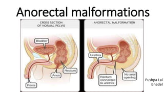
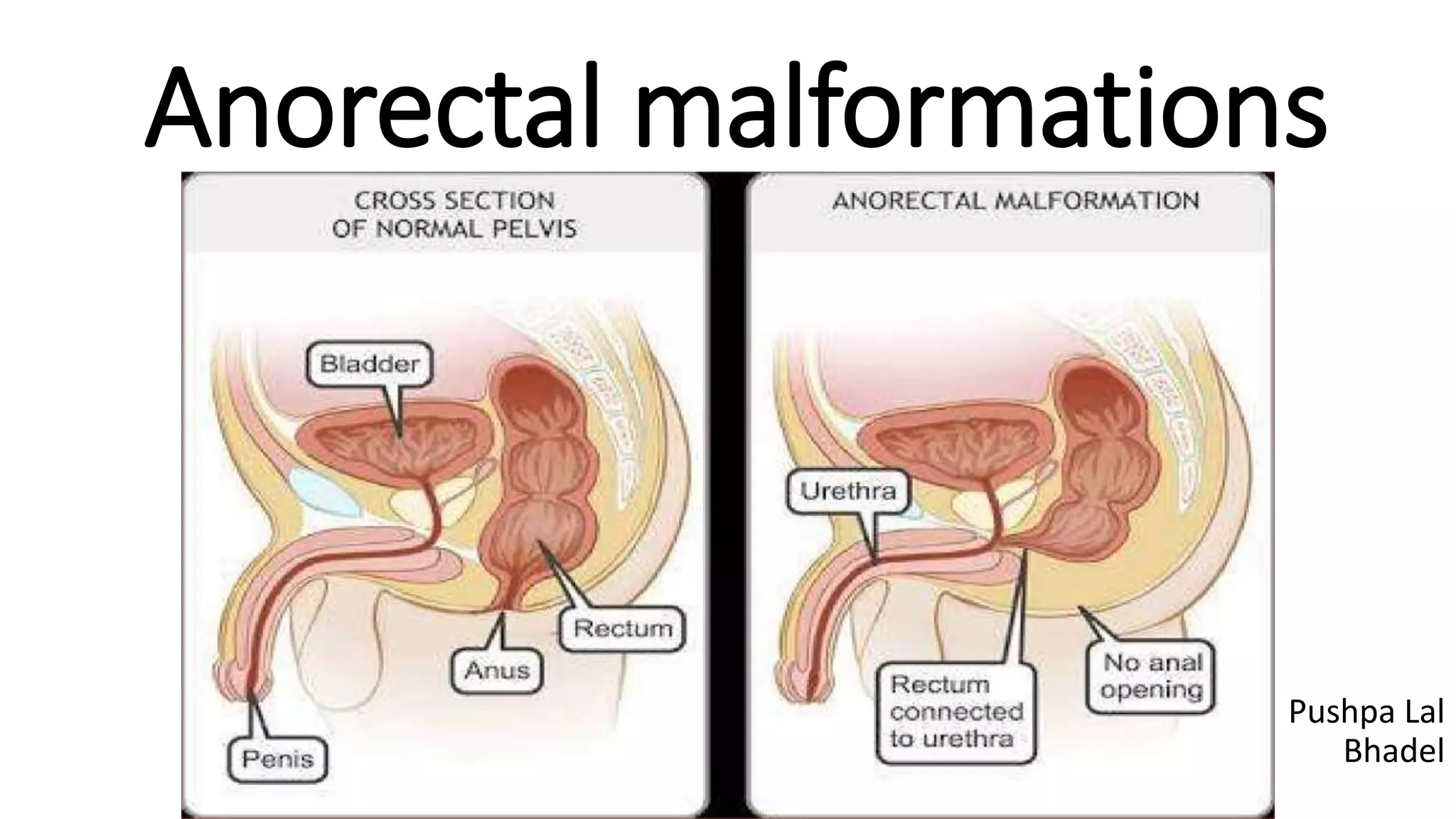
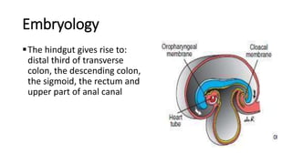
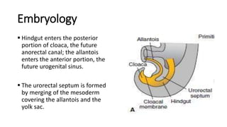
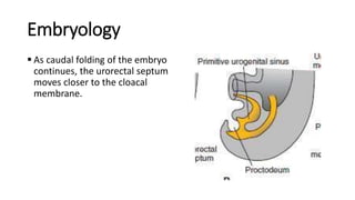
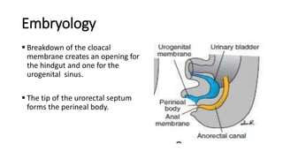
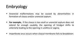
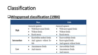
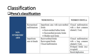
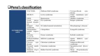
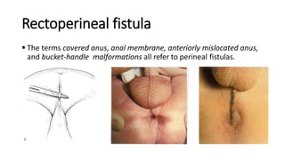
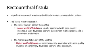
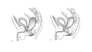
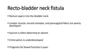
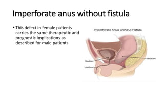
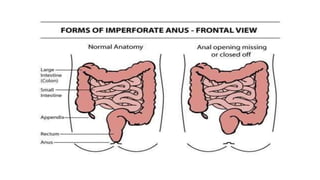
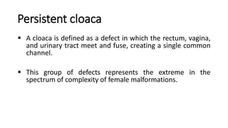
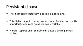
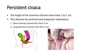
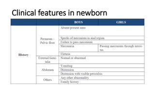
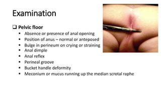
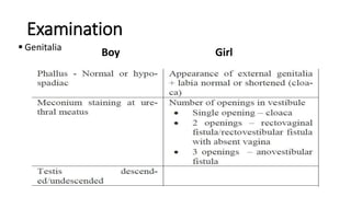
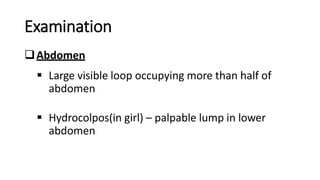
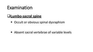
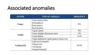
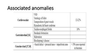
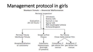
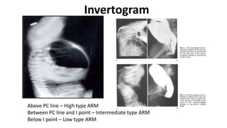
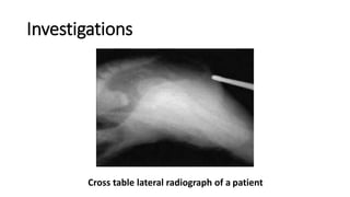
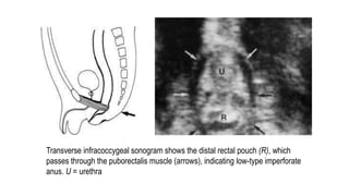
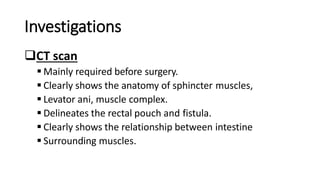
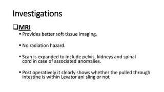
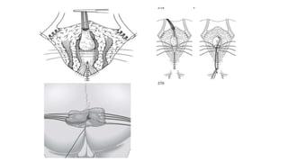
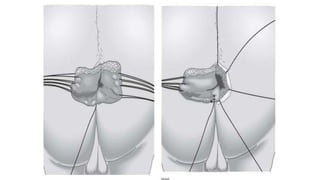
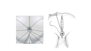
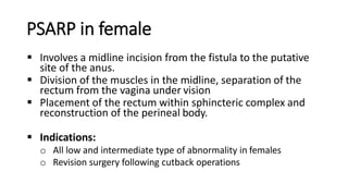
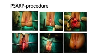
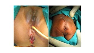
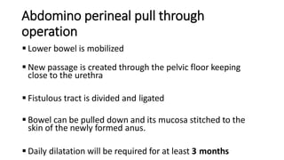
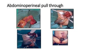
SlideShare a Scribd company logo
Anorectal malformations
Pushpa Lal
Bhadel
Introduction
 Birth defects in which the anus and rectum don’t develop properly.
 More frequent congenital anomalies encountered in pediatric
surgery.
 Incidence : 1 in 4000 live births
 Male > female
Introduction
 In female: Rectovestibular fistula is the most common
 In male: Rectourethral fistula is the most common
 Imperforate anus without fistula: 5%
 Estimated risk of 2nd child being affected is 1.4%
 Risk increases to 3% if first child is born with perineal or vestibular
fistula
Introduction
 Most female have low or intermediate anorectal malformations
while the reverse is true for males.
 Female babies usually have a fistula from the terminal end of the
bowel opening externally
 While in male this fistula is usually well hidden.
Embryology
The hindgut gives rise to:
distal third of transverse
colon, the descending colon,
the sigmoid, the rectum and
upper part of anal canal
Embryology
 Hindgut enters the posterior
portion of cloaca, the future
anorectal canal; the allantois
enters the anterior portion, the
future urogenital sinus.
 The urorectal septum is formed
by merging of the mesoderm
covering the allantois and the
yolk sac.
Embryology
 As caudal folding of the embryo
continues, the urorectal septum
moves closer to the cloacal
membrane.
Embryology
 Breakdown of the cloacal
membrane creates an opening for
the hindgut and one for the
urogenital sinus.
 The tip of the urorectal septum
forms the perineal body.
Embryology
 Anorectal malformations may be caused by abnormalities in
formation of cloaca and/or urorectal septum.
 For example, if the cloaca is too small or urorectal septum does not
extend far enough caudally, the opening of hindgut shifts to
anteriorly leading to the opening in urethra or vagina.
 Imperforate anus occurs when cloacal membrane fails to breakdown.
Classification
 Wingspread classification
 Pena’s classification
 Krickenbeck classification
 Anatomical classification
Classification
Wingspread classification (1984)
Classification
Pena’s classification
Classification
Krickenbeck classification
Classification
Anatomical classification
Rectoperineal fistula
 Traditionally was known as a “low
defect.”
 The rectum is located within most
of the sphincter mechanism.
 Only the lowest part of the rectum
is anteriorly mislocated .
Rectoperineal fistula
Sometimes, the fistula does not open into the perineum but
rather follows a subepithelial midline tract, opening
somewhere along the midline perineal raphe, scrotum, or
even at the base of the penis.
Diagnosis established by perineal inspection
No further investigations required.
Rectoperineal fistula
 The terms covered anus, anal membrane, anteriorly mislocated anus,
and bucket-handle malformations all refer to perineal fistulas.
Rectourethral fistula
 Imperforate anus with a rectourethral fistula is most common defect in boys.
 The fistula may be located at
o The lower (bulbar) part of the urethra
• Lower urethral fistulas are usually associated with good-quality
muscles, a well-developed sacrum, a prominent midline groove, and a
prominent anal dimple.
o The higher (prostatic) part of the urethra
• Higher urethral fistulas are more frequently associated with poor-quality
muscles, an abnormally developed sacrum, a flat perineum.
Recto-bladder neck fistula
 Rectum opens into the bladder neck
 Levator muscle, muscle complex, and parasagittal fibers are poorly
developed
 Sacrum is often deformed or absent
 Entire pelvis is underdeveloped
 Prognosis for bowel function is poor
Recto-bladder neck fistula
 The perineum is often flat,
which is evidence of poor
muscle development.
 About 10% of males with
anorectal atresia fall into this
category.
Imperforate anus without fistula
 Have a well-developed sacrum and good muscles and have
a good prognosis in terms of bowel function.
 The rectum usually terminates approximately 2 cm from the
perineal skin.
 Common in patients with Down syndrome.
Rectal atresia
 Occur in 1 % of case
 Born with normal appearing anal canal
 Often discovered during an attempt to take rectal temperature
 ~2 cm from the anal verge, there is an atretic or stenotic area
 The sacrum is normal, the sphincter mechanism is excellent
 Prognosis is good.
Rectal atresia
 These two structures may be
separated by a thin membrane or
by dense fibrous tissue.
Rectoperineal fistula
 From the therapeutic and prognostic viewpoint, this common defect
is equivalent to the perineal fistula described in the male patient.
 The rectum is well positioned within the sphincter mechanism, except
for its lower portion, which is anteriorly located
 The rectum and vagina are well separated.
Rectovestibular fistula
 Rectovestibular fistula is the most common defect in girls and has
an excellent functional prognosis.
 The diagnosis is based on clinical examination.
 Meticulous examination of neonatal genitalia allows clinician to
observe normal urethral meatus and a normal vagina, with a third
hole in the vestibule which is the recto vestibular fistula
Imperforate anus without fistula
 This defect in female patients
carries the same therapeutic and
prognostic implications as
described for male patients.
Persistent cloaca
 A cloaca is defined as a defect in which the rectum, vagina,
and urinary tract meet and fuse, creating a single common
channel.
 This group of defects represents the extreme in the
spectrum of complexity of female malformations.
Persistent cloaca
 The diagnosis of persistent cloaca is a clinical one.
 This defect should be suspected in a female born with
imperforate anus and small-looking genitalia.
 Careful separation of the labia discloses a single perineal
orifice.
Persistent cloaca
 The length of the common channel varies from 1 to 7 cm.
 This distance has technical and prognostic implications.
1. Short common channel less than 3 cm
2. Long common channel more than 3 cm
Clinical features in newborn
Examination
 Pelvic floor
 Absence or presence of anal opening
 Position of anus – normal or anteposed
 Bulge in perineum on crying or straining
 Anal dimple
 Anal reflex
 Perineal groove
 Bucket handle deformity
 Meconium or mucus running up the median scrotal raphe
Examination
 Genitalia
Boy Girl
Examination
Abdomen
 Large visible loop occupying more than half of
abdomen
 Hydrocolpos(in girl) – palpable lump in lower
abdomen
Examination
Lumbo-sacral spine
 Occult or obvious spinal dysraphism
 Absent sacral vertebrae of variable levels
Associated anomalies
Management protocol in boys
Management protocol in girls
Approach to case of ARM
 History of neonate
 Clinical examination
 Investigation
Investigations
 Invertogram
 Cross table lateral radiography
 USG abdomen & pelvis(to rule out genitourinary anomalies)
 Ultrasonography
o a)transperineal
o b)infracoccygeal
 MCU,IVP
 CT Scan, MRI
 High-pressure distal colostography
Invertogram
Above PC line – High type ARM
Between PC line and I point – Intermediate type ARM
Below I point – Low type ARM
Investigations
Cross table lateral radiograph of a patient
Transverse infracoccygeal sonogram shows the distal rectal pouch (R), which
passes through the puborectalis muscle (arrows), indicating low-type imperforate
anus. U = urethra
Investigations
CT scan
 Mainly required before surgery.
 Clearly shows the anatomy of sphincter muscles,
 Levator ani, muscle complex.
 Delineates the rectal pouch and fistula.
 Clearly shows the relationship between intestine
 Surrounding muscles.
Investigations
MRI
 Provides better soft tissue imaging.
 No radiation hazard.
 Scan is expanded to include pelvis, kidneys and spinal
cord in case of associated anomalies.
 Post operatively it clearly shows whether the pulled through
intestine is within Levator ani sling or not
Investigations
High pressure distal colostography
 Before the definitive repair, distal colostography
is performed.
 It is the most valuable and accurate diagnostic study to
define the anatomy of the anorectal malformation.
Investigations
High pressure distal colostography
 Water-soluble contrast medium is instilled into the distal stoma,
which fills the distal intestine and enables demonstration of the
location of the blind rectum and the precise site of a rectourinary
fistula.
 The contrast medium must be injected with considerable
hydrostatic pressure under fluoroscopic control.
Investigations
High pressure distal colostography
 The use of a Foley catheter is recommended; it is passed through
the distal stoma, the balloon is inflated (2–5 mL), and it is pulled
back as far as possible to occlude the stoma during the injection
of the contrast medium.
 This maneuver permits to overcome the muscle tone of the
striated muscle mechanism, fill the rectum, and demonstrate the
urinary fistula when present.
Operative procedures
 Colostomy
 Posterior sagittal anorectoplasty
 Pull through procedures
 Laparoscopic assisted procedures
Colostomy
 Descending colostomy is preferred.
 The colostomy is constructed through a left lower quadrant
oblique or transverse incision.
 The proximal stoma is exteriorized through the upper and
lateral part of the wound and the mucous fistula is placed
in the medial or lower part of the wound.
Colostomy
 The colostomy should be made in the mobile portion of
the colon, immediately distal to the descending colon
taking advantage of its retroperitoneal attachments, and
the mucous fistula is made very small to avoid prolapse.
 During the opening of the colostomy, the distal intestine
must be irrigated to remove all the meconium, preventing
the formation of a mega-sigmoid.
Colostomy
 Advantages of descending colostomy
o Mechanical preparation of the distal colon before the definitive
repair is easy due small length of remaining segment.
o Due to shorter distal segment in patient with recto-urethral or
recto-vesical fistula urine is not accumulate in distal segment of
colon.(which leads to development of metabolic acidosis)
o Less chance of development of megarectosigmoid.
o The incidence of prolapse in the proximal limb of descending
colostomies is almost zero.
Posterior sagittal anorectoplasty (PSARP)
 All anorectal malformations benefit from the use of the
posterior sagittal approach.
 The length of the incision depends on the specific
defect.
 The patient is placed in the prone position with the
pelvis elevated.
Posterior sagittal anorectoplasty (PSARP)
Position
Posterior sagittal anorectoplasty (PSARP)
An incision that starts in the lower portion of the
sacrum and extends anteriorly to the anal sphincter.
Recto vestibular and recto perineal fistula requires
smaller incision so, called limited posterior sagittal ano
rectoplasty and minimal posterior sagittal ano
rectoplasty respectively.
Repair in Boys
Recto perineal fistula
 The repair of these defects consists of a small anoplasty
with minimal mobilization of the rectum, sufficient for it
to be transposed and placed within the limits of the
sphincter.
 It is done during the neonatal period without a
colostomy.
Repair in Boys
 These patients have an excellent prognosis.
 If they have significant associated spinal or sacral
problems an alternative approach, a Pott’s transplant
anoplasty, whereby the majority of the perineal
body is preserved, the mobilized fistula is brought
through a separate incision which is confined to the
size of the future neoanal canal.
Repair in Boys
Rectourethral fistula
 Most important thing in these cases are to put per
urethral catheter.
 Toavoid the catheter to be entered in rectum, the
catheter must be intentionally directed anteriorly by the
use of a lacrimal probe inserted in the distal tip of the
catheter to find its correct path.
Repair in Boys
Rectobladder neck fistula
As it is very high defect both approach (through perineum
and through abdomen) is needed.
 A plasty of the distal dilated portion of the rectum is
necessary in some cases to reach the perineal skin.
 It is also called as abdomino perineal pull through
operation.
Repair in Boys
Imperforate anus without fistula
 About 5 percent of patients have imperforate anus without a fistula.
 In both boys and girls, the rectum lies about 2 cm from the perineal
skin.
 The rectum must be carefully separated from the urethra because the
two structures have a common wall.
 The rest of the repair must be performed as described for the
rectourethral fistula type of defect
Repair in Boys
Rectal atresia and stenosis
 These defects are repaired through a posterior sagittal
approach.
 The entire sphincteric mechanism is divided in the
midline.
 The narrowed area of the distal rectum is opened
posteriorly.
Repair in Boys
Rectal atresia and stenosis
 The posterior rectum is mobilized to reach the anal skin.
No anterior dissection is needed.
 The sphincter mechanism posterior to the rectum is
reconstructed.
 Any presacral mass is dealt simultaneously in the
same operation.
Repair in Girls
Recto perineal fistula
 The treatment of rectoperineal fistula in girls is the same
as that discussed for boys, except of course that the
anterior rectal wall is mobilized off the area behind the
vagina.
Repair in Girls
Rectovestibular fistula
 Incision in this defect is usually shorter as compare to
recto urethral fistula in boys.
 Starting from posteriorly sphincter mechanism is
divided till reach rectal fascia which is helpful in
indenting the plane of dissection.
 Then we go laterally and then anteriorly using this
plane.
Repair in Girls
Rectovestibular fistula
 Because the rectum and vagina have single wall it is
divided using needle cautery.
 The most common error in performing this operation is
incomplete separation of the vagina and rectum.
 This may create a tense anastomosis between the
rectum and the skin, which may provoke dehiscence
and recurrence of the fistula
Repair in Girls
Rectovestibular fistula
The anterior limit of the external sphincter is identified
using electrical stimulation and the anterior edge of the
muscle complex are reapproximated as previously
described, creating the perineal body.
Repair in Girls
Rectovaginal fistula
 Imperforate anus with a true rectovaginal fistula is extremely
rare.
 A true rectovaginal fistula requires a full posterior sagittal
incision.
 The operation is essentially the same as that described for
a rectovestibular fistula, except that it is necessary to
dissect much more of the rectum to gain enough length to
pull it down to the perineum.
PSARP in female
 Involves a midline incision from the fistula to the putative
site of the anus.
 Division of the muscles in the midline, separation of the
rectum from the vagina under vision
 Placement of the rectum within sphincteric complex and
reconstruction of the perineal body.
 Indications:
o All low and intermediate type of abnormality in females
o Revision surgery following cutback operations
PSARP-procedure
95
96
Abdomino perineal pull through
operation
 Lower bowel is mobilized
 New passage is created through the pelvic floor keeping
close to the urethra
 Fistulous tract is divided and ligated
 Bowel can be pulled down and its mucosa stitched to the
skin of the newly formed anus.
 Daily dilatation will be required for at least 3 months
Abdominoperineal pull through
Laparoscopically assisted anorectal pull
through (LAARP) for highARM
 Advantages:
 LAARP allows the surgeon to treat a high lesion like a low lesion.
 No need to divide the muscle complex from below.
 Immediately after the procedure strong and symmetric contraction of the
sphincter around the neo anus can be seen.
 It also avoids the complication and multiple procedures associated with
colostomy.
 More rapid return of bowel function
 Improved cosmetic appearance
 Shorter postoperative recovery
 Decreased postoperative complications
Post operative care
 In cases of rectourethral fistula in boys, the urethral
catheter is left in place for 7 days.
 If the urethral catheter is accidentally dislodged, the patient
can be observed for spontaneous voiding, which usually
occurs.
 Attempts to reintroduce a urethral catheter is avoided.
Post operative care
 Intravenous antibiotics are administered for 24 hours. An antibiotic ointment
is applied to the anoplasty for 5 days.
 The patient is discharged after 2 days in cases of a posterior approach without a
laparotomy or laparoscopy, and after 3–5 days in cases of an abdominal
approach.
 The parents are instructed to keep the incision clean, not to wipe, and to apply
antibiotic ointment for 1 week.
 Two weeks after the operations, anal dilatations are started. On the first
occasion, a dilator that fits loosely into the anus is used to instruct the parents,
who must carry out dilatation twice daily.
Post operative care
 Every week, the size of the dilator is increased until the rectum
reaches the desired size, which depends on the patient’s age.
 Once the desired size is reached, the colostomy can be closed.
 Frequency of dilatation should be reduced in following
schedule:
o At least once a day for one month; every third day for one month;
twice a week for one month; once a week for one month; and every
2 weeks for three months.
Post operative care
Post operative care
Initially patient may have diaper rash due to multiple bowel
movement which is converted to 2 or 3 bowel movement per
day in 6 months.
Patient with 2 to 3 bowel movement in day, with some feeling
on having bowel movement have good prognosis and
responds to toilet training.
Complication
 Wound infection
 Anal strictures
 Constipation
 Transient femoral nerve pressure
 Neurogenic bladder
 Fecal incontinence
Thank you

More Related Content

What's hot

Undescended testis
Undescended testisUndescended testis
Undescended testis
GAURAV NAHAR
 
anorectal malformation
anorectal malformationanorectal malformation
anorectal malformation
Pushpa Latha
 
ANORECTAL-MALFORMATIONS.pptx
ANORECTAL-MALFORMATIONS.pptxANORECTAL-MALFORMATIONS.pptx
ANORECTAL-MALFORMATIONS.pptx
DR MUKESH SAH
 
Duodenal atresia stenosis PRANAYA PPT
Duodenal atresia stenosis PRANAYA PPTDuodenal atresia stenosis PRANAYA PPT
Duodenal atresia stenosis PRANAYA PPT
PRANAYA PANIGRAHI
 
Internal hernia
Internal herniaInternal hernia
Internal hernia
Asif Ansari
 
Anorectal malformations
Anorectal malformations Anorectal malformations
Anorectal malformations
Dr.Manish Kumar
 
Anorectal malformation seminar
Anorectal malformation seminarAnorectal malformation seminar
Anorectal malformation seminar
Dr. Dixit
 
Congenital anamolies of pancrease
Congenital anamolies of pancreaseCongenital anamolies of pancrease
Congenital anamolies of pancrease
Dr Dipesh K.K
 
Management and surgical procedures of Hirschsprung disease
Management and surgical procedures of Hirschsprung disease Management and surgical procedures of Hirschsprung disease
Management and surgical procedures of Hirschsprung disease
Thorlikonda Sasidhar
 
Mesenteric cyst - Journal club
Mesenteric cyst - Journal clubMesenteric cyst - Journal club
Mesenteric cyst - Journal club
Priyadarshan Konar
 
The exstrophy epispadias complex
The exstrophy epispadias complexThe exstrophy epispadias complex
The exstrophy epispadias complex
Elsayed Salih
 
Testicular tumors - ramu
Testicular tumors  - ramuTesticular tumors  - ramu
Testicular tumors - ramu
damuluri ramu
 
Ureteric injury (1)
Ureteric injury (1)Ureteric injury (1)
Ureteric injury (1)
Rojan Adhikari
 
Sigmoid volvulus (2)
Sigmoid volvulus (2)Sigmoid volvulus (2)
Sigmoid volvulus (2)Todd Peterson
 
Cloaca approach to decision making
Cloaca approach to decision makingCloaca approach to decision making
Cloaca approach to decision making
Faheem Andrabi
 
Ventral hernia management
Ventral hernia managementVentral hernia management
Ventral hernia management
manjil malla
 
Duodenal atresia
Duodenal atresiaDuodenal atresia
Duodenal atresia
Shambhavi Sharma
 
Anorectal Malformations
Anorectal MalformationsAnorectal Malformations
Anorectal Malformations
Kurnool Medical College, Kurnool
 
Anorectal malformations
Anorectal malformationsAnorectal malformations
Anorectal malformations
Dr.Manish Kumar
 

What's hot (20)

Undescended testis
Undescended testisUndescended testis
Undescended testis
 
anorectal malformation
anorectal malformationanorectal malformation
anorectal malformation
 
ANORECTAL-MALFORMATIONS.pptx
ANORECTAL-MALFORMATIONS.pptxANORECTAL-MALFORMATIONS.pptx
ANORECTAL-MALFORMATIONS.pptx
 
Duodenal atresia stenosis PRANAYA PPT
Duodenal atresia stenosis PRANAYA PPTDuodenal atresia stenosis PRANAYA PPT
Duodenal atresia stenosis PRANAYA PPT
 
Internal hernia
Internal herniaInternal hernia
Internal hernia
 
Anorectal malformations
Anorectal malformations Anorectal malformations
Anorectal malformations
 
Anorectal malformation seminar
Anorectal malformation seminarAnorectal malformation seminar
Anorectal malformation seminar
 
Congenital anamolies of pancrease
Congenital anamolies of pancreaseCongenital anamolies of pancrease
Congenital anamolies of pancrease
 
Management and surgical procedures of Hirschsprung disease
Management and surgical procedures of Hirschsprung disease Management and surgical procedures of Hirschsprung disease
Management and surgical procedures of Hirschsprung disease
 
Mesenteric cyst - Journal club
Mesenteric cyst - Journal clubMesenteric cyst - Journal club
Mesenteric cyst - Journal club
 
The exstrophy epispadias complex
The exstrophy epispadias complexThe exstrophy epispadias complex
The exstrophy epispadias complex
 
Testicular tumors - ramu
Testicular tumors  - ramuTesticular tumors  - ramu
Testicular tumors - ramu
 
Ureteric injury (1)
Ureteric injury (1)Ureteric injury (1)
Ureteric injury (1)
 
Sigmoid volvulus (2)
Sigmoid volvulus (2)Sigmoid volvulus (2)
Sigmoid volvulus (2)
 
Cloaca approach to decision making
Cloaca approach to decision makingCloaca approach to decision making
Cloaca approach to decision making
 
Ventral hernia management
Ventral hernia managementVentral hernia management
Ventral hernia management
 
Duodenal atresia
Duodenal atresiaDuodenal atresia
Duodenal atresia
 
Anorectal Malformations
Anorectal MalformationsAnorectal Malformations
Anorectal Malformations
 
Anorectal malformations
Anorectal malformationsAnorectal malformations
Anorectal malformations
 
Orchiectomy
OrchiectomyOrchiectomy
Orchiectomy
 

Similar to Anorectal malformations.pptx

Presentation1 3 (4).ppt
Presentation1 3 (4).pptPresentation1 3 (4).ppt
Presentation1 3 (4).ppt
ssuser8eb265
 
Anorectal malformations in children
Anorectal malformations in childrenAnorectal malformations in children
Anorectal malformations in children
dhanyav54
 
Imperforate Anus
Imperforate Anus Imperforate Anus
ANORECTAL MALFORMATIONS
ANORECTAL MALFORMATIONSANORECTAL MALFORMATIONS
ANORECTAL MALFORMATIONS
karrar adil
 
Benign anorectal disorders 2
Benign anorectal disorders 2Benign anorectal disorders 2
Benign anorectal disorders 2
Dr. Azhar
 
Urethral anomalies
Urethral anomaliesUrethral anomalies
Urethral anomalies
indumathibalakrishna
 
Uterine Fistula
Uterine FistulaUterine Fistula
Uterine Fistula
Susmita Halder
 
Anorectal malformation ppt 5
Anorectal malformation ppt 5Anorectal malformation ppt 5
Anorectal malformation ppt 5
RamanUppal3
 
Surgical Anatomy of Urinary bladder.pptx
Surgical Anatomy of Urinary bladder.pptxSurgical Anatomy of Urinary bladder.pptx
Surgical Anatomy of Urinary bladder.pptx
DurgaPrasadM10
 
undescended testis.pptx
undescended testis.pptxundescended testis.pptx
undescended testis.pptx
AllenDavid32
 
Inguinal hernia
Inguinal herniaInguinal hernia
Inguinal hernia
Dr Mohamed Ruvais
 
Anorectal malformation
Anorectal malformationAnorectal malformation
Anorectal malformation
manahrsinh rajput
 
Undescended Testis
Undescended TestisUndescended Testis
Undescended Testis
Junish Bagga
 
Undescended testes
Undescended testes Undescended testes
Undescended testes
racheetha
 
Vcu ppt
Vcu pptVcu ppt
Vcu ppt
dypradio
 
Ano-rectal Malformations copy.pptx
Ano-rectal Malformations copy.pptxAno-rectal Malformations copy.pptx
Ano-rectal Malformations copy.pptx
bishwokunwar3
 
Antegrade & retrograde urethrogram
Antegrade & retrograde urethrogramAntegrade & retrograde urethrogram
Antegrade & retrograde urethrogram
Amlendu Kumar
 
evaluation of Undescended testes
evaluation of Undescended testesevaluation of Undescended testes
evaluation of Undescended testes
Vernon Pashi
 
Inguinal hernia
Inguinal herniaInguinal hernia
Inguinal hernia
karrar adil
 
Pelvic organ prolapse
Pelvic organ prolapsePelvic organ prolapse
Pelvic organ prolapse
Sourav Chowdhury
 

Similar to Anorectal malformations.pptx (20)

Presentation1 3 (4).ppt
Presentation1 3 (4).pptPresentation1 3 (4).ppt
Presentation1 3 (4).ppt
 
Anorectal malformations in children
Anorectal malformations in childrenAnorectal malformations in children
Anorectal malformations in children
 
Imperforate Anus
Imperforate Anus Imperforate Anus
Imperforate Anus
 
ANORECTAL MALFORMATIONS
ANORECTAL MALFORMATIONSANORECTAL MALFORMATIONS
ANORECTAL MALFORMATIONS
 
Benign anorectal disorders 2
Benign anorectal disorders 2Benign anorectal disorders 2
Benign anorectal disorders 2
 
Urethral anomalies
Urethral anomaliesUrethral anomalies
Urethral anomalies
 
Uterine Fistula
Uterine FistulaUterine Fistula
Uterine Fistula
 
Anorectal malformation ppt 5
Anorectal malformation ppt 5Anorectal malformation ppt 5
Anorectal malformation ppt 5
 
Surgical Anatomy of Urinary bladder.pptx
Surgical Anatomy of Urinary bladder.pptxSurgical Anatomy of Urinary bladder.pptx
Surgical Anatomy of Urinary bladder.pptx
 
undescended testis.pptx
undescended testis.pptxundescended testis.pptx
undescended testis.pptx
 
Inguinal hernia
Inguinal herniaInguinal hernia
Inguinal hernia
 
Anorectal malformation
Anorectal malformationAnorectal malformation
Anorectal malformation
 
Undescended Testis
Undescended TestisUndescended Testis
Undescended Testis
 
Undescended testes
Undescended testes Undescended testes
Undescended testes
 
Vcu ppt
Vcu pptVcu ppt
Vcu ppt
 
Ano-rectal Malformations copy.pptx
Ano-rectal Malformations copy.pptxAno-rectal Malformations copy.pptx
Ano-rectal Malformations copy.pptx
 
Antegrade & retrograde urethrogram
Antegrade & retrograde urethrogramAntegrade & retrograde urethrogram
Antegrade & retrograde urethrogram
 
evaluation of Undescended testes
evaluation of Undescended testesevaluation of Undescended testes
evaluation of Undescended testes
 
Inguinal hernia
Inguinal herniaInguinal hernia
Inguinal hernia
 
Pelvic organ prolapse
Pelvic organ prolapsePelvic organ prolapse
Pelvic organ prolapse
 

More from Pushpa Lal Bhadel

Gall Bladder Cancer.pptx
Gall Bladder Cancer.pptxGall Bladder Cancer.pptx
Gall Bladder Cancer.pptx
Pushpa Lal Bhadel
 
Hepatobiliary Imaging.pptx
Hepatobiliary Imaging.pptxHepatobiliary Imaging.pptx
Hepatobiliary Imaging.pptx
Pushpa Lal Bhadel
 
Surgical anatomy of Liver, Concept of liver.pptx
Surgical anatomy of Liver, Concept of liver.pptxSurgical anatomy of Liver, Concept of liver.pptx
Surgical anatomy of Liver, Concept of liver.pptx
Pushpa Lal Bhadel
 
Malignant Neoplasms of Stomach.pptx
Malignant Neoplasms of Stomach.pptxMalignant Neoplasms of Stomach.pptx
Malignant Neoplasms of Stomach.pptx
Pushpa Lal Bhadel
 
Rectal and Umbilical polyps.pptx
Rectal and Umbilical polyps.pptxRectal and Umbilical polyps.pptx
Rectal and Umbilical polyps.pptx
Pushpa Lal Bhadel
 
Hypospadias.pptx
Hypospadias.pptxHypospadias.pptx
Hypospadias.pptx
Pushpa Lal Bhadel
 
Liver resection and Metastasectomy.pptx
Liver resection and Metastasectomy.pptxLiver resection and Metastasectomy.pptx
Liver resection and Metastasectomy.pptx
Pushpa Lal Bhadel
 
Radical Cholecystectomy.pptx
Radical Cholecystectomy.pptxRadical Cholecystectomy.pptx
Radical Cholecystectomy.pptx
Pushpa Lal Bhadel
 
Complete mesocolic excision (CME) Vs D2.pptx
Complete mesocolic excision (CME) Vs D2.pptxComplete mesocolic excision (CME) Vs D2.pptx
Complete mesocolic excision (CME) Vs D2.pptx
Pushpa Lal Bhadel
 
Diagnostic modalities for Gastric diseases.pptx
Diagnostic modalities for Gastric diseases.pptxDiagnostic modalities for Gastric diseases.pptx
Diagnostic modalities for Gastric diseases.pptx
Pushpa Lal Bhadel
 
CLEFT LIP AND PALATE.pptx
CLEFT LIP AND PALATE.pptxCLEFT LIP AND PALATE.pptx
CLEFT LIP AND PALATE.pptx
Pushpa Lal Bhadel
 
Carcinoma Urinary Bladder.pptx
Carcinoma Urinary Bladder.pptxCarcinoma Urinary Bladder.pptx
Carcinoma Urinary Bladder.pptx
Pushpa Lal Bhadel
 
risk factor for breast cancer.pptx
risk factor for breast cancer.pptxrisk factor for breast cancer.pptx
risk factor for breast cancer.pptx
Pushpa Lal Bhadel
 
Cholecystectomy in patient with Liver Cirrhosis.pptx
Cholecystectomy in patient with Liver Cirrhosis.pptxCholecystectomy in patient with Liver Cirrhosis.pptx
Cholecystectomy in patient with Liver Cirrhosis.pptx
Pushpa Lal Bhadel
 
Lateral lymph nodes in rectal cancer.pptx
Lateral lymph nodes in rectal cancer.pptxLateral lymph nodes in rectal cancer.pptx
Lateral lymph nodes in rectal cancer.pptx
Pushpa Lal Bhadel
 

More from Pushpa Lal Bhadel (15)

Gall Bladder Cancer.pptx
Gall Bladder Cancer.pptxGall Bladder Cancer.pptx
Gall Bladder Cancer.pptx
 
Hepatobiliary Imaging.pptx
Hepatobiliary Imaging.pptxHepatobiliary Imaging.pptx
Hepatobiliary Imaging.pptx
 
Surgical anatomy of Liver, Concept of liver.pptx
Surgical anatomy of Liver, Concept of liver.pptxSurgical anatomy of Liver, Concept of liver.pptx
Surgical anatomy of Liver, Concept of liver.pptx
 
Malignant Neoplasms of Stomach.pptx
Malignant Neoplasms of Stomach.pptxMalignant Neoplasms of Stomach.pptx
Malignant Neoplasms of Stomach.pptx
 
Rectal and Umbilical polyps.pptx
Rectal and Umbilical polyps.pptxRectal and Umbilical polyps.pptx
Rectal and Umbilical polyps.pptx
 
Hypospadias.pptx
Hypospadias.pptxHypospadias.pptx
Hypospadias.pptx
 
Liver resection and Metastasectomy.pptx
Liver resection and Metastasectomy.pptxLiver resection and Metastasectomy.pptx
Liver resection and Metastasectomy.pptx
 
Radical Cholecystectomy.pptx
Radical Cholecystectomy.pptxRadical Cholecystectomy.pptx
Radical Cholecystectomy.pptx
 
Complete mesocolic excision (CME) Vs D2.pptx
Complete mesocolic excision (CME) Vs D2.pptxComplete mesocolic excision (CME) Vs D2.pptx
Complete mesocolic excision (CME) Vs D2.pptx
 
Diagnostic modalities for Gastric diseases.pptx
Diagnostic modalities for Gastric diseases.pptxDiagnostic modalities for Gastric diseases.pptx
Diagnostic modalities for Gastric diseases.pptx
 
CLEFT LIP AND PALATE.pptx
CLEFT LIP AND PALATE.pptxCLEFT LIP AND PALATE.pptx
CLEFT LIP AND PALATE.pptx
 
Carcinoma Urinary Bladder.pptx
Carcinoma Urinary Bladder.pptxCarcinoma Urinary Bladder.pptx
Carcinoma Urinary Bladder.pptx
 
risk factor for breast cancer.pptx
risk factor for breast cancer.pptxrisk factor for breast cancer.pptx
risk factor for breast cancer.pptx
 
Cholecystectomy in patient with Liver Cirrhosis.pptx
Cholecystectomy in patient with Liver Cirrhosis.pptxCholecystectomy in patient with Liver Cirrhosis.pptx
Cholecystectomy in patient with Liver Cirrhosis.pptx
 
Lateral lymph nodes in rectal cancer.pptx
Lateral lymph nodes in rectal cancer.pptxLateral lymph nodes in rectal cancer.pptx
Lateral lymph nodes in rectal cancer.pptx
 

Recently uploaded

Synthetic Fiber Construction in lab .pptx
Synthetic Fiber Construction in lab .pptxSynthetic Fiber Construction in lab .pptx
Synthetic Fiber Construction in lab .pptx
Pavel ( NSTU)
 
The French Revolution Class 9 Study Material pdf free download
The French Revolution Class 9 Study Material pdf free downloadThe French Revolution Class 9 Study Material pdf free download
The French Revolution Class 9 Study Material pdf free download
Vivekanand Anglo Vedic Academy
 
Template Jadual Bertugas Kelas (Boleh Edit)
Template Jadual Bertugas Kelas (Boleh Edit)Template Jadual Bertugas Kelas (Boleh Edit)
Template Jadual Bertugas Kelas (Boleh Edit)
rosedainty
 
Fish and Chips - have they had their chips
Fish and Chips - have they had their chipsFish and Chips - have they had their chips
Fish and Chips - have they had their chips
GeoBlogs
 
How libraries can support authors with open access requirements for UKRI fund...
How libraries can support authors with open access requirements for UKRI fund...How libraries can support authors with open access requirements for UKRI fund...
How libraries can support authors with open access requirements for UKRI fund...
Jisc
 
Introduction to Quality Improvement Essentials
Introduction to Quality Improvement EssentialsIntroduction to Quality Improvement Essentials
Introduction to Quality Improvement Essentials
Excellence Foundation for South Sudan
 
Students, digital devices and success - Andreas Schleicher - 27 May 2024..pptx
Students, digital devices and success - Andreas Schleicher - 27 May 2024..pptxStudents, digital devices and success - Andreas Schleicher - 27 May 2024..pptx
Students, digital devices and success - Andreas Schleicher - 27 May 2024..pptx
EduSkills OECD
 
Thesis Statement for students diagnonsed withADHD.ppt
Thesis Statement for students diagnonsed withADHD.pptThesis Statement for students diagnonsed withADHD.ppt
Thesis Statement for students diagnonsed withADHD.ppt
EverAndrsGuerraGuerr
 
Sha'Carri Richardson Presentation 202345
Sha'Carri Richardson Presentation 202345Sha'Carri Richardson Presentation 202345
Sha'Carri Richardson Presentation 202345
beazzy04
 
Welcome to TechSoup New Member Orientation and Q&A (May 2024).pdf
Welcome to TechSoup   New Member Orientation and Q&A (May 2024).pdfWelcome to TechSoup   New Member Orientation and Q&A (May 2024).pdf
Welcome to TechSoup New Member Orientation and Q&A (May 2024).pdf
TechSoup
 
Unit 8 - Information and Communication Technology (Paper I).pdf
Unit 8 - Information and Communication Technology (Paper I).pdfUnit 8 - Information and Communication Technology (Paper I).pdf
Unit 8 - Information and Communication Technology (Paper I).pdf
Thiyagu K
 
Polish students' mobility in the Czech Republic
Polish students' mobility in the Czech RepublicPolish students' mobility in the Czech Republic
Polish students' mobility in the Czech Republic
Anna Sz.
 
PART A. Introduction to Costumer Service
PART A. Introduction to Costumer ServicePART A. Introduction to Costumer Service
PART A. Introduction to Costumer Service
PedroFerreira53928
 
The Art Pastor's Guide to Sabbath | Steve Thomason
The Art Pastor's Guide to Sabbath | Steve ThomasonThe Art Pastor's Guide to Sabbath | Steve Thomason
The Art Pastor's Guide to Sabbath | Steve Thomason
Steve Thomason
 
special B.ed 2nd year old paper_20240531.pdf
special B.ed 2nd year old paper_20240531.pdfspecial B.ed 2nd year old paper_20240531.pdf
special B.ed 2nd year old paper_20240531.pdf
Special education needs
 
The Roman Empire A Historical Colossus.pdf
The Roman Empire A Historical Colossus.pdfThe Roman Empire A Historical Colossus.pdf
The Roman Empire A Historical Colossus.pdf
kaushalkr1407
 
aaaaaaaaaaaaaaaaaaaaaaaaaaaaaaaaaaaaaaaaaaaaaaaaaaaaaaa
aaaaaaaaaaaaaaaaaaaaaaaaaaaaaaaaaaaaaaaaaaaaaaaaaaaaaaaaaaaaaaaaaaaaaaaaaaaaaaaaaaaaaaaaaaaaaaaaaaaaaaaaaaaaaa
aaaaaaaaaaaaaaaaaaaaaaaaaaaaaaaaaaaaaaaaaaaaaaaaaaaaaaa
siemaillard
 
Unit 2- Research Aptitude (UGC NET Paper I).pdf
Unit 2- Research Aptitude (UGC NET Paper I).pdfUnit 2- Research Aptitude (UGC NET Paper I).pdf
Unit 2- Research Aptitude (UGC NET Paper I).pdf
Thiyagu K
 
1.4 modern child centered education - mahatma gandhi-2.pptx
1.4 modern child centered education - mahatma gandhi-2.pptx1.4 modern child centered education - mahatma gandhi-2.pptx
1.4 modern child centered education - mahatma gandhi-2.pptx
JosvitaDsouza2
 
How to Split Bills in the Odoo 17 POS Module
How to Split Bills in the Odoo 17 POS ModuleHow to Split Bills in the Odoo 17 POS Module
How to Split Bills in the Odoo 17 POS Module
Celine George
 

Recently uploaded (20)

Synthetic Fiber Construction in lab .pptx
Synthetic Fiber Construction in lab .pptxSynthetic Fiber Construction in lab .pptx
Synthetic Fiber Construction in lab .pptx
 
The French Revolution Class 9 Study Material pdf free download
The French Revolution Class 9 Study Material pdf free downloadThe French Revolution Class 9 Study Material pdf free download
The French Revolution Class 9 Study Material pdf free download
 
Template Jadual Bertugas Kelas (Boleh Edit)
Template Jadual Bertugas Kelas (Boleh Edit)Template Jadual Bertugas Kelas (Boleh Edit)
Template Jadual Bertugas Kelas (Boleh Edit)
 
Fish and Chips - have they had their chips
Fish and Chips - have they had their chipsFish and Chips - have they had their chips
Fish and Chips - have they had their chips
 
How libraries can support authors with open access requirements for UKRI fund...
How libraries can support authors with open access requirements for UKRI fund...How libraries can support authors with open access requirements for UKRI fund...
How libraries can support authors with open access requirements for UKRI fund...
 
Introduction to Quality Improvement Essentials
Introduction to Quality Improvement EssentialsIntroduction to Quality Improvement Essentials
Introduction to Quality Improvement Essentials
 
Students, digital devices and success - Andreas Schleicher - 27 May 2024..pptx
Students, digital devices and success - Andreas Schleicher - 27 May 2024..pptxStudents, digital devices and success - Andreas Schleicher - 27 May 2024..pptx
Students, digital devices and success - Andreas Schleicher - 27 May 2024..pptx
 
Thesis Statement for students diagnonsed withADHD.ppt
Thesis Statement for students diagnonsed withADHD.pptThesis Statement for students diagnonsed withADHD.ppt
Thesis Statement for students diagnonsed withADHD.ppt
 
Sha'Carri Richardson Presentation 202345
Sha'Carri Richardson Presentation 202345Sha'Carri Richardson Presentation 202345
Sha'Carri Richardson Presentation 202345
 
Welcome to TechSoup New Member Orientation and Q&A (May 2024).pdf
Welcome to TechSoup   New Member Orientation and Q&A (May 2024).pdfWelcome to TechSoup   New Member Orientation and Q&A (May 2024).pdf
Welcome to TechSoup New Member Orientation and Q&A (May 2024).pdf
 
Unit 8 - Information and Communication Technology (Paper I).pdf
Unit 8 - Information and Communication Technology (Paper I).pdfUnit 8 - Information and Communication Technology (Paper I).pdf
Unit 8 - Information and Communication Technology (Paper I).pdf
 
Polish students' mobility in the Czech Republic
Polish students' mobility in the Czech RepublicPolish students' mobility in the Czech Republic
Polish students' mobility in the Czech Republic
 
PART A. Introduction to Costumer Service
PART A. Introduction to Costumer ServicePART A. Introduction to Costumer Service
PART A. Introduction to Costumer Service
 
The Art Pastor's Guide to Sabbath | Steve Thomason
The Art Pastor's Guide to Sabbath | Steve ThomasonThe Art Pastor's Guide to Sabbath | Steve Thomason
The Art Pastor's Guide to Sabbath | Steve Thomason
 
special B.ed 2nd year old paper_20240531.pdf
special B.ed 2nd year old paper_20240531.pdfspecial B.ed 2nd year old paper_20240531.pdf
special B.ed 2nd year old paper_20240531.pdf
 
The Roman Empire A Historical Colossus.pdf
The Roman Empire A Historical Colossus.pdfThe Roman Empire A Historical Colossus.pdf
The Roman Empire A Historical Colossus.pdf
 
aaaaaaaaaaaaaaaaaaaaaaaaaaaaaaaaaaaaaaaaaaaaaaaaaaaaaaa
aaaaaaaaaaaaaaaaaaaaaaaaaaaaaaaaaaaaaaaaaaaaaaaaaaaaaaaaaaaaaaaaaaaaaaaaaaaaaaaaaaaaaaaaaaaaaaaaaaaaaaaaaaaaaa
aaaaaaaaaaaaaaaaaaaaaaaaaaaaaaaaaaaaaaaaaaaaaaaaaaaaaaa
 
Unit 2- Research Aptitude (UGC NET Paper I).pdf
Unit 2- Research Aptitude (UGC NET Paper I).pdfUnit 2- Research Aptitude (UGC NET Paper I).pdf
Unit 2- Research Aptitude (UGC NET Paper I).pdf
 
1.4 modern child centered education - mahatma gandhi-2.pptx
1.4 modern child centered education - mahatma gandhi-2.pptx1.4 modern child centered education - mahatma gandhi-2.pptx
1.4 modern child centered education - mahatma gandhi-2.pptx
 
How to Split Bills in the Odoo 17 POS Module
How to Split Bills in the Odoo 17 POS ModuleHow to Split Bills in the Odoo 17 POS Module
How to Split Bills in the Odoo 17 POS Module
 

Anorectal malformations.pptx

  • 2. Introduction  Birth defects in which the anus and rectum don’t develop properly.  More frequent congenital anomalies encountered in pediatric surgery.  Incidence : 1 in 4000 live births  Male > female
  • 3. Introduction  In female: Rectovestibular fistula is the most common  In male: Rectourethral fistula is the most common  Imperforate anus without fistula: 5%  Estimated risk of 2nd child being affected is 1.4%  Risk increases to 3% if first child is born with perineal or vestibular fistula
  • 4. Introduction  Most female have low or intermediate anorectal malformations while the reverse is true for males.  Female babies usually have a fistula from the terminal end of the bowel opening externally  While in male this fistula is usually well hidden.
  • 5. Embryology The hindgut gives rise to: distal third of transverse colon, the descending colon, the sigmoid, the rectum and upper part of anal canal
  • 6. Embryology  Hindgut enters the posterior portion of cloaca, the future anorectal canal; the allantois enters the anterior portion, the future urogenital sinus.  The urorectal septum is formed by merging of the mesoderm covering the allantois and the yolk sac.
  • 7. Embryology  As caudal folding of the embryo continues, the urorectal septum moves closer to the cloacal membrane.
  • 8. Embryology  Breakdown of the cloacal membrane creates an opening for the hindgut and one for the urogenital sinus.  The tip of the urorectal septum forms the perineal body.
  • 9. Embryology  Anorectal malformations may be caused by abnormalities in formation of cloaca and/or urorectal septum.  For example, if the cloaca is too small or urorectal septum does not extend far enough caudally, the opening of hindgut shifts to anteriorly leading to the opening in urethra or vagina.  Imperforate anus occurs when cloacal membrane fails to breakdown.
  • 10. Classification  Wingspread classification  Pena’s classification  Krickenbeck classification  Anatomical classification
  • 16. Rectoperineal fistula  Traditionally was known as a “low defect.”  The rectum is located within most of the sphincter mechanism.  Only the lowest part of the rectum is anteriorly mislocated .
  • 17. Rectoperineal fistula Sometimes, the fistula does not open into the perineum but rather follows a subepithelial midline tract, opening somewhere along the midline perineal raphe, scrotum, or even at the base of the penis. Diagnosis established by perineal inspection No further investigations required.
  • 18. Rectoperineal fistula  The terms covered anus, anal membrane, anteriorly mislocated anus, and bucket-handle malformations all refer to perineal fistulas.
  • 19. Rectourethral fistula  Imperforate anus with a rectourethral fistula is most common defect in boys.  The fistula may be located at o The lower (bulbar) part of the urethra • Lower urethral fistulas are usually associated with good-quality muscles, a well-developed sacrum, a prominent midline groove, and a prominent anal dimple. o The higher (prostatic) part of the urethra • Higher urethral fistulas are more frequently associated with poor-quality muscles, an abnormally developed sacrum, a flat perineum.
  • 20.
  • 21. Recto-bladder neck fistula  Rectum opens into the bladder neck  Levator muscle, muscle complex, and parasagittal fibers are poorly developed  Sacrum is often deformed or absent  Entire pelvis is underdeveloped  Prognosis for bowel function is poor
  • 22. Recto-bladder neck fistula  The perineum is often flat, which is evidence of poor muscle development.  About 10% of males with anorectal atresia fall into this category.
  • 23. Imperforate anus without fistula  Have a well-developed sacrum and good muscles and have a good prognosis in terms of bowel function.  The rectum usually terminates approximately 2 cm from the perineal skin.  Common in patients with Down syndrome.
  • 24. Rectal atresia  Occur in 1 % of case  Born with normal appearing anal canal  Often discovered during an attempt to take rectal temperature  ~2 cm from the anal verge, there is an atretic or stenotic area  The sacrum is normal, the sphincter mechanism is excellent  Prognosis is good.
  • 25. Rectal atresia  These two structures may be separated by a thin membrane or by dense fibrous tissue.
  • 26. Rectoperineal fistula  From the therapeutic and prognostic viewpoint, this common defect is equivalent to the perineal fistula described in the male patient.  The rectum is well positioned within the sphincter mechanism, except for its lower portion, which is anteriorly located  The rectum and vagina are well separated.
  • 27.
  • 28. Rectovestibular fistula  Rectovestibular fistula is the most common defect in girls and has an excellent functional prognosis.  The diagnosis is based on clinical examination.  Meticulous examination of neonatal genitalia allows clinician to observe normal urethral meatus and a normal vagina, with a third hole in the vestibule which is the recto vestibular fistula
  • 29.
  • 30. Imperforate anus without fistula  This defect in female patients carries the same therapeutic and prognostic implications as described for male patients.
  • 31.
  • 32. Persistent cloaca  A cloaca is defined as a defect in which the rectum, vagina, and urinary tract meet and fuse, creating a single common channel.  This group of defects represents the extreme in the spectrum of complexity of female malformations.
  • 33. Persistent cloaca  The diagnosis of persistent cloaca is a clinical one.  This defect should be suspected in a female born with imperforate anus and small-looking genitalia.  Careful separation of the labia discloses a single perineal orifice.
  • 34. Persistent cloaca  The length of the common channel varies from 1 to 7 cm.  This distance has technical and prognostic implications. 1. Short common channel less than 3 cm 2. Long common channel more than 3 cm
  • 36. Examination  Pelvic floor  Absence or presence of anal opening  Position of anus – normal or anteposed  Bulge in perineum on crying or straining  Anal dimple  Anal reflex  Perineal groove  Bucket handle deformity  Meconium or mucus running up the median scrotal raphe
  • 38. Examination Abdomen  Large visible loop occupying more than half of abdomen  Hydrocolpos(in girl) – palpable lump in lower abdomen
  • 39. Examination Lumbo-sacral spine  Occult or obvious spinal dysraphism  Absent sacral vertebrae of variable levels
  • 44. Approach to case of ARM  History of neonate  Clinical examination  Investigation
  • 45. Investigations  Invertogram  Cross table lateral radiography  USG abdomen & pelvis(to rule out genitourinary anomalies)  Ultrasonography o a)transperineal o b)infracoccygeal  MCU,IVP  CT Scan, MRI  High-pressure distal colostography
  • 46. Invertogram Above PC line – High type ARM Between PC line and I point – Intermediate type ARM Below I point – Low type ARM
  • 47. Investigations Cross table lateral radiograph of a patient
  • 48. Transverse infracoccygeal sonogram shows the distal rectal pouch (R), which passes through the puborectalis muscle (arrows), indicating low-type imperforate anus. U = urethra
  • 49. Investigations CT scan  Mainly required before surgery.  Clearly shows the anatomy of sphincter muscles,  Levator ani, muscle complex.  Delineates the rectal pouch and fistula.  Clearly shows the relationship between intestine  Surrounding muscles.
  • 50. Investigations MRI  Provides better soft tissue imaging.  No radiation hazard.  Scan is expanded to include pelvis, kidneys and spinal cord in case of associated anomalies.  Post operatively it clearly shows whether the pulled through intestine is within Levator ani sling or not
  • 51. Investigations High pressure distal colostography  Before the definitive repair, distal colostography is performed.  It is the most valuable and accurate diagnostic study to define the anatomy of the anorectal malformation.
  • 52. Investigations High pressure distal colostography  Water-soluble contrast medium is instilled into the distal stoma, which fills the distal intestine and enables demonstration of the location of the blind rectum and the precise site of a rectourinary fistula.  The contrast medium must be injected with considerable hydrostatic pressure under fluoroscopic control.
  • 53. Investigations High pressure distal colostography  The use of a Foley catheter is recommended; it is passed through the distal stoma, the balloon is inflated (2–5 mL), and it is pulled back as far as possible to occlude the stoma during the injection of the contrast medium.  This maneuver permits to overcome the muscle tone of the striated muscle mechanism, fill the rectum, and demonstrate the urinary fistula when present.
  • 54. Operative procedures  Colostomy  Posterior sagittal anorectoplasty  Pull through procedures  Laparoscopic assisted procedures
  • 55. Colostomy  Descending colostomy is preferred.  The colostomy is constructed through a left lower quadrant oblique or transverse incision.  The proximal stoma is exteriorized through the upper and lateral part of the wound and the mucous fistula is placed in the medial or lower part of the wound.
  • 56. Colostomy  The colostomy should be made in the mobile portion of the colon, immediately distal to the descending colon taking advantage of its retroperitoneal attachments, and the mucous fistula is made very small to avoid prolapse.  During the opening of the colostomy, the distal intestine must be irrigated to remove all the meconium, preventing the formation of a mega-sigmoid.
  • 57.
  • 58. Colostomy  Advantages of descending colostomy o Mechanical preparation of the distal colon before the definitive repair is easy due small length of remaining segment. o Due to shorter distal segment in patient with recto-urethral or recto-vesical fistula urine is not accumulate in distal segment of colon.(which leads to development of metabolic acidosis) o Less chance of development of megarectosigmoid. o The incidence of prolapse in the proximal limb of descending colostomies is almost zero.
  • 59. Posterior sagittal anorectoplasty (PSARP)  All anorectal malformations benefit from the use of the posterior sagittal approach.  The length of the incision depends on the specific defect.  The patient is placed in the prone position with the pelvis elevated.
  • 60. Posterior sagittal anorectoplasty (PSARP) Position
  • 61. Posterior sagittal anorectoplasty (PSARP) An incision that starts in the lower portion of the sacrum and extends anteriorly to the anal sphincter. Recto vestibular and recto perineal fistula requires smaller incision so, called limited posterior sagittal ano rectoplasty and minimal posterior sagittal ano rectoplasty respectively.
  • 62. Repair in Boys Recto perineal fistula  The repair of these defects consists of a small anoplasty with minimal mobilization of the rectum, sufficient for it to be transposed and placed within the limits of the sphincter.  It is done during the neonatal period without a colostomy.
  • 63. Repair in Boys  These patients have an excellent prognosis.  If they have significant associated spinal or sacral problems an alternative approach, a Pott’s transplant anoplasty, whereby the majority of the perineal body is preserved, the mobilized fistula is brought through a separate incision which is confined to the size of the future neoanal canal.
  • 64. Repair in Boys Rectourethral fistula  Most important thing in these cases are to put per urethral catheter.  Toavoid the catheter to be entered in rectum, the catheter must be intentionally directed anteriorly by the use of a lacrimal probe inserted in the distal tip of the catheter to find its correct path.
  • 65. Repair in Boys Rectobladder neck fistula As it is very high defect both approach (through perineum and through abdomen) is needed.  A plasty of the distal dilated portion of the rectum is necessary in some cases to reach the perineal skin.  It is also called as abdomino perineal pull through operation.
  • 66. Repair in Boys Imperforate anus without fistula  About 5 percent of patients have imperforate anus without a fistula.  In both boys and girls, the rectum lies about 2 cm from the perineal skin.  The rectum must be carefully separated from the urethra because the two structures have a common wall.  The rest of the repair must be performed as described for the rectourethral fistula type of defect
  • 67. Repair in Boys Rectal atresia and stenosis  These defects are repaired through a posterior sagittal approach.  The entire sphincteric mechanism is divided in the midline.  The narrowed area of the distal rectum is opened posteriorly.
  • 68. Repair in Boys Rectal atresia and stenosis  The posterior rectum is mobilized to reach the anal skin. No anterior dissection is needed.  The sphincter mechanism posterior to the rectum is reconstructed.  Any presacral mass is dealt simultaneously in the same operation.
  • 69. Repair in Girls Recto perineal fistula  The treatment of rectoperineal fistula in girls is the same as that discussed for boys, except of course that the anterior rectal wall is mobilized off the area behind the vagina.
  • 70. Repair in Girls Rectovestibular fistula  Incision in this defect is usually shorter as compare to recto urethral fistula in boys.  Starting from posteriorly sphincter mechanism is divided till reach rectal fascia which is helpful in indenting the plane of dissection.  Then we go laterally and then anteriorly using this plane.
  • 71. Repair in Girls Rectovestibular fistula  Because the rectum and vagina have single wall it is divided using needle cautery.  The most common error in performing this operation is incomplete separation of the vagina and rectum.  This may create a tense anastomosis between the rectum and the skin, which may provoke dehiscence and recurrence of the fistula
  • 72. Repair in Girls Rectovestibular fistula The anterior limit of the external sphincter is identified using electrical stimulation and the anterior edge of the muscle complex are reapproximated as previously described, creating the perineal body.
  • 73. Repair in Girls Rectovaginal fistula  Imperforate anus with a true rectovaginal fistula is extremely rare.  A true rectovaginal fistula requires a full posterior sagittal incision.  The operation is essentially the same as that described for a rectovestibular fistula, except that it is necessary to dissect much more of the rectum to gain enough length to pull it down to the perineum.
  • 74.
  • 75.
  • 76.
  • 77.
  • 78. PSARP in female  Involves a midline incision from the fistula to the putative site of the anus.  Division of the muscles in the midline, separation of the rectum from the vagina under vision  Placement of the rectum within sphincteric complex and reconstruction of the perineal body.  Indications: o All low and intermediate type of abnormality in females o Revision surgery following cutback operations
  • 80. 96
  • 81. Abdomino perineal pull through operation  Lower bowel is mobilized  New passage is created through the pelvic floor keeping close to the urethra  Fistulous tract is divided and ligated  Bowel can be pulled down and its mucosa stitched to the skin of the newly formed anus.  Daily dilatation will be required for at least 3 months
  • 83. Laparoscopically assisted anorectal pull through (LAARP) for highARM  Advantages:  LAARP allows the surgeon to treat a high lesion like a low lesion.  No need to divide the muscle complex from below.  Immediately after the procedure strong and symmetric contraction of the sphincter around the neo anus can be seen.  It also avoids the complication and multiple procedures associated with colostomy.  More rapid return of bowel function  Improved cosmetic appearance  Shorter postoperative recovery  Decreased postoperative complications
  • 84.
  • 85. Post operative care  In cases of rectourethral fistula in boys, the urethral catheter is left in place for 7 days.  If the urethral catheter is accidentally dislodged, the patient can be observed for spontaneous voiding, which usually occurs.  Attempts to reintroduce a urethral catheter is avoided.
  • 86. Post operative care  Intravenous antibiotics are administered for 24 hours. An antibiotic ointment is applied to the anoplasty for 5 days.  The patient is discharged after 2 days in cases of a posterior approach without a laparotomy or laparoscopy, and after 3–5 days in cases of an abdominal approach.  The parents are instructed to keep the incision clean, not to wipe, and to apply antibiotic ointment for 1 week.  Two weeks after the operations, anal dilatations are started. On the first occasion, a dilator that fits loosely into the anus is used to instruct the parents, who must carry out dilatation twice daily.
  • 87. Post operative care  Every week, the size of the dilator is increased until the rectum reaches the desired size, which depends on the patient’s age.  Once the desired size is reached, the colostomy can be closed.  Frequency of dilatation should be reduced in following schedule: o At least once a day for one month; every third day for one month; twice a week for one month; once a week for one month; and every 2 weeks for three months.
  • 89. Post operative care Initially patient may have diaper rash due to multiple bowel movement which is converted to 2 or 3 bowel movement per day in 6 months. Patient with 2 to 3 bowel movement in day, with some feeling on having bowel movement have good prognosis and responds to toilet training.
  • 90. Complication  Wound infection  Anal strictures  Constipation  Transient femoral nerve pressure  Neurogenic bladder  Fecal incontinence

Editor's Notes

  1. Females Rectovestibular followed by recto perineal Males : rectourethral followed by rectoperineal
  2. -Excessive pressure on the groin during a posterior sagittal operation can lead to this problem, which can be avoided by adequate cushioning of the patient’s pelvis while in the prone position. -Neurogenic bladder following a posterior sagittal approach in patients with favorable anatomy can occur due to nerve damage during the rectal dissection.