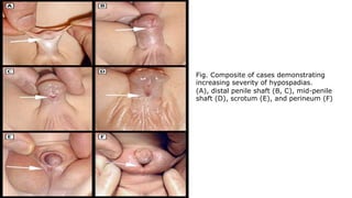
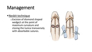
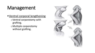
Hypospadias Pushpa Lal Bhadel
Department of Pediatric surgery
Kathmandu Model Hospital
Introduction
Congenital anomaly of the male urethra that results in
abnormal ventral placement of the urethral opening
Defined as a combination of any or all of the following
associated penile anomalies
oEctopic urethral meatus
oPenile curvature (chordee)
oDorsal hooded prepuce
Fig. Composite of cases demonstrating
increasing severity of hypospadias.
(A), distal penile shaft (B, C), mid-penile
shaft (D), scrotum (E), and perineum (F)
Embryology
elongation and enlargement + fusion of urethral
folds
Testosterone
Penis
Genital tubercle
Embryology
 Cloacal ectoderm primitive urethral plate, ventrally
 Medial endoderm male urethra, ventrally
 Lateral ectoderm skin of penile shaft and prepuce
 Layers fuse posteriorly to anteriorly, forming median raphe
 Arrest in fusion hypospadias
 Abnormal development of growth plate, disproportionate corpora
and fibrosis leads to penile curvature
Etiology
 Multifactorial
 Proposed mechanism is disruption of androgenic stimulation required
for development of normal male external genitalia 1
 Genetic defects:
oMutation in genes that affect androgen metabolism, and estrogen and
androgen response
oHOXA4, IRX5, IRX6, EYA1, DGKK
 Associated genetic syndromes
oDenys-Drash syndrome
oWAGR syndrome
oOpitz syndrome
1 Choudhry S, Baskin LS, Lammer EJ, et al. Genetic polymorphisms in ESR1 and ESR2 genes, and risk of hypospadias in a multiethnic study population. J Urol. 2015;193(5):1625-31.
Etiology
Environmental exposures
oPrenatal exposure to estrogenic compounds 1
• Maternal progestin, diethylstilbestrol
oExposure to paints/solvents/adhesives, detergents, pesticides,
cosmetics and industrial chemicals 2
oMaternal high dietary intake of phytoestrogens associated with
decreased risk 3
1 Brouwers MM, Feitz WF, Roelofs LA, Kiemeney LA, De gier RP, Roeleveld N. Risk factors for hypospadias. Eur J Pediatr. 2007;166(7):671-8.
3 Carmichael SL, Cogswell ME, Ma C, Gonzalez-Feliciano A, Olney RS, Correa A, Shaw GM. Hypospadias and maternal intake of phytoestrogens.
American journal of epidemiology 2013;178(3):434-40.
2 Poon S, Koren G, Carnevale A, et al. Association of In Utero Exposure to Polybrominated Diphenyl Ethers With the Risk of Hypospadias. JAMA Pediatr.
2018;172(9):851-856.
Etiology
 Other risk factors 1
oAdvanced maternal age
oPreexisting maternal diabetes mellitus
oGestational age before 37 weeks
oHistory of paternal hypospadias
oExposure to smoking and pesticides
oPlacental insufficiency (low placental weight and pathology)
oPrematurity
oFetal growth restriction
oIn-vitro fertilization
1 Brouwers MM, Feitz WF, Roelofs LA, Kiemeney LA, De gier RP, Roeleveld N. Risk factors for hypospadias. Eur J Pediatr. 2007;166(7):671-8.
Incidence
 Most common congenital anomalies
 Ranges from 0.3-0.7 percent live births 1
 Risk is 13-fold in 1st degreerelatives
 Risk of hypospadias in next child, when one child is affected withhypospadias:
o 12% with negative family history
o 19% if cousin or uncle has hypospadias
o 26% if father or sibling has hypospadias
 8.5 times more risk in monozygotic twins
1 Nassar N, Bower C, Barker A. Increasing prevalence of hypospadias in Western Australia, 1980-2000. Arch Dis Child. 2007;92(7):580-4.
CLASSIFICATION
ANTERIOR
•GRANULAR
•CORONAL
•SUBCORONAL
MIDDLE
DISTAL PENILE
MID SHAFT
PROXIMALPENILE
POSTERIOR
•PENOSCROTAL
•SCROTAL
•PERINEAL
Classification
Diagnosis
Generally made during the newborn genital examination
Physical findings consistent with the diagnosis include:
oAbnormal foreskin resulting in an incomplete closure around the
glans : dorsal hooded prepuce.
oAbnormal penile curvature (chordee).
oThe appearance of "two urethral openings";
• First in the normal position at the end of the glans, which is usually a blind
ending urethral pit
• Second, the abnormally located true urethral meatus
Initial evaluation
 Includes focused history, close examination of genitalia and
identification of other congenital anomalies
 History: positive family history, maternal or infant risk factors
 Physical examination:
oIdentifying other associated anomalies and genital examination determining
the presence and degree of hypospadias
Initial evaluation
 Genital examination:
 Assessment of stretched penile length: 2.5-3.5 cm in full term male
 Assessment regarding evidence of penile curvature if so, degree of curvature
 Severity:
• Normal – No curvature to 15 degrees
• Mild – 15 to 40 degrees
• Moderate – 40 to 80 degrees
• Severe – >80 degrees
 Assessment of foreskin to ensure complete and circumferential development
without ant ventral asymmetry or deficiency
 Determination of the presence of both testicles in the normal dependent position
in the scrotum.
Management
Indications of surgery
Functional indications:
 Proximally located meatus
 Ventrally deflected urinary stream
 Meatal stenosis
 Curved penis
Management
Cosmetic indications : strongly linked patient’s future
psychology are:
oAbnormally located meatus
oCleft glans
oRotated penis with abnormal cutaneous raphe
oPreputial hood
oPenoscrotal transposition
oSplit scrotum
Management
Timing of surgery:
oRecommendations from American Academy of Pediatrics (AAP)
oBetween 6 months and 1 year of age
oAllows ample time for completion of two-stage procedure in
patient with severe hypospadias
oPrior to beginning of gender identification
oTime between two stages: six months
 Preoperative Hormonal Stimulation:
HCG 250-500 U sc twice a week for 3 weeks. Increase in penile
size and length
Decrease in hypospadias and chordee severity
Increased vascularity and thickness of corpus spongiosum
Allowance of more simple repairs
IM testosterone enanthate – 2mg/kg/dose given for a total of 2
or 3 doses before hypospadias repair
Testosterone propionate cream – 2% three times daily for 3
weeks
Management
General principles of hypospadias repair
oOrthoplasty
oUrethroplasty
oMeatoplasty
oGranuloplasty
oSkin coverage
Management
Orthoplasty
oCorrection of ventral curvature
oVentral tissues—including shaft skin, dartos, corpus
spongiosum, urethral plate, and overlying tunics of the
corpora cavernosa may be shortened relative to the dorsal
surface.
oVC occurred in 11% of primary distal cases, 30% midshaft, and
81% proximal hypospadias.
Management
Orthoplasty
oPreoperative assessment cannot accurately predict either the
extent of curvature or the means required for straightening.
oIntraoperative assessment of penile curvature by either
artificial or pharmacologic methods is a critical step in
hypospadias repair.
oPerformed after degloving of penile shaft skin
Management
 Artificial erection induced by saline injection remains the
most used means to assess presence and severity of VC.
 Pharmacologic erection allows for a more accurate and
continued assessment of penile curvature before, during,
and after its correction.
 Intracorporal injection of the arterial vasodilator
prostaglandin E1.
Management
Curvature up to 30 degrees
can be corrected by Midline
Dorsal Plication into the
tunica albuginea of the
corpora cavernosa directly
opposite the area of greatest
bending.
Management
Nesbit technique
oExcision of diamond shaped
wedge/s at the point of
maximum curvature and
closing the tunica transversely
with absorbable sutures.
Management
Ventral corporal lengthening
oVentral corporotomy with
grafting.
oMultiple corporotomy
without grafting.
Management
Dermal graft – Devine and Horton
Management
Urethroplasty
Distal hypospadias :
oTIP repair
oOthers like MAGPI, Mathieu flip-flap, and Urethral advancement.
Midshaft hypospadias :
oTIP repair
oOnlay preputial flap
Management
 Proximal hypospadias :
o TIP repair
o Onlay preputial flap
o Single stage urethroplasty with preputial flap or the
Koyanagi flap.
o Two stage repair with Byars flaps or grafts.
Management
Distal hypospadias
Tubularized Incised Plate (TIP) repair
oCircumscribing incision is made approximately 2 mm below meatus
oVentral V incision
oPenis degloved
oMidline incision of the urethral plate
oUrethral plate tubularization begins distally approximately 3 mm
from the end of the plate, ensuring an oval, not rounded, meatus.
Management
 Dartos flap is dissected from the dorsal prepuce and shaft
skin, buttonholed, and transposed ventrally to cover the
neourethra.
 Glansplasty begins distally, and a 7-0 polyglactin suture.
Management
Midshaft hypospadias
TIP repair
Onlay preputial flap
oThin skin proximal to the urethral meatus is incised to the midline
convergence of corpus spongiosum wings.
oThen inner prepuce is harvested on its vascular pedicle from
either the dorsal hood or dartos flap.
oThe flap should be gently stretched to fit the urethral plate without
redundancy.
Management
Proximal hypospadias
 The greatest controversy in primary hypospadias surgery concerns
decision making for proximal cases.
 Options depend on whether the urethral plate is available for
urethroplasty after associated VC is straightened.
 If so, then either TIP repair or an onlay preputial flap can be used.
 When the urethral plate is transected a one-stage urethroplasty
can be accomplished by tubularized preputial flaps or the Koyanagi
flap or a two-stage repair done with Byars flaps or preputial grafts.
Management
Proximal tubularized incised plate repair
oCircumscribing incision preserves urethral plate in patient desiring
circumcision.
oAfter degloving, glans wings are separated from the urethral plate.
oCorpus spongiosum is dissected from the cavernosal bodies
oMidline urethral plate incision.
oSpongioplasty over the neourethra.
Management
Tunica vaginalis flap
Management
Koyanagi flap
 Proposed lines of incisions to create flap
 The flap can be divided into two wings as shown or maintained in
one piece with a central buttonhole to transpose it ventrally.
Management
The urethral plate in the center of the flap
is dissected from the corpora to near the
meatus, and the glanular portion of the
plate is excised as glans wings are made.
Inner flap margins are reapproximated,
and excess flap skin is excised.
The outer margins are closed to complete
tubularization
Management
Byars flap
 After degloving and release of ventral dartos, persisting
ventral curvature greater than 30 degrees led to
excision of the urethral plate.
 The dorsal preputial hood is incised in the midline and
the two flaps transposed ventrally on either side of the
penis.
 The prepuce is advanced into the glans; alternatively,
the urethral plate can be maintained within the glans
Management
 Flap edges are approximated
in the midline.
 Six months later a U-shaped
incision is made
approximately 10 mm wide.
Management
The resultant strip is tubularized in two layers.
Complications
 Bleeding/hematoma,
 Meatal stenosis,
 Urethrocutaneous fistula,
 Urethral stricture,
 Urethral diverticulum,
 Wound infection,
 Impaired healing, and
 Breakdown of the repair
THANK YOU

Hypospadias.pptx

  • 1.
    Hypospadias Pushpa LalBhadel Department of Pediatric surgery Kathmandu Model Hospital
  • 2.
    Introduction Congenital anomaly ofthe male urethra that results in abnormal ventral placement of the urethral opening Defined as a combination of any or all of the following associated penile anomalies oEctopic urethral meatus oPenile curvature (chordee) oDorsal hooded prepuce
  • 3.
    Fig. Composite ofcases demonstrating increasing severity of hypospadias. (A), distal penile shaft (B, C), mid-penile shaft (D), scrotum (E), and perineum (F)
  • 4.
    Embryology elongation and enlargement+ fusion of urethral folds Testosterone Penis Genital tubercle
  • 5.
    Embryology  Cloacal ectodermprimitive urethral plate, ventrally  Medial endoderm male urethra, ventrally  Lateral ectoderm skin of penile shaft and prepuce  Layers fuse posteriorly to anteriorly, forming median raphe  Arrest in fusion hypospadias  Abnormal development of growth plate, disproportionate corpora and fibrosis leads to penile curvature
  • 6.
    Etiology  Multifactorial  Proposedmechanism is disruption of androgenic stimulation required for development of normal male external genitalia 1  Genetic defects: oMutation in genes that affect androgen metabolism, and estrogen and androgen response oHOXA4, IRX5, IRX6, EYA1, DGKK  Associated genetic syndromes oDenys-Drash syndrome oWAGR syndrome oOpitz syndrome 1 Choudhry S, Baskin LS, Lammer EJ, et al. Genetic polymorphisms in ESR1 and ESR2 genes, and risk of hypospadias in a multiethnic study population. J Urol. 2015;193(5):1625-31.
  • 7.
    Etiology Environmental exposures oPrenatal exposureto estrogenic compounds 1 • Maternal progestin, diethylstilbestrol oExposure to paints/solvents/adhesives, detergents, pesticides, cosmetics and industrial chemicals 2 oMaternal high dietary intake of phytoestrogens associated with decreased risk 3 1 Brouwers MM, Feitz WF, Roelofs LA, Kiemeney LA, De gier RP, Roeleveld N. Risk factors for hypospadias. Eur J Pediatr. 2007;166(7):671-8. 3 Carmichael SL, Cogswell ME, Ma C, Gonzalez-Feliciano A, Olney RS, Correa A, Shaw GM. Hypospadias and maternal intake of phytoestrogens. American journal of epidemiology 2013;178(3):434-40. 2 Poon S, Koren G, Carnevale A, et al. Association of In Utero Exposure to Polybrominated Diphenyl Ethers With the Risk of Hypospadias. JAMA Pediatr. 2018;172(9):851-856.
  • 8.
    Etiology  Other riskfactors 1 oAdvanced maternal age oPreexisting maternal diabetes mellitus oGestational age before 37 weeks oHistory of paternal hypospadias oExposure to smoking and pesticides oPlacental insufficiency (low placental weight and pathology) oPrematurity oFetal growth restriction oIn-vitro fertilization 1 Brouwers MM, Feitz WF, Roelofs LA, Kiemeney LA, De gier RP, Roeleveld N. Risk factors for hypospadias. Eur J Pediatr. 2007;166(7):671-8.
  • 9.
    Incidence  Most commoncongenital anomalies  Ranges from 0.3-0.7 percent live births 1  Risk is 13-fold in 1st degreerelatives  Risk of hypospadias in next child, when one child is affected withhypospadias: o 12% with negative family history o 19% if cousin or uncle has hypospadias o 26% if father or sibling has hypospadias  8.5 times more risk in monozygotic twins 1 Nassar N, Bower C, Barker A. Increasing prevalence of hypospadias in Western Australia, 1980-2000. Arch Dis Child. 2007;92(7):580-4.
  • 10.
  • 11.
  • 12.
    Diagnosis Generally made duringthe newborn genital examination Physical findings consistent with the diagnosis include: oAbnormal foreskin resulting in an incomplete closure around the glans : dorsal hooded prepuce. oAbnormal penile curvature (chordee). oThe appearance of "two urethral openings"; • First in the normal position at the end of the glans, which is usually a blind ending urethral pit • Second, the abnormally located true urethral meatus
  • 13.
    Initial evaluation  Includesfocused history, close examination of genitalia and identification of other congenital anomalies  History: positive family history, maternal or infant risk factors  Physical examination: oIdentifying other associated anomalies and genital examination determining the presence and degree of hypospadias
  • 14.
    Initial evaluation  Genitalexamination:  Assessment of stretched penile length: 2.5-3.5 cm in full term male  Assessment regarding evidence of penile curvature if so, degree of curvature  Severity: • Normal – No curvature to 15 degrees • Mild – 15 to 40 degrees • Moderate – 40 to 80 degrees • Severe – >80 degrees  Assessment of foreskin to ensure complete and circumferential development without ant ventral asymmetry or deficiency  Determination of the presence of both testicles in the normal dependent position in the scrotum.
  • 15.
    Management Indications of surgery Functionalindications:  Proximally located meatus  Ventrally deflected urinary stream  Meatal stenosis  Curved penis
  • 16.
    Management Cosmetic indications :strongly linked patient’s future psychology are: oAbnormally located meatus oCleft glans oRotated penis with abnormal cutaneous raphe oPreputial hood oPenoscrotal transposition oSplit scrotum
  • 17.
    Management Timing of surgery: oRecommendationsfrom American Academy of Pediatrics (AAP) oBetween 6 months and 1 year of age oAllows ample time for completion of two-stage procedure in patient with severe hypospadias oPrior to beginning of gender identification oTime between two stages: six months
  • 18.
     Preoperative HormonalStimulation: HCG 250-500 U sc twice a week for 3 weeks. Increase in penile size and length Decrease in hypospadias and chordee severity Increased vascularity and thickness of corpus spongiosum Allowance of more simple repairs IM testosterone enanthate – 2mg/kg/dose given for a total of 2 or 3 doses before hypospadias repair Testosterone propionate cream – 2% three times daily for 3 weeks
  • 19.
    Management General principles ofhypospadias repair oOrthoplasty oUrethroplasty oMeatoplasty oGranuloplasty oSkin coverage
  • 20.
    Management Orthoplasty oCorrection of ventralcurvature oVentral tissues—including shaft skin, dartos, corpus spongiosum, urethral plate, and overlying tunics of the corpora cavernosa may be shortened relative to the dorsal surface. oVC occurred in 11% of primary distal cases, 30% midshaft, and 81% proximal hypospadias.
  • 21.
    Management Orthoplasty oPreoperative assessment cannotaccurately predict either the extent of curvature or the means required for straightening. oIntraoperative assessment of penile curvature by either artificial or pharmacologic methods is a critical step in hypospadias repair. oPerformed after degloving of penile shaft skin
  • 24.
    Management  Artificial erectioninduced by saline injection remains the most used means to assess presence and severity of VC.  Pharmacologic erection allows for a more accurate and continued assessment of penile curvature before, during, and after its correction.  Intracorporal injection of the arterial vasodilator prostaglandin E1.
  • 25.
    Management Curvature up to30 degrees can be corrected by Midline Dorsal Plication into the tunica albuginea of the corpora cavernosa directly opposite the area of greatest bending.
  • 26.
    Management Nesbit technique oExcision ofdiamond shaped wedge/s at the point of maximum curvature and closing the tunica transversely with absorbable sutures.
  • 27.
    Management Ventral corporal lengthening oVentralcorporotomy with grafting. oMultiple corporotomy without grafting.
  • 28.
  • 29.
    Management Urethroplasty Distal hypospadias : oTIPrepair oOthers like MAGPI, Mathieu flip-flap, and Urethral advancement. Midshaft hypospadias : oTIP repair oOnlay preputial flap
  • 30.
    Management  Proximal hypospadias: o TIP repair o Onlay preputial flap o Single stage urethroplasty with preputial flap or the Koyanagi flap. o Two stage repair with Byars flaps or grafts.
  • 31.
    Management Distal hypospadias Tubularized IncisedPlate (TIP) repair oCircumscribing incision is made approximately 2 mm below meatus oVentral V incision oPenis degloved oMidline incision of the urethral plate oUrethral plate tubularization begins distally approximately 3 mm from the end of the plate, ensuring an oval, not rounded, meatus.
  • 32.
    Management  Dartos flapis dissected from the dorsal prepuce and shaft skin, buttonholed, and transposed ventrally to cover the neourethra.  Glansplasty begins distally, and a 7-0 polyglactin suture.
  • 33.
    Management Midshaft hypospadias TIP repair Onlaypreputial flap oThin skin proximal to the urethral meatus is incised to the midline convergence of corpus spongiosum wings. oThen inner prepuce is harvested on its vascular pedicle from either the dorsal hood or dartos flap. oThe flap should be gently stretched to fit the urethral plate without redundancy.
  • 36.
    Management Proximal hypospadias  Thegreatest controversy in primary hypospadias surgery concerns decision making for proximal cases.  Options depend on whether the urethral plate is available for urethroplasty after associated VC is straightened.  If so, then either TIP repair or an onlay preputial flap can be used.  When the urethral plate is transected a one-stage urethroplasty can be accomplished by tubularized preputial flaps or the Koyanagi flap or a two-stage repair done with Byars flaps or preputial grafts.
  • 37.
    Management Proximal tubularized incisedplate repair oCircumscribing incision preserves urethral plate in patient desiring circumcision. oAfter degloving, glans wings are separated from the urethral plate. oCorpus spongiosum is dissected from the cavernosal bodies oMidline urethral plate incision. oSpongioplasty over the neourethra.
  • 39.
  • 40.
    Management Koyanagi flap  Proposedlines of incisions to create flap  The flap can be divided into two wings as shown or maintained in one piece with a central buttonhole to transpose it ventrally.
  • 41.
    Management The urethral platein the center of the flap is dissected from the corpora to near the meatus, and the glanular portion of the plate is excised as glans wings are made. Inner flap margins are reapproximated, and excess flap skin is excised. The outer margins are closed to complete tubularization
  • 42.
    Management Byars flap  Afterdegloving and release of ventral dartos, persisting ventral curvature greater than 30 degrees led to excision of the urethral plate.  The dorsal preputial hood is incised in the midline and the two flaps transposed ventrally on either side of the penis.  The prepuce is advanced into the glans; alternatively, the urethral plate can be maintained within the glans
  • 43.
    Management  Flap edgesare approximated in the midline.  Six months later a U-shaped incision is made approximately 10 mm wide.
  • 44.
    Management The resultant stripis tubularized in two layers.
  • 45.
    Complications  Bleeding/hematoma,  Meatalstenosis,  Urethrocutaneous fistula,  Urethral stricture,  Urethral diverticulum,  Wound infection,  Impaired healing, and  Breakdown of the repair
  • 46.

Editor's Notes

  • #3 Ventral foreskin deficiency with incomplete foreskin closure around the glans
  • #4 Location of displaced urethral meatus may range anywhere: Glans, Shaft of penis, The scrotum, perineum
  • #7 Denys-Drash (renal insufficiency, male pseudohermaphroditism, and Wilms tumor) WAGR (Wilms tumor, aniridia, genitourinary malformations, and mental retardation) Opitz (ocular hypertelorism, asymmetry of the skull, and laryngoesophageal defects)
  • #15 •If the foreskin is normally circumferential, it is unlikely that a significant hypospadias requiring surgery is present. As a result, it is not necessary to retract the foreskin, which in the neonate is typically adherent to the glans. In addition, the terminal meatus may be seen through the tiny opening in the normal newborn foreskin. •If the foreskin is not properly formed, the presence of an ectopic urethral meatus along the ventral shaft of the penis should be sought after and identified.
  • #30 MAGPI- Meatal Advancement and Granuloplasty Incorporation