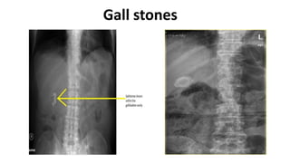
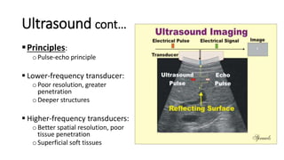
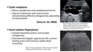
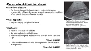
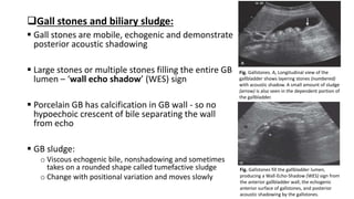
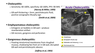
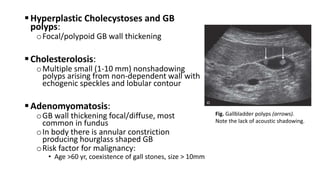
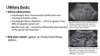
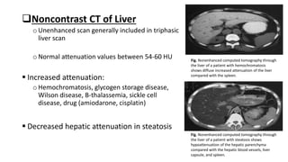
Hepatobiliary Imaging
Pushpa Lal Bhadel
FCPS Resident
Department of Surgery
Kathmandu Model Hospital
Moderator:
Dr. Udaya Koirala
HOD
Department of General Surgery
 Acute conditions are often inflammatory
 Pain is a predominant syndrome
 Jaundice: benign and malignant HPB disease
 Thorough and complete history and clinical examination
 Avoid ‘scan first, clinic later’
Imaging modalities
 X-ray
 Ultrasound
 Computed Tomography (CT)
 Magnetic Resonance Imaging(MRI) and Magnetic Resonance
Cholangiopancreatography (MRCP)
 Endoscopic Retrograde Cholangiopancreatography (ERCP)
 Hepatic Iminodiacetic acid (HIDA) scan
 Percutaneous Transhepatic Cholangiography (PTC)
 Nuclear Medicine
 Direct cholangiography
 Intraoperative diagnostic techniques
Plain abdominal X-ray
Used in past, but has been widely replaced by US
Can be used to visualize:
oCalcified stones
oEmphysematous cholecystitis
oBiliary fistula (gas within biliary system)
oPorcelain gall bladder
Gall stones
Fig. Abdominal radiograph (frontal projection) shows
intraluminal air (arrow) with air– fluid levels –
Emphysematous cholecystitis
Fig. Plain x-ray showing air in the biliary tree
Well defined globular coarse calcification of gall bladder wall in right hypochonrium with an oval calcified area adjacent to it suggestive
Fig. Well defined globular
coarse calcification of gall
bladder wall in right
hypochonrium with an oval
calcified area adjacent to it
suggestive of GB calculus.
Ultrasound
 Favored imaging modality
oVersatility
oLow cost
oReal-time capability and portability
oNo harmful biologic effects on operator or patient
oDoppler US – assessment of blood flow dynamics
 Limitations:
oWaves unable to penetrate bone/air
oOperator dependent
oInfluenced by skill and experience
Ultrasound cont…
Principles:
oPulse-echo principle
 Lower-frequency transducer:
oPoor resolution, greater
penetration
oDeeper structures
 Higher-frequency transducers:
oBetter spatial resolution, poor
tissue penetration
oSuperficial soft tissues
Ultrasound cont…
 Echogenicity: reflection of transmission of
US waves relative to surrounding tissue
 Based on gray scale imaging, structure
displayed characterized as:
oAnechoic (uniformly black)
oHypoechoic (dark gray)
oHyperechoic (light gray)
Fig. Transverse gray scale ultrasound image of
the liver shows the right hepatic vein (hv) as an
anechoic fluid-containing structure. Note that
the walls of the veins are hyperechoic.
Ultrasound cont…
Doppler ultrasound:
oTo identify and evaluate blood flow in vessels
oAssessment of vessel patency, direction of blood flow, flow velocity
 Doppler effect: change in frequency proportional to the velocity of
reflector relative to transducer
 Three different Doppler displays:
oColor doppler
oPower doppler
oSpectral doppler
Fig. Doppler ultrasound of the liver.
A, Color Doppler mode shows mean flow velocity and direction of flow
toward or away from the transducer, as indicated in the bar display at left
of the image. The portal vein (PV) indicates hepatopetal flow. The inferior
vena cava (IVC) shows flow away from the transducer. The hepatic artery
(arrowhead) has mixed signals due to high-velocity flow and aliasing.
B, Power Doppler shows amplitude of flow but not direction. It is useful for
small vessels and low flow, as shown in this image of vessels in the
gallbladder (GB) wall (arrowheads).
C, Spectral Doppler shows flow pattern over time; when angle corrected, it
can measure true velocity. A sample cursor is placed within the vessel, as
shown in this transverse image of the middle hepatic vein (MHV).
Liver ultrasound:
 Hepatic parenchyma is hypoechoic to spleen, either isoechoic or minimally
hyperechoic to renal parenchyma
 Commonly determined by longitudinal image of right lobe
Fig. Normal liver longitudinal ultrasound images.
A, Left hepatic lobe. Segments are numbered. LH, Left hepatic vein.
B, Left lobe segment IV. Hepatic segments are numbered. GB, Gallbladder; IVC, inferior vena cava; PV, portal vein.
C, Right lobe. Hepatic segments are numbered. RHV, Right hepatic vein; RK, right kidney.
Fig. Transverse images of the liver.
A, Transverse sonogram at the level of
the hepatic veins. Hepatic segments
are numbered. IVC, inferior vena cava;
L, left hepatic vein; M, middle hepatic
vein; R, right hepatic vein.
B, Transverse section of the left lobe
at the level of the portal
vein bifurcation. L, Left portal vein; M,
uppermost main portal vein; R, right
portal vein;.
C, Transverse image of the left portal
vein (LPV) and right portal vein (RPV)
at the bifurcation. Segments IVB and I
are indicated. A, Aorta; IVC,inferior
vena cava; L, left hepatic vein; R, right
hepatic vein; RK, right kidney;.
D, Transverse image of the inferior
right lobe segments V and VI. IVC,
inferior vena cava; K, Right kidney; RA,
anterior right portal vein; RP, posterior
right portal vein.
Hepatic masses:
 On gray scale US liver masses differentiated by internal architecture
and described as cystic, hypoechoic or hyperechoic
 Hypoechogenicity: feature of malignancies
 Hyperechoic masses: benign etiologies like hemangioma, focal fat,
HCC, metastases
Role of contrast-enhanced US(CEUS) in evaluation of focal liver lesion:
 Non-invasive real-time imaging
 US contrast agent:
o Microbubbles that are highly echogenic and oscillate in US field
o Enhance US signal in gray scale, color and spectral Doppler
o Bubbles capable of transpulmonary passage
 Improves characterization of focal liver mass and liver lesion
 Improved ability to see greater number and smaller lesions
 Diagnostic performance is similar to CT and MRI in recognition of
malignancy.
(Trillaud et. al, 2009)
 Vascular phases:
oArterial (10-40 seconds)
oPortal venous (40-120 seconds)
oLate parenchymal (>120 seconds)
(Wilson & Burns, 2006)
Benign liver lesion:
 Cystic masses:
o Demonstrate smooth, barely perceptible walls,
posterior acoustic enhancement and no internal
echoes
o Partial septation, thin septation or puckered wall may
be present
 Lymphomatous mass:
o Markedly hypoechoic, may mimic simple cyst,
demonstrate internal vascularity on color doppler
 Abscess:
o Solid initially, then cystic with debris, wall is usually
vascular
 Echinococcal cyst:
o Variable appearance, simple fluid-filled cyst, may
contain wavy membranes from rupture and detached
endocyst, may contain daughter cysts or may show
calcification
(Lewall & McCorkell, 1985)
Fig. Benign hepatic cyst with a thin,
smooth wall. Note the posterior acoustic
enhancement (arrows) that is a
characteristic feature of a cystic lesion.
Fig. Hepatic abscess in a patient after embolization
for hepatic neuroendocrine metastases
(arrowheads). The abscess (asterisk) contains
complex fluid and has posterior acoustic
enhancement (arrows).
 Cystic neoplasm:
oBiliary cystadenoma and cystadenocarcinoma
oMay be multilocular with cystic locules
demonstrating different echogenicities depending
on fluid content
(Levy et. Al, 2002)
 Focal nodular Hyperplasia:
oSmooth lobulated contour and variable
echogenicity
oCharacteristic Doppler appearance off a central
feeding artery with tortuous spoke wheel
vascularity
Fig. The images show a large and well-defined
multiloculated cystic lesion in the IV-hepatic-
segment with enhancing septations without any
solid pole or calcifications in the wall.
Fig. Focal nodular hyperplasia (FNH). Gray scale image of a
hypoechoic FNH. FNH lesions are often subtle.
 Malignant liver neoplasm:
oSensitivity 58-89%
(Bolondi, 2003)
oHCC: solitary, multiple or diffuse extending to bile
ducts causing biliary obstruction or hemobilia
(Kojiro et. Al, 1982)
oSmall HCC(<5cm): hypoechoic, may be hyperechoic
d/t fatty metamorphosis
(Caturelli et. Al, 2001)
oLarger HCC: heterogeneous d/t liquefaction necrosis
and fibrosis
(Tanaka et. Al, 1983)
oCEUS improves HCC characterization
oRegional lymphadenopathy is common
Fig. Hepatocellular carcinoma (HCC) with
portal vein thrombosis. A, Transverse image
shows a hypoechoic HCC in the right hepatic
lobe (arrows).
 Liver metastases:
oVary according to primary malignancy
oCommon appearance is hypoechoic lesions or lesions
with peripheral hypoechoic halos
oHypoechoic halo: fibrosis, compressed sinusoids,
tumor neovascularity surrounding the metastatic
deposit
(Kruskal et. Al, 2000)
oHypoechoic mets: metastatic breast, lung,
pancreatic, gastric and esophageal carcinoma,
lymphoma and HCC
oEchogenic mets: HCC or GI malignancies
oHyperechoic mets: vascular mets, mets from RCC
islet cell tumor, carcinoids and cholangiocarcinoma
(Tanaka et. Al, 1990, Marchal et. Atl, 1985)
Fig. Liver metastases with hypoechoic
halo. Metastatic neuroendocrine tumors
(arrows) in the liver have a hypoechoic
halo peripherally and a target
appearance. A hypoechoic halo usually
indicates malignant lesion or, less
commonly, an infectious etiology.
Sonography of diffuse liver disease
 Fatty liver disease:
o Fat deposition within hepatocytes results in increased
echogenicity of liver, decreased acoustic penetration and loss
of echogenic borders of portal vessels
 Viral hepatitis:
o Hepatomegaly, peripheral edema
 Cirrhosis:
o Neither sensitive nor specific
o Surface nodularity- reliable sign
o Nodularity along the deep surfaces or liver- more sensitive
sign
(Filly et. Al, 2002)
o Coarsened echotexture and heterogeneous parenchymal
echogenicity
(Caturelliet. Al, 2003)
Fig. Diffuse liver disease. A, Hepatic
steatosis, longitudinal right lobe. Hepatic
parenchyma of fatty liver is very
echogenic relative to the right kidney.
Fig. Cirrhosis. The liver is small with
coarsened echogenicity and a nodular
surface (arrows). a, Aorta.
US of gall bladder and biliary tree
 Sonographically GB is best visualized from the
right intercostal approach with patient supine or
left lateral decubitus
 Best evaluated when distended, thus patient
should fast before scan
 CBD best seen from right intercostal approach
with the patient in left lateral decubitus position
 The right and left hepatic duct lie anterior to
portal vein confluence and are best visualized on
transverse subcostal US image
Fig. Gallbladder. A, Normal longitudinal
gallbladder image with visualization of the
gallbladder neck.
Gall stones and biliary sludge:
 Gall stones are mobile, echogenic and demonstrate
posterior acoustic shadowing
 Large stones or multiple stones filling the entire GB
lumen – ‘wall echo shadow’ (WES) sign
 Porcelain GB has calcification in GB wall - so no
hypoechoic crescent of bile separating the wall
from echo
 GB sludge:
o Viscous echogenic bile, nonshadowing and sometimes
takes on a rounded shape called tumefactive sludge
o Change with positional variation and moves slowly
Fig. Gallstones. A, Longitudinal view of the
gallbladder shows layering stones (numbered)
with acoustic shadow. A small amount of sludge
(arrow) is also seen in the dependent portion of
the gallbladder.
Fig. Gallstones fill the gallbladder lumen,
producing a Wall-Echo-Shadow (WES) sign from
the anterior gallbladder wall, the echogenic
anterior surface of gallstones, and posterior
acoustic shadowing by the gallstones.
 Cholecystitis:
o Sensitivity: 80-100%, specificity: 60-100%, PPV: 90-94%
(Harvey & Miller, 1999)
o GB wall thickening > 3mm, pericholecystic fluid,
positive sonographic Murphy sign
(Smith et.al, 2009)
 Emphysematous cholecystitis:
o Echogenic air bubbles in GB wall – produce
reverberation artifact
o GB necrosis, gangrene and perforation
 Gangrenous cholecystitis:
o Floating intraluminal membranes from sloughed
mucosa, shadowing foci from air in GB wall, disrupted
GB wall and pericholecystic abscess
(Jeffrey et.al, 1983)
Hyperplastic Cholecystoses and GB
polyps:
oFocal/polypoid GB wall thickening
Cholesterolosis:
oMultiple small (1-10 mm) nonshadowing
polyps arising from non-dependent wall with
echogenic speckles and lobular contour
Adenomyomatosis:
oGB wall thickening focal/diffuse, most
common in fundus
oIn body there is annular constriction
producing hourglass shaped GB
oRisk factor for malignancy:
• Age >60 yr, coexistence of gall stones, size > 10mm
Fig. Gallbladder polyps (arrows).
Note the lack of acoustic shadowing.
Gall bladder carcinoma:
 Sessile/polypoid mass, thickened GB
wall/infiltrative mass that fills the GB lumen and
extends into the adjacent liver
(Wibbernmeyer et.at, 1995)
 Secondary signs: discontinuity of echogenic
mucosal lining, absence of echogenic specks seen
in cholesterol crystals and high velocity arterial
flow greater than 60 cm/s
 Selective mucosal calcification is significantly
associated with GB cancer but diffuse intramural
calcification is not
(Stephen & Berger 2001)
Fig. Gallbladder carcinoma. A, Gray scale
longitudinal image of the gallbladder
shows a solid irregular mass in the
fundus.
Biliary Ducts:
 Biliary obstruction:
oExtrahepatic duct measurement performed near
crossing of hepatic artery
oIntrahepatic biliary dilatation - >2mm or greater than
40% of adjacent portal vein
oDouble track sign – caused by dilated bile ducts parallel
to the portal vein branches
 Bile duct calculi: appear as intraluminal filling
defects
Fig. Biliary obstruction from choledocholithiasis.
Transverse sonogram of the liver reveals the
“double-track” sign (circled areas), consistent
with intrahepatic biliary dilatation.
Fig. B, Longitudinal view of the common bile duct
(cbd) shows an echogenic stone (arrow) that
produces acoustic shadowing (arrowheads). gb,
Gallbladder; v, portal vein.
Cholangiocarcinoma:
 Intrahepatic peripheral form:
oFocal mass, peripheral biliary ductal dilatation(25%),
capsular retraction, lack of hypoechoic halo, lack
venous thrombosis
(Chung et.al, 2009)
 Hilar cholangiocarcinoma:
oSegmental upstream dilatation with abrupt cutoff
and nonunion of right and left dilated ducts at porta
hepatis
oIsoechoic, larger tumors have hypoechoic rim
(Hann et.al, 1997)
 Extrahepatic cholangiocarcinoma:
oInfiltrative spreading along the duct walls, nodular
mural thickening or papillary
(Hann et.al, 1997)
Fig. Hilar cholangiocarcinoma
Computed tomography (CT)
 Traditionally primarily been used for hepatic lesion
detection and characterization
 Identifies peritoneal disease involvement, distant
lymphadenopathy and other distant metastatic disease, key
vessel involvement
 Role in initial staging of patients with malignant disease and
preprocedural vascular evaluation
 Portal and hepatic veins can be identified as anatomic
landmarks to localize tumors to specific hepatic segments
 To assess the proximity of lesions to the inflow and
outflow vessels
Fig. Volume-rendered 3D model of the
liver created from axially acquired
computed tomographic data.
Noncontrast CT of Liver
oUnenhanced scan generally included in triphasic
liver scan
oNormal attenuation values between 54-60 HU
 Increased attenuation:
oHemochromatosis, glycogen storage disease,
Wilson disease, B-thalassemia, sickle cell
disease, drug (amiodarone, cisplatin)
 Decreased hepatic attenuation in steatosis
Fig. Nonenhanced computed tomography through
the liver of a patient with hemochromatosis
shows diffuse increased attenuation of the liver
compared with the spleen.
Fig. Nonenhanced computed tomography through
the liver of a patient with steatosis shows
hypoattenuation of the hepatic parenchyma
compared with the hepatic blood vessels, liver
capsule, and spleen.
Contrast enhanced CT of Liver:
 Different enhancement of various types of
lesion relative to background hepatic
parenchyma dictates timing/phases
 Arterial phase imaging:
oHigher injection rate (4-6 ml/s) for early arterial
imaging
oImages acquired at ~20-30 seconds
oLate arterial phase used to evaluate
hypervascular lesion (about 40 seconds): HCC,
metastatic NET
Fig. A, Late arterial-phase image reveals
multiple hypervascular enhancing foci of
metastatic renal cell carcinoma (arrows).
B, Portal venous phase image at the same
level does not reveal the numerous masses.
 Portal venous phase imaging:
oNormal hepatic parenchyma enhances
maximally at approx. 70 sec
oMaximal contrast differential between typical
hypovascular liver lesion and surrounding
parenchyma is achieved
oClear delineation of portal and hepatic veins
 Delayed imaging phase:
oHypervascular tumors becomes
hypoattenuation after late arterial phase
oIf suspicious of HCC ~3-5 min delayed phase
(late venous/equilibrium phase) may be added
Fig. A, Arterial phase image through the inferior right
hepatic lobe reveals branches of the right hepatic
artery. Faint enhancement of a hypervascular mass is
seen in segment VI, which proved to be focal nodular
hyperplasia (arrow).
B, Portal venous phase image through the same
level reveals enhancement of the right portal vein
(long arrow) and the sectoral branches. The mass
(short arrow) is nearly hypointense to the liver.
Systematic approach to segmentation
Benign tumors and tumor like conditions
of liver
 Cysts:
oOn CT they are round or ovoid, sharply-marginated,
bounded by imperceptible wall and they do not
enhance after administration of IV contrast
 Hemangioma:
oHypoattenuating to surrounding liver, after IV
contrast on arterial phase - peripheral
discontinuous nodular enhancement
(Quinn & Benjamin, 1992)
oPortal and late phase demonstrate progressive
centripetal enhancement, a ‘filling in’ of
enhancement towards center of lesion
(Leslie et.al, 1995)
Fig. Multiple simple hepatic cysts in a
patient with polycystic kidneys.
Fig. Cavernous hemangioma of the right
liver showing typical enhancement
starting at the periphery of the lesion
Inflammatory conditions:
Pyogenic abscess:
oUnilocular cavities with smooth outer
margins to highly complex and septated
structures with internal debris and irregular
contour
Echinococcus:
oLarge solitary mass or multiple well-defined
cystic lesion often containing internal
daughter cyst
oCoarse calcification(50%)
Fig. Contrast-enhanced computed tomography of a
pyogenic liver abscess shows enhancement of the
walls and internal septa.
Fig. Contrast-enhanced axial CT reveals a
multiseptated echinococcal cyst in the right
hepatic lobe. Note eccentric calcifications along
the wall of one of the cysts (arrow).
 Cirrhosis:
oBands or regions of confluent fibrosis, relative
left lateral segment and caudate lobe
hypertrophy with right lobe and left medial
segment atrophy, nodular hepatic contour
oElevation of caudate-to-right lobe ratio >0.65
(Harbin et.al, 1980)
Malignant lesions
 HCC:
oLiver cirrhosis (70%)
oHypervascular in arterial phase, subsequently
becoming hypoattenuating to surrounding liver
in later phase (washout appearance)
Fig. Cirrhosis. The interlobar fissure, delineated by the
gallbladder, is deviated toward the right as a result of
atrophy of the right liver with hypertrophy of the left
liver and the caudate lobe.
Fig. Precontrast, arterial-phase, and portal venous phase images in a patient with a large hepatocellular carcinoma.
A, Unenhanced imaging reveals a hypoattenuating mass.
B, Arterial-phase images reveal the mass is heterogeneously hypervascular with supply from small arteries (arrow).
C, Portal venous phase image reveals the “washout appearance” with tumor now enhancing to a lesser degree than
surrounding parenchyma.
 Hepatic metastases:
oLesion morphology: round, ovoid or irregular and borders may be sharp,
poorly defined or nodular
oAttenuation lower than that of surrounding hepatic parenchyma
oAttenuation decrease as they undergo necrosis
Fig. Contrast-enhanced computed tomography in a patient with metastatic gastrointestinal stromal tumor.
A, Before treatment, heterogeneous solid enhancing masses are seen in the right and left hepatic lobes (arrows).
B, After treatment, lesions have lower attenuation and appear more cystic (arrows).
 Cholecystitis:
oCT not as a screening technique
oCT findings of GB distension, wall thickening and gall
stones
oPresence of ill-defined pericholecystic lucency within
hepatic parenchyma adjacent to GB
 Mirizzi syndrome:
oIdentification of impacted stone in adjacent
structures, associated dilatation of proximal biliary
system with a normal caliber downstream system
oIrregular cavity with surrounding edema and
inflammation adjacent to GB neck
Fig. Acute cholecystitis. CT shows a distended,
thick-walled gallbladder with pericholecystic
fluid. No gallstones are seen.
Fig. Mirrizi syndrome. CECT reveals a large
calcified gallstone associated with gallbladder
wall thickening and extensive pericholecystic
inflammatory change (arrowheads).
Choledocholithiasis :
oCT detection rate: 76%
(Baron, 1987)
oPresence of dense intraluminal
calcification or target sign
GB carcinoma
oMass replacing the GB (40-65%)
oFocal/diffuse GB wall thickening (20-30%)
oIntraluminal polypoid mass (15-25%)
Fig. target sign
Fig. Gallbladder carcinoma. The gallbladder is
distended and contains calcified stones.
Nodular soft tissue emanates from the GB
wall into the lumen (arrow).
Biliary cystic tumors (cystadenoma and
cystadenocarcinoma)
oMultiloculated cystic lesions with internal
septation, rarely unilocular
oAttenuation depends on its content
(hemorrhagic, mucinous, proteinaceous or
bilious)
oMultiple calcifications may be present
within wall or septa
Fig. Biliary cystadenoma. CECT reveals a
large cystic mass with a very subtle
internal septation (curved arrows).
Magnetic Resonance Imaging (MRI)
 Cross sectional multiplanar imaging technique
 Principle:
Factor
CT (CT abdo
used as
example)
MRI
X-ray (CXR
used as
example)
Ultrasound
Duration 3-7 minutes 30-45 min 2-3 min 5-10 minutes
Cost Cheaper Expensive Cheap Cheap
Dimensions 3 3 2 2
Soft tissue Poor detail Excellent detail Poor detail Poor detail
Bone Excellent detail Poor detail Excellent detail Poor detail
Radiation 10mSv None 0.15mSv None
 Free water has long T1 relaxation, is
low signal on standard T1-weighted
imaging and high signal on standard
T2-weighted imaging
 Diffusion weighted MRI(DWI):
alternative tissue contrast mechanism
that produce images dependent on
local magnitude of water diffusion
oTo detect focal hepatic lesions, esp.
mets.
oTo assess liver parenchyma like liver
fibrosis
Appearance
T1 Weighted
Image
T2 Weighted
Image
White
FatProtein
Rich Fluid
Water
Content E.g.
Inflammation,
Tumour,
Haemorrhage,
Infection
Intermediate
Gray Spinal
Matter darker
than White
White Spinal
matter darker
than gray
spinal matter.
Dark
BoneAir
Water
Content e.g.
Inflamation,
Tumour,
Haemorrhage
Bone, Air
Fat
It can help to remember that a T tWo weighted image shows Water as White.
Fig. Multiplanar T2-weighted images
through the liver in a patient with
multiple hepatic metastases. A,
Axial image. B, Sagittal image. C,
Coronal image.
A variety of techniques can be used
to obtain these images. Note that
fluid-containing structures, including
small bowel (curved arrow, A),
gallbladder, biliary tree, and
pancreatic duct (arrow, C), are
bright
Magnetic resonance Imaging
Cholangiopancreatography(MRCP)
 Imaging technique to evaluate the bile and pancreatic ducts
 Important role in imaging benign disorders
 Comprehensive evaluation of malignancies of biliary system
(Mandelia et.al, 2013; Singh et.al, 2014)
 Heavily T2-weighted images used to provide an overview of biliary and
pancreatic ductal anatomy
 Diagnose ductal dilatation, strictures and intraductal abnormalities
(Sandrasegaran et.al, 2010; Palmucci et.al, 2010)
Fig. "Normal hepatic ductal anatomy". Coronal
oblique MIP reformat image reveals the
confluence (circle) between the right posterior
duct (RPD) and the right anterior duct (RAD),
originating the right hepatic duct (RHD). Note
that the RPD has a more horizontal route while
the RAD is more vertical. By its turn the RHD
joins the left hepatic duct (LHD), originating the
common hepatic duct. The LHD results from the
confluence of the ducts of the left hepatic lobe
segments, here only represented by segments II
(S2) and III (S3). Cystic duct (CD), common bile
duct (CBD), main pancreatic duct (MPD),
gallbladder (GB).
Fig. Normal hepatic magnetic resonance
image.
A, T1-weighted. The liver is brighter
(hyperintense) relative to the signal of the
spleen.(straight arrow, intrahepatic portion
of the inferior venacava; curved arrow,
aorta).
B, T2-weighted image. Because of reversal
of the liver/spleen contrast, the normal
spleen is brighter than the liver (straight
arrow, intrahepatic portion of the inferior
vena cava; curved arrow, aorta).
C, Postcontrast T1-weighted gradient-echo
image. Normal enhancement in the liver
and spleen is evident, and the parenchyma
of both is about equal in this phase of the
injection. All the vessels are bright as a
result of the T1 shortening effect of
gadolinium (straight arrow, intrahepatic
portion of the inferior vena cava; curved
arrow, aorta).
 Fatty liver:
oFat accumulation in patient with alcohol intake, DM, medications and obesity
Fig. D, T1-weighted fat saturation precontrast image also shows mild signal loss in this area (arrow).
E, T1-weighted fat saturation postcontrast image. The area in question has normal vascularity coursing through it; no
mass was present—the typical appearance of fatty infiltration.
Cysts:
 Small cysts(<1cm) which are
difficult to characterize on CT are
easily diagnosed by MRI
 Simple cyst:
oNonenhancing lesion that is
homogeneously low in signal
intensity on T1-weighted
oHomogeneous very bright on T2-
weighted
Fig. Hepatic cysts associated with polycystic kidney
disease. A, Axial T2-weighted image at the level of
the kidneys shows bilaterally enlarged kidneys with
multiple hyperintense cysts. Little normal renal
parenchyma is present at this level, and multiple
small hepatic cysts (arrow) are seen
Hepatic metastases
 Mildly hypointense on T1-weighted image
 Mildly hyperintense on T2-weighted image
 Hypervascular mets from NET, HCC, RCC
o Best seen on arterial phase imaging
 Most mets are hypovascular having less well-defined borders
 Larger mets: thick irregular rim of enhancement with areas of central necrosis
Fig. Hepatic metastasis from colorectal
carcinoma.
A, T1-weighted image shows a low signal
intensity metastasis (arrow) within segment
IVB of the liver.
B, T2-weighted image shows the mass is
mildly brighter than background hepatic
parenchyma. C
Hepatocellular carcinoma
 Hypointense on T1-weighted
 Hyperintense on T2-weighted images
 Arterial phase hyperenhancement with delayed dynamic phase washout
Fig. Large hepatocellular carcinoma showing the mosaic pattern.
A, T1-weighted image shows a large heterogeneous mass (m) in the liver. Note the hypertrophied left hepatic lobe and caudate.
B, T2-weighted image also shows the mass (m) to be heterogeneous, with islands of tissue that are hyperintense with respect to
other portions of the same mass.
Gallbladder carcinoma:
 Improved characterization of GB cancer compared to
others
 Differentiation from inflammatory conditions is
difficult
 Shows irregular intermediate to high T2 signal
thickening of GB wall
 Early and prolonged heterogeneous enhancement
(Tan et.al, 2013)
 Evidence of liver invasion and spread to regional LN
(Dai et.al, 2009)
Bile duct cancer:
 May be intrahepatic, hilar or extrahepatic
 Seen as intermediate, mildly increased T2 signal
Fig. Hilar mass. A, Axial T2-weighted
image shows dilatation of the left hepatic
duct and a low to intermediate–intensity
mass expanding the right hepatic duct
(arrow).
Cholelithiasis and choledocholithiasis:
 Gallstones – well identified on T2-weighted images
 Appear as low signal intensity structures in fluid filled GB
 Coronal T2-weighted imaging – readily identifies CBD stones
Fig. Choledocholithiasis.
A, Coronal T2-weighted single-shot
fast spin-echo image through the
common duct in a patient after
cholecystectomy shows multiple
stones within the common bile
duct. Note the distal stone
impacted at the level of the ampulla
(arrow).
B, Axial T2-weighted image with
the same technique also shows a
stone, surrounded by bile, in the
distal common bile duct (arrow).
Choledochal cyst:
Thank you
To be continued…..
Hepatobiliary Imaging
Part II
Pushpa Lal Bhadel
FCPS Resident
Department of Surgery
Kathmandu Model Hospital
Moderator:
Dr. Udaya Koirala
HOD
Department of General Surgery
Imaging modalities
 X-ray
 Ultrasound
 Computed Tomography (CT)
 Magnetic Resonance Imaging(MRI) and Magnetic Resonance
Cholangiopancreatography (MRCP)
 Nuclear Medicine
 Direct cholangiography
 Endoscopic Retrograde Cholangiopancreatography (ERCP)
 Percutaneous Transhepatic Cholangiography (PTC)
 Intraoperative diagnostic techniques
Endoscopic Ultrasound
 Transducer in duodenum:
oPancreatic head and uncinate process, ampulla of Vater, pancreatic ducts,
common bile duct(CBD), surrounding vascular and nodal structures
 Transducer in stomach:
oPancreatic body and tail, gallbladder, left lobe of liver
 Celiac, splenic, hepatic and SMA as well as splenic, SMV and portal
veins are seen in detail
 Cysts differentiated from vascular structures using doppler flow
Detecting benign causes of biliary
obstruction like CBD stones:
 Sensitivity 89-94%; specificity 94-100%
(Garrow et.al, 2007)
 If biliary stricture visualized, malignancy
suggested by presence of irregular, thickened
(>3mm) bile duct wall
 EUS guided FNA in diagnosis of bile duct
strictures sensitivity: 43-86%
(De witt et.al, 2006, Rosch et.al, 2004)
Fig. Common bile duct stone with sludge
seen on linear endoscopic ultrasound from
the duodenal bulb
Diagnosis and staging of
cholangiocarcinoma
 Sensitivity 73%
 EUS determined resectability: sensitivity
53%; specificity 97%
 Staging on basis of depth of invasion,
maximal longitudinal extent of lesion,
presence of invasion into other organs and
major blood vessels esp. portal vein
invasion with accuracy of 60-80%
(Inui & Miyoshi, 2005)
Fig. Hypoechoic mass appearance of
cholangiocarcinoma of the common bile duct
(solid arrow) with biliary stent visible (open
arrow).
Novel therapeutics:
oEUS-guided ethanol ablation of cysts
(DeWitt et.al, 2009)
oIn cases with duodenal and biliary obstruction associated with
abdominal malignancies:
• EUS-guided transduodenal or transgastric biliary drainage
(Yamao et.al, 2008, Park et.al, 2009)
oEUS guided biliary drainage equally effective with fewer adverse
events, reduced cost as compared to ERCP
Nuclear Medicine
 Radiopharmaceuticals:
oRadioactive compound containing a radionuclide, also referred as
radioisotope
 Energy decay
oMakes elemental atom either a different isotope of same element (e.g., the
radioisotope of technetium 99m decays to stable isotope of technetium 99)
oOr become different element by transmutation (e.g., the radioisotope 18F
decays to elemental oxygen)
 Can be placed into three major categories of application:
oDetection and evaluation of HPB tumors
oTreatment of HPB cancers
oEvaluation of HPB organ function
Fluorodeoxyglucose Positron Emission Tomography
 FDG (2-deoxy-2-fluoro-D-glucose): analogue of glucose
 Blood FDG concentration decreased to low levels (45-90 mins after
injection)
 Most tissues have relatively minor FDG uptake whereas most tumors have
relatively stable uptake
 Liver concentration of FDG begin to decline approx. 1 hr. after injection
 In tissues affected by infectious or inflammatory disease, concentration of
tracer often decline significantly with time
 Warburg effect: In cancer cells, glucose metabolism occurs predominantly
by glycolysis in cytosol, regardless of whether or not the tumor cells were
well oxygenated
Colorectal cancer metastasis to Liver:
 FDG PET/CT diagnostic adjunct to dedicated
structural CT and MRI
 Sensitivity 90-92%
(van de Velde et.al, 2014)
Cholangiocarcinoma
 FDG-avid PET-positive disease
 Sensitivity 82%, specificity 75%
 Sensitive for intrahepatic and extrahepatic
metastases Fig. (FDG) PET scan of colon cancer patient
with liver metastases Two known liver
metastases demonstrate focal
hypermetabolic activity (arrowheads)
Hepatobiliary scintigraphy/
Cholescintigraphy
 Diagnostic imaging of radiotracers that
assess hepatic perfusion, hepatocellular
function and hepatobiliary drainage
 Detection of cholecystitis:
oAcute d/t cystic duct obstruction and
oChronic d/t impaired GB contractility
 Sensitivity and specificity of 90-95%
 Cholescintigraphy uses iminodiacetic acid (IDA)- derivative
pharmaceutical labelled with 99mTc: HIDA scan
o99mTc disofenin or mebrofenin
 Marked decrease in IDA radiotracer and bile excretion and associated lack
of bile (tracer) flow through biliary tree: sign of severe hepatic
dysfunction
Fig. Normal hepatobiliary iminodiacetic acid study. Serial images of the liver, in anterior projection, acquired beginning immediately after
tracer injection; each image is 5 minutes in duration. The serial images show prompt systemic clearance of radiotracer (e.g., disappearing
cardiac blood pool, gray arrowhead) via hepatic uptake, followed by prompt hepatobiliary excretion of tracer. Excreted tracer flows promptly
through the extrahepatic biliary tree, including into the gallbladder lumen (large arrow). Excreted tracer passes from biliary tree into the
duodenum, carried away into distal bowel loops by peristalsis (black arrowhead). One atypical incidental finding is enterogastric reflux of the
biliary tracer (small arrow).
Pathology in hepatobiliary scintigraphy is indicated by one or
more of following
oAn abnormally low amount of hepatic tracer uptake
oAn abnormally prolonged hepatic retention of tracer
oAn abnormally delayed appearance of excreted tracer in biliary
tree
oAn abnormally delayed appearance of excreted tracer in the
intestines
Acute cholecystitis:
 Sensitivity 87-98%; specificity 81-100%
(Ziessman, 2003)
 Nubbin sign (cystic duct sign): focal accumulation of biliary tracer
within proximal cystic duct
Chronic cholecystitis:
 Hallmark: GB dyskinesia, abnormally low GB ejection fraction of
<38%
(Ziessmann, 2014)
 Acute high-grade extrahepatic bile duct obstruction diagnosed if:
oPatient has good hepatic function, as indicated by blood test and in
Cholescintigraphy by rapid hepatic tracer-uptake and rapid tracer clearance
from the blood pool
oExcreted biliary tracer is not detectable or scantly present in extrahepatic
biliary tree by 1 hour after injection
 Partial obstruction of extrahepatic bile duct is associated with:
oHepatic uptake is normally prompt
oExcreted-tracer appears promptly within extrahepatic biliary tree
oClearance of excreted tracer from extrahepatic biliary tree is absent or scant
during the 1st hour after injection
oFurther clearance of excreted tracer from the extrahepatic biliary tree into
bowels, after Sincalide administration or at delayed time points, is less than
expected.
Direct cholangiography
 Introduction of contrast medium into the biliary system
 Can be performed under fluoroscopic guidance percutaneously,
endoscopically or intraoperatively
 Nonoperative techniques:
oPercutaneous transhepatic cholangiography (PTC)
oEndoscopic retrograde Cholangiopancreatography (ERCP)
Percutaneous Transhepatic Cholangiography
History:
 First reported fluoroscopic imaging of biliary tree: Burckhardt and
Muller (1921)
 First reported PTC by Huard and Do-Xuan-Hop (1937)
 Transhepatic cholangiography by Carter and Saypol’s (1952)
 Fine needle technique developed at Chiba university, Ohto and
Tsuchiya (1969)
Preprocedural preparation:
All patients should undergo prior CECT or MRI
oDepicting the level of obstruction
oPosition of liver
oPortal vein patency
oRelationship of liver and bile ducts to other structures
oPresence of tumors or lobar atrophy
Coagulation parameters and platelets count
Informed consent
Broad spectrum antibiotics 1 hour prior
Procedure:
Right-sided puncture:
 Site: right mid axillary line, 1-2 interspace below costophrenic angle
 1% lidocaine infused s/c, f/b small dermatotomy with no. 11 scalpel
 21- or 22-gauge, 15- to 20-cm Chiba-style needle advanced under
fluoroscopic guidance
 Care taken to avoid puncture of GB or extrahepatic bile duct
 Small amount of water-soluble contrast is injected while slowly
withdrawing the needle until bile duct is identified
 Appearance of oil being dropped in water
 Failed to enter bile duct, needle withdrawn and reintroduce in a
slightly different direction
Left-sided puncture:
 Considerable variation in size and anatomic position of left lateral
segments of liver
 Site: subxiphoid approach to a bile duct in segment II or III
 Through right liver via anterior axillary line approach to segment IV
 Percutaneous access can be simplified by using real time US guidance
 Success rate:
o95-100% for biliary obstruction
(Mueller et.al, 1981)
o60-95% for non-dilated system
(Wetter et.al, 1991)
Pitfalls:
Lack of opacification:
oFailure to inject adequate volume
of contrast agent
Ductal dilatation:
oAbsence of duct dilatation doesn’t
exclude biliary obstruction
• Sclerosing cholangitis, AIDS,
chemotherapy induced biliary
sclerosis
oPresence of dilatation not imply
presence of obstructed biliary
system
• Caroli disease, choledochal cyst
Fig. Ampullary carcinoma.
A, With the patient supine, contrast pools proximally, giving a
false impression of a high bile duct obstruction. The spurious
nature of the level is suggested by the hazy inferior margin to
the contrast column.
B, With the patient sitting semierect, the contrast pools at the
true point of obstruction, which is sharply defined.
Complications:
 Bile leak (1-2%)
 Sepsis (2-3%)
 Hemorrhage (0.2-0.4%)
 Pneumothorax
 Biliothorax
 Injury to colon
 Abscess formation
Endoscopic Retrograde Cholangiopancreatography (ERCP)
History:
 First described in 1968
(McCune et.al, 1968)
 Rapidly became accepted as diagnostic modality for pt with
hepatobiliary and pancreatic diseases
(Freeney, 1988; Oi et.al, 1970)
 Improvement of side-viewing duodenoscope with elevator: facilitated
cannulation of papilla of Vater
(Takagi et.al, 1970)
 Development of therapeutic application: sphincterotomy
(Soehendra et.al, 1980)
Indications:
 Obstructive jaundice,
 Biliary or pancreatic ductal system disease treatment or
tissue sampling,
 Suspicion for pancreatic cancer,
 Pancreatitis of unknown cause,
 Manometry for sphincter of Oddi,
 Nasobiliary drainage,
 Biliary stenting for strictures and leakage,
 Drainage of pancreatic pseudocysts,
 Balloon dilation of the duodenal papilla and ductal
strictures.
 Sphincterotomy is indicated in cases of the sphincter of
Oddi dysfunction or stenosis
Fig. Side viewing endoscope.
Fig. A: Normal biliary and pancreatic ducts during an ERCP;
B: ERCP image
Contraindications:
 Lack of evidence for biliary or pancreatic disease,
 Safer diagnostic tools are available,
 Cases of abdominal pain of unknown cause,
 ERCP would not change the plan of action
Complications:
 Associated with endoscopic procedures
 Pancreatitis (3-5%) (Andriulli et.al, 2007)
 Infection (1.4%) (Andriulli et.al, 2007)
 Bleeding
 Perforation (0.5-2.1%) (Cotton et.al, 1991)
Cholangioscopy:
 Miniature endoscopes through
the channel of duodenoscope:
direct visualization of the bile
ducts
 Diagnostic cholangioscopy:
evaluate indeterminate biliary
stricture and filling defects
Fig. Peroral cholangioscopy
Direct cholangiography by PTC or ERCP:
 Knowledge of common variation of ductal
branching essential
 In right lobe:
oPosterior segments lie more laterally than anterior
segments
oMost lateral ducts on cholangiogram are segments VI
inferiorly and VII superiorly
oConfluence of right and left duct takes the form of
trifurcation rather than bifurcation in 12%
 In left lobe:
oSuperior and inferior lateral segment ducts (segment
II & III) unite in line of or to the right of umbilical
fissure (92%)
Fig. Standard intrahepatic ductal anatomy. The
segments are numbered according to
Couinaud’s description (see CHD, Common
hepatic duct; LHD, left hepatic duct; RASD, right
anterior sectoral duct; RHD, right hepatic duct;
RPSD, right posterior sectoral duct.
 Left hepatic duct (ave. length 17 mm) considerably longer than right
hepatic duct (ave. length 9 mm)
Interpretations:
 Bile leaks: extravasation of contrast agent at the site of bile duct
injury
 Filling defects: air bubbles, blood clots, calculi, primary and
secondary bile duct cancers and parasitic diseases
oAir bubbles: perfectly circular and distribution to non dependent structures
oBlood clots: cast like and mask other filling defects
oCalculous disease: seen as discrete filling defects/ cast like structures filling
entire ducts
oDifferentiating GB stones and bile duct stones: faceted appearance and
disposition to move to gravity dependent positions
oPrimary bile duct cancers: T-shaped filling defects
oParasitic infections: intraductal filling defects, widely dilated extrahepatic bile
ducts, filled with sludges and stones
Intraoperative diagnostic technique
Intraoperative Ultrasonography (IOUS)
 Both open and laparoscopic procedures
 Assessment of (Jakimowicz, 1993)
o Hepatic and biliary anatomy
o Localization of tumors
o Determination and extent of and /or resectability of disease
o Determination of the extent of mesenteric vasculature involvement of pancreatic
tumors
Intraoperative cholangiography:
First described by Mirizzi in 1937
To assess (Sotiropoulos et.al, 2004)
oCholedocholithiasis
oProvide clarification of the biliary anatomy
oAssess extent of biliary tumors and hepatic resection
Indications:
 Choledocholithiasis: suspicion on clinical grounds or after serologic testing for
elevated liver enzymes, preop visualization in US, ERCP or MRI
 Bile duct injuries
 Clinical history of jaundice, pancreatitis, increased amylase levels, high lipase
level
 Dilated CBD on preoperative US
 Unclear anatomy during laparoscopic dissection
 Unsuccessful preoperative ERCP for choledocholithiasis
Fig. Intraoperative cholangiography showing
cystic duct (white arrow) draining in the right
hepatic duct
Fig. No filling defect within the extra-hepatic
bile ducts. Free passage of contrast into the
duodenum. No contrast extravasation to
suggest bile duct injury.
Fig. T-tube cholangiogram
Reference
 Blumgarts Surgery of the Liver, Biliary Tract and Pancreas; 6th edition
 Internet sources
Hepatobiliary Imaging.pptx

Hepatobiliary Imaging.pptx

  • 1.
    Hepatobiliary Imaging Pushpa LalBhadel FCPS Resident Department of Surgery Kathmandu Model Hospital Moderator: Dr. Udaya Koirala HOD Department of General Surgery
  • 2.
     Acute conditionsare often inflammatory  Pain is a predominant syndrome  Jaundice: benign and malignant HPB disease  Thorough and complete history and clinical examination  Avoid ‘scan first, clinic later’
  • 3.
    Imaging modalities  X-ray Ultrasound  Computed Tomography (CT)  Magnetic Resonance Imaging(MRI) and Magnetic Resonance Cholangiopancreatography (MRCP)  Endoscopic Retrograde Cholangiopancreatography (ERCP)  Hepatic Iminodiacetic acid (HIDA) scan  Percutaneous Transhepatic Cholangiography (PTC)  Nuclear Medicine  Direct cholangiography  Intraoperative diagnostic techniques
  • 4.
    Plain abdominal X-ray Usedin past, but has been widely replaced by US Can be used to visualize: oCalcified stones oEmphysematous cholecystitis oBiliary fistula (gas within biliary system) oPorcelain gall bladder
  • 5.
  • 6.
    Fig. Abdominal radiograph(frontal projection) shows intraluminal air (arrow) with air– fluid levels – Emphysematous cholecystitis Fig. Plain x-ray showing air in the biliary tree
  • 7.
    Well defined globularcoarse calcification of gall bladder wall in right hypochonrium with an oval calcified area adjacent to it suggestive Fig. Well defined globular coarse calcification of gall bladder wall in right hypochonrium with an oval calcified area adjacent to it suggestive of GB calculus.
  • 8.
    Ultrasound  Favored imagingmodality oVersatility oLow cost oReal-time capability and portability oNo harmful biologic effects on operator or patient oDoppler US – assessment of blood flow dynamics  Limitations: oWaves unable to penetrate bone/air oOperator dependent oInfluenced by skill and experience
  • 9.
    Ultrasound cont… Principles: oPulse-echo principle Lower-frequency transducer: oPoor resolution, greater penetration oDeeper structures  Higher-frequency transducers: oBetter spatial resolution, poor tissue penetration oSuperficial soft tissues
  • 10.
    Ultrasound cont…  Echogenicity:reflection of transmission of US waves relative to surrounding tissue  Based on gray scale imaging, structure displayed characterized as: oAnechoic (uniformly black) oHypoechoic (dark gray) oHyperechoic (light gray) Fig. Transverse gray scale ultrasound image of the liver shows the right hepatic vein (hv) as an anechoic fluid-containing structure. Note that the walls of the veins are hyperechoic.
  • 11.
    Ultrasound cont… Doppler ultrasound: oToidentify and evaluate blood flow in vessels oAssessment of vessel patency, direction of blood flow, flow velocity  Doppler effect: change in frequency proportional to the velocity of reflector relative to transducer  Three different Doppler displays: oColor doppler oPower doppler oSpectral doppler
  • 12.
    Fig. Doppler ultrasoundof the liver. A, Color Doppler mode shows mean flow velocity and direction of flow toward or away from the transducer, as indicated in the bar display at left of the image. The portal vein (PV) indicates hepatopetal flow. The inferior vena cava (IVC) shows flow away from the transducer. The hepatic artery (arrowhead) has mixed signals due to high-velocity flow and aliasing. B, Power Doppler shows amplitude of flow but not direction. It is useful for small vessels and low flow, as shown in this image of vessels in the gallbladder (GB) wall (arrowheads). C, Spectral Doppler shows flow pattern over time; when angle corrected, it can measure true velocity. A sample cursor is placed within the vessel, as shown in this transverse image of the middle hepatic vein (MHV).
  • 13.
    Liver ultrasound:  Hepaticparenchyma is hypoechoic to spleen, either isoechoic or minimally hyperechoic to renal parenchyma  Commonly determined by longitudinal image of right lobe Fig. Normal liver longitudinal ultrasound images. A, Left hepatic lobe. Segments are numbered. LH, Left hepatic vein. B, Left lobe segment IV. Hepatic segments are numbered. GB, Gallbladder; IVC, inferior vena cava; PV, portal vein. C, Right lobe. Hepatic segments are numbered. RHV, Right hepatic vein; RK, right kidney.
  • 14.
    Fig. Transverse imagesof the liver. A, Transverse sonogram at the level of the hepatic veins. Hepatic segments are numbered. IVC, inferior vena cava; L, left hepatic vein; M, middle hepatic vein; R, right hepatic vein. B, Transverse section of the left lobe at the level of the portal vein bifurcation. L, Left portal vein; M, uppermost main portal vein; R, right portal vein;. C, Transverse image of the left portal vein (LPV) and right portal vein (RPV) at the bifurcation. Segments IVB and I are indicated. A, Aorta; IVC,inferior vena cava; L, left hepatic vein; R, right hepatic vein; RK, right kidney;. D, Transverse image of the inferior right lobe segments V and VI. IVC, inferior vena cava; K, Right kidney; RA, anterior right portal vein; RP, posterior right portal vein.
  • 15.
    Hepatic masses:  Ongray scale US liver masses differentiated by internal architecture and described as cystic, hypoechoic or hyperechoic  Hypoechogenicity: feature of malignancies  Hyperechoic masses: benign etiologies like hemangioma, focal fat, HCC, metastases
  • 16.
    Role of contrast-enhancedUS(CEUS) in evaluation of focal liver lesion:  Non-invasive real-time imaging  US contrast agent: o Microbubbles that are highly echogenic and oscillate in US field o Enhance US signal in gray scale, color and spectral Doppler o Bubbles capable of transpulmonary passage  Improves characterization of focal liver mass and liver lesion  Improved ability to see greater number and smaller lesions  Diagnostic performance is similar to CT and MRI in recognition of malignancy. (Trillaud et. al, 2009)
  • 17.
     Vascular phases: oArterial(10-40 seconds) oPortal venous (40-120 seconds) oLate parenchymal (>120 seconds) (Wilson & Burns, 2006)
  • 18.
    Benign liver lesion: Cystic masses: o Demonstrate smooth, barely perceptible walls, posterior acoustic enhancement and no internal echoes o Partial septation, thin septation or puckered wall may be present  Lymphomatous mass: o Markedly hypoechoic, may mimic simple cyst, demonstrate internal vascularity on color doppler  Abscess: o Solid initially, then cystic with debris, wall is usually vascular  Echinococcal cyst: o Variable appearance, simple fluid-filled cyst, may contain wavy membranes from rupture and detached endocyst, may contain daughter cysts or may show calcification (Lewall & McCorkell, 1985) Fig. Benign hepatic cyst with a thin, smooth wall. Note the posterior acoustic enhancement (arrows) that is a characteristic feature of a cystic lesion. Fig. Hepatic abscess in a patient after embolization for hepatic neuroendocrine metastases (arrowheads). The abscess (asterisk) contains complex fluid and has posterior acoustic enhancement (arrows).
  • 19.
     Cystic neoplasm: oBiliarycystadenoma and cystadenocarcinoma oMay be multilocular with cystic locules demonstrating different echogenicities depending on fluid content (Levy et. Al, 2002)  Focal nodular Hyperplasia: oSmooth lobulated contour and variable echogenicity oCharacteristic Doppler appearance off a central feeding artery with tortuous spoke wheel vascularity Fig. The images show a large and well-defined multiloculated cystic lesion in the IV-hepatic- segment with enhancing septations without any solid pole or calcifications in the wall. Fig. Focal nodular hyperplasia (FNH). Gray scale image of a hypoechoic FNH. FNH lesions are often subtle.
  • 20.
     Malignant liverneoplasm: oSensitivity 58-89% (Bolondi, 2003) oHCC: solitary, multiple or diffuse extending to bile ducts causing biliary obstruction or hemobilia (Kojiro et. Al, 1982) oSmall HCC(<5cm): hypoechoic, may be hyperechoic d/t fatty metamorphosis (Caturelli et. Al, 2001) oLarger HCC: heterogeneous d/t liquefaction necrosis and fibrosis (Tanaka et. Al, 1983) oCEUS improves HCC characterization oRegional lymphadenopathy is common Fig. Hepatocellular carcinoma (HCC) with portal vein thrombosis. A, Transverse image shows a hypoechoic HCC in the right hepatic lobe (arrows).
  • 21.
     Liver metastases: oVaryaccording to primary malignancy oCommon appearance is hypoechoic lesions or lesions with peripheral hypoechoic halos oHypoechoic halo: fibrosis, compressed sinusoids, tumor neovascularity surrounding the metastatic deposit (Kruskal et. Al, 2000) oHypoechoic mets: metastatic breast, lung, pancreatic, gastric and esophageal carcinoma, lymphoma and HCC oEchogenic mets: HCC or GI malignancies oHyperechoic mets: vascular mets, mets from RCC islet cell tumor, carcinoids and cholangiocarcinoma (Tanaka et. Al, 1990, Marchal et. Atl, 1985) Fig. Liver metastases with hypoechoic halo. Metastatic neuroendocrine tumors (arrows) in the liver have a hypoechoic halo peripherally and a target appearance. A hypoechoic halo usually indicates malignant lesion or, less commonly, an infectious etiology.
  • 22.
    Sonography of diffuseliver disease  Fatty liver disease: o Fat deposition within hepatocytes results in increased echogenicity of liver, decreased acoustic penetration and loss of echogenic borders of portal vessels  Viral hepatitis: o Hepatomegaly, peripheral edema  Cirrhosis: o Neither sensitive nor specific o Surface nodularity- reliable sign o Nodularity along the deep surfaces or liver- more sensitive sign (Filly et. Al, 2002) o Coarsened echotexture and heterogeneous parenchymal echogenicity (Caturelliet. Al, 2003) Fig. Diffuse liver disease. A, Hepatic steatosis, longitudinal right lobe. Hepatic parenchyma of fatty liver is very echogenic relative to the right kidney. Fig. Cirrhosis. The liver is small with coarsened echogenicity and a nodular surface (arrows). a, Aorta.
  • 23.
    US of gallbladder and biliary tree  Sonographically GB is best visualized from the right intercostal approach with patient supine or left lateral decubitus  Best evaluated when distended, thus patient should fast before scan  CBD best seen from right intercostal approach with the patient in left lateral decubitus position  The right and left hepatic duct lie anterior to portal vein confluence and are best visualized on transverse subcostal US image Fig. Gallbladder. A, Normal longitudinal gallbladder image with visualization of the gallbladder neck.
  • 24.
    Gall stones andbiliary sludge:  Gall stones are mobile, echogenic and demonstrate posterior acoustic shadowing  Large stones or multiple stones filling the entire GB lumen – ‘wall echo shadow’ (WES) sign  Porcelain GB has calcification in GB wall - so no hypoechoic crescent of bile separating the wall from echo  GB sludge: o Viscous echogenic bile, nonshadowing and sometimes takes on a rounded shape called tumefactive sludge o Change with positional variation and moves slowly Fig. Gallstones. A, Longitudinal view of the gallbladder shows layering stones (numbered) with acoustic shadow. A small amount of sludge (arrow) is also seen in the dependent portion of the gallbladder. Fig. Gallstones fill the gallbladder lumen, producing a Wall-Echo-Shadow (WES) sign from the anterior gallbladder wall, the echogenic anterior surface of gallstones, and posterior acoustic shadowing by the gallstones.
  • 25.
     Cholecystitis: o Sensitivity:80-100%, specificity: 60-100%, PPV: 90-94% (Harvey & Miller, 1999) o GB wall thickening > 3mm, pericholecystic fluid, positive sonographic Murphy sign (Smith et.al, 2009)  Emphysematous cholecystitis: o Echogenic air bubbles in GB wall – produce reverberation artifact o GB necrosis, gangrene and perforation  Gangrenous cholecystitis: o Floating intraluminal membranes from sloughed mucosa, shadowing foci from air in GB wall, disrupted GB wall and pericholecystic abscess (Jeffrey et.al, 1983)
  • 26.
    Hyperplastic Cholecystoses andGB polyps: oFocal/polypoid GB wall thickening Cholesterolosis: oMultiple small (1-10 mm) nonshadowing polyps arising from non-dependent wall with echogenic speckles and lobular contour Adenomyomatosis: oGB wall thickening focal/diffuse, most common in fundus oIn body there is annular constriction producing hourglass shaped GB oRisk factor for malignancy: • Age >60 yr, coexistence of gall stones, size > 10mm Fig. Gallbladder polyps (arrows). Note the lack of acoustic shadowing.
  • 27.
    Gall bladder carcinoma: Sessile/polypoid mass, thickened GB wall/infiltrative mass that fills the GB lumen and extends into the adjacent liver (Wibbernmeyer et.at, 1995)  Secondary signs: discontinuity of echogenic mucosal lining, absence of echogenic specks seen in cholesterol crystals and high velocity arterial flow greater than 60 cm/s  Selective mucosal calcification is significantly associated with GB cancer but diffuse intramural calcification is not (Stephen & Berger 2001) Fig. Gallbladder carcinoma. A, Gray scale longitudinal image of the gallbladder shows a solid irregular mass in the fundus.
  • 28.
    Biliary Ducts:  Biliaryobstruction: oExtrahepatic duct measurement performed near crossing of hepatic artery oIntrahepatic biliary dilatation - >2mm or greater than 40% of adjacent portal vein oDouble track sign – caused by dilated bile ducts parallel to the portal vein branches  Bile duct calculi: appear as intraluminal filling defects Fig. Biliary obstruction from choledocholithiasis. Transverse sonogram of the liver reveals the “double-track” sign (circled areas), consistent with intrahepatic biliary dilatation. Fig. B, Longitudinal view of the common bile duct (cbd) shows an echogenic stone (arrow) that produces acoustic shadowing (arrowheads). gb, Gallbladder; v, portal vein.
  • 29.
    Cholangiocarcinoma:  Intrahepatic peripheralform: oFocal mass, peripheral biliary ductal dilatation(25%), capsular retraction, lack of hypoechoic halo, lack venous thrombosis (Chung et.al, 2009)  Hilar cholangiocarcinoma: oSegmental upstream dilatation with abrupt cutoff and nonunion of right and left dilated ducts at porta hepatis oIsoechoic, larger tumors have hypoechoic rim (Hann et.al, 1997)  Extrahepatic cholangiocarcinoma: oInfiltrative spreading along the duct walls, nodular mural thickening or papillary (Hann et.al, 1997) Fig. Hilar cholangiocarcinoma
  • 30.
    Computed tomography (CT) Traditionally primarily been used for hepatic lesion detection and characterization  Identifies peritoneal disease involvement, distant lymphadenopathy and other distant metastatic disease, key vessel involvement  Role in initial staging of patients with malignant disease and preprocedural vascular evaluation  Portal and hepatic veins can be identified as anatomic landmarks to localize tumors to specific hepatic segments  To assess the proximity of lesions to the inflow and outflow vessels Fig. Volume-rendered 3D model of the liver created from axially acquired computed tomographic data.
  • 31.
    Noncontrast CT ofLiver oUnenhanced scan generally included in triphasic liver scan oNormal attenuation values between 54-60 HU  Increased attenuation: oHemochromatosis, glycogen storage disease, Wilson disease, B-thalassemia, sickle cell disease, drug (amiodarone, cisplatin)  Decreased hepatic attenuation in steatosis Fig. Nonenhanced computed tomography through the liver of a patient with hemochromatosis shows diffuse increased attenuation of the liver compared with the spleen. Fig. Nonenhanced computed tomography through the liver of a patient with steatosis shows hypoattenuation of the hepatic parenchyma compared with the hepatic blood vessels, liver capsule, and spleen.
  • 32.
    Contrast enhanced CTof Liver:  Different enhancement of various types of lesion relative to background hepatic parenchyma dictates timing/phases  Arterial phase imaging: oHigher injection rate (4-6 ml/s) for early arterial imaging oImages acquired at ~20-30 seconds oLate arterial phase used to evaluate hypervascular lesion (about 40 seconds): HCC, metastatic NET Fig. A, Late arterial-phase image reveals multiple hypervascular enhancing foci of metastatic renal cell carcinoma (arrows). B, Portal venous phase image at the same level does not reveal the numerous masses.
  • 33.
     Portal venousphase imaging: oNormal hepatic parenchyma enhances maximally at approx. 70 sec oMaximal contrast differential between typical hypovascular liver lesion and surrounding parenchyma is achieved oClear delineation of portal and hepatic veins  Delayed imaging phase: oHypervascular tumors becomes hypoattenuation after late arterial phase oIf suspicious of HCC ~3-5 min delayed phase (late venous/equilibrium phase) may be added Fig. A, Arterial phase image through the inferior right hepatic lobe reveals branches of the right hepatic artery. Faint enhancement of a hypervascular mass is seen in segment VI, which proved to be focal nodular hyperplasia (arrow). B, Portal venous phase image through the same level reveals enhancement of the right portal vein (long arrow) and the sectoral branches. The mass (short arrow) is nearly hypointense to the liver.
  • 34.
  • 35.
  • 36.
  • 37.
  • 38.
  • 39.
  • 40.
    Benign tumors andtumor like conditions of liver  Cysts: oOn CT they are round or ovoid, sharply-marginated, bounded by imperceptible wall and they do not enhance after administration of IV contrast  Hemangioma: oHypoattenuating to surrounding liver, after IV contrast on arterial phase - peripheral discontinuous nodular enhancement (Quinn & Benjamin, 1992) oPortal and late phase demonstrate progressive centripetal enhancement, a ‘filling in’ of enhancement towards center of lesion (Leslie et.al, 1995) Fig. Multiple simple hepatic cysts in a patient with polycystic kidneys. Fig. Cavernous hemangioma of the right liver showing typical enhancement starting at the periphery of the lesion
  • 41.
    Inflammatory conditions: Pyogenic abscess: oUnilocularcavities with smooth outer margins to highly complex and septated structures with internal debris and irregular contour Echinococcus: oLarge solitary mass or multiple well-defined cystic lesion often containing internal daughter cyst oCoarse calcification(50%) Fig. Contrast-enhanced computed tomography of a pyogenic liver abscess shows enhancement of the walls and internal septa. Fig. Contrast-enhanced axial CT reveals a multiseptated echinococcal cyst in the right hepatic lobe. Note eccentric calcifications along the wall of one of the cysts (arrow).
  • 42.
     Cirrhosis: oBands orregions of confluent fibrosis, relative left lateral segment and caudate lobe hypertrophy with right lobe and left medial segment atrophy, nodular hepatic contour oElevation of caudate-to-right lobe ratio >0.65 (Harbin et.al, 1980) Malignant lesions  HCC: oLiver cirrhosis (70%) oHypervascular in arterial phase, subsequently becoming hypoattenuating to surrounding liver in later phase (washout appearance) Fig. Cirrhosis. The interlobar fissure, delineated by the gallbladder, is deviated toward the right as a result of atrophy of the right liver with hypertrophy of the left liver and the caudate lobe.
  • 43.
    Fig. Precontrast, arterial-phase,and portal venous phase images in a patient with a large hepatocellular carcinoma. A, Unenhanced imaging reveals a hypoattenuating mass. B, Arterial-phase images reveal the mass is heterogeneously hypervascular with supply from small arteries (arrow). C, Portal venous phase image reveals the “washout appearance” with tumor now enhancing to a lesser degree than surrounding parenchyma.
  • 44.
     Hepatic metastases: oLesionmorphology: round, ovoid or irregular and borders may be sharp, poorly defined or nodular oAttenuation lower than that of surrounding hepatic parenchyma oAttenuation decrease as they undergo necrosis Fig. Contrast-enhanced computed tomography in a patient with metastatic gastrointestinal stromal tumor. A, Before treatment, heterogeneous solid enhancing masses are seen in the right and left hepatic lobes (arrows). B, After treatment, lesions have lower attenuation and appear more cystic (arrows).
  • 45.
     Cholecystitis: oCT notas a screening technique oCT findings of GB distension, wall thickening and gall stones oPresence of ill-defined pericholecystic lucency within hepatic parenchyma adjacent to GB  Mirizzi syndrome: oIdentification of impacted stone in adjacent structures, associated dilatation of proximal biliary system with a normal caliber downstream system oIrregular cavity with surrounding edema and inflammation adjacent to GB neck Fig. Acute cholecystitis. CT shows a distended, thick-walled gallbladder with pericholecystic fluid. No gallstones are seen. Fig. Mirrizi syndrome. CECT reveals a large calcified gallstone associated with gallbladder wall thickening and extensive pericholecystic inflammatory change (arrowheads).
  • 46.
    Choledocholithiasis : oCT detectionrate: 76% (Baron, 1987) oPresence of dense intraluminal calcification or target sign GB carcinoma oMass replacing the GB (40-65%) oFocal/diffuse GB wall thickening (20-30%) oIntraluminal polypoid mass (15-25%) Fig. target sign Fig. Gallbladder carcinoma. The gallbladder is distended and contains calcified stones. Nodular soft tissue emanates from the GB wall into the lumen (arrow).
  • 47.
    Biliary cystic tumors(cystadenoma and cystadenocarcinoma) oMultiloculated cystic lesions with internal septation, rarely unilocular oAttenuation depends on its content (hemorrhagic, mucinous, proteinaceous or bilious) oMultiple calcifications may be present within wall or septa Fig. Biliary cystadenoma. CECT reveals a large cystic mass with a very subtle internal septation (curved arrows).
  • 48.
    Magnetic Resonance Imaging(MRI)  Cross sectional multiplanar imaging technique  Principle:
  • 49.
    Factor CT (CT abdo usedas example) MRI X-ray (CXR used as example) Ultrasound Duration 3-7 minutes 30-45 min 2-3 min 5-10 minutes Cost Cheaper Expensive Cheap Cheap Dimensions 3 3 2 2 Soft tissue Poor detail Excellent detail Poor detail Poor detail Bone Excellent detail Poor detail Excellent detail Poor detail Radiation 10mSv None 0.15mSv None
  • 50.
     Free waterhas long T1 relaxation, is low signal on standard T1-weighted imaging and high signal on standard T2-weighted imaging  Diffusion weighted MRI(DWI): alternative tissue contrast mechanism that produce images dependent on local magnitude of water diffusion oTo detect focal hepatic lesions, esp. mets. oTo assess liver parenchyma like liver fibrosis Appearance T1 Weighted Image T2 Weighted Image White FatProtein Rich Fluid Water Content E.g. Inflammation, Tumour, Haemorrhage, Infection Intermediate Gray Spinal Matter darker than White White Spinal matter darker than gray spinal matter. Dark BoneAir Water Content e.g. Inflamation, Tumour, Haemorrhage Bone, Air Fat It can help to remember that a T tWo weighted image shows Water as White.
  • 51.
    Fig. Multiplanar T2-weightedimages through the liver in a patient with multiple hepatic metastases. A, Axial image. B, Sagittal image. C, Coronal image. A variety of techniques can be used to obtain these images. Note that fluid-containing structures, including small bowel (curved arrow, A), gallbladder, biliary tree, and pancreatic duct (arrow, C), are bright
  • 52.
    Magnetic resonance Imaging Cholangiopancreatography(MRCP) Imaging technique to evaluate the bile and pancreatic ducts  Important role in imaging benign disorders  Comprehensive evaluation of malignancies of biliary system (Mandelia et.al, 2013; Singh et.al, 2014)  Heavily T2-weighted images used to provide an overview of biliary and pancreatic ductal anatomy  Diagnose ductal dilatation, strictures and intraductal abnormalities (Sandrasegaran et.al, 2010; Palmucci et.al, 2010)
  • 53.
    Fig. "Normal hepaticductal anatomy". Coronal oblique MIP reformat image reveals the confluence (circle) between the right posterior duct (RPD) and the right anterior duct (RAD), originating the right hepatic duct (RHD). Note that the RPD has a more horizontal route while the RAD is more vertical. By its turn the RHD joins the left hepatic duct (LHD), originating the common hepatic duct. The LHD results from the confluence of the ducts of the left hepatic lobe segments, here only represented by segments II (S2) and III (S3). Cystic duct (CD), common bile duct (CBD), main pancreatic duct (MPD), gallbladder (GB).
  • 54.
    Fig. Normal hepaticmagnetic resonance image. A, T1-weighted. The liver is brighter (hyperintense) relative to the signal of the spleen.(straight arrow, intrahepatic portion of the inferior venacava; curved arrow, aorta). B, T2-weighted image. Because of reversal of the liver/spleen contrast, the normal spleen is brighter than the liver (straight arrow, intrahepatic portion of the inferior vena cava; curved arrow, aorta). C, Postcontrast T1-weighted gradient-echo image. Normal enhancement in the liver and spleen is evident, and the parenchyma of both is about equal in this phase of the injection. All the vessels are bright as a result of the T1 shortening effect of gadolinium (straight arrow, intrahepatic portion of the inferior vena cava; curved arrow, aorta).
  • 55.
     Fatty liver: oFataccumulation in patient with alcohol intake, DM, medications and obesity Fig. D, T1-weighted fat saturation precontrast image also shows mild signal loss in this area (arrow). E, T1-weighted fat saturation postcontrast image. The area in question has normal vascularity coursing through it; no mass was present—the typical appearance of fatty infiltration.
  • 56.
    Cysts:  Small cysts(<1cm)which are difficult to characterize on CT are easily diagnosed by MRI  Simple cyst: oNonenhancing lesion that is homogeneously low in signal intensity on T1-weighted oHomogeneous very bright on T2- weighted Fig. Hepatic cysts associated with polycystic kidney disease. A, Axial T2-weighted image at the level of the kidneys shows bilaterally enlarged kidneys with multiple hyperintense cysts. Little normal renal parenchyma is present at this level, and multiple small hepatic cysts (arrow) are seen
  • 57.
    Hepatic metastases  Mildlyhypointense on T1-weighted image  Mildly hyperintense on T2-weighted image  Hypervascular mets from NET, HCC, RCC o Best seen on arterial phase imaging  Most mets are hypovascular having less well-defined borders  Larger mets: thick irregular rim of enhancement with areas of central necrosis Fig. Hepatic metastasis from colorectal carcinoma. A, T1-weighted image shows a low signal intensity metastasis (arrow) within segment IVB of the liver. B, T2-weighted image shows the mass is mildly brighter than background hepatic parenchyma. C
  • 58.
    Hepatocellular carcinoma  Hypointenseon T1-weighted  Hyperintense on T2-weighted images  Arterial phase hyperenhancement with delayed dynamic phase washout Fig. Large hepatocellular carcinoma showing the mosaic pattern. A, T1-weighted image shows a large heterogeneous mass (m) in the liver. Note the hypertrophied left hepatic lobe and caudate. B, T2-weighted image also shows the mass (m) to be heterogeneous, with islands of tissue that are hyperintense with respect to other portions of the same mass.
  • 59.
    Gallbladder carcinoma:  Improvedcharacterization of GB cancer compared to others  Differentiation from inflammatory conditions is difficult  Shows irregular intermediate to high T2 signal thickening of GB wall  Early and prolonged heterogeneous enhancement (Tan et.al, 2013)  Evidence of liver invasion and spread to regional LN (Dai et.al, 2009) Bile duct cancer:  May be intrahepatic, hilar or extrahepatic  Seen as intermediate, mildly increased T2 signal Fig. Hilar mass. A, Axial T2-weighted image shows dilatation of the left hepatic duct and a low to intermediate–intensity mass expanding the right hepatic duct (arrow).
  • 60.
    Cholelithiasis and choledocholithiasis: Gallstones – well identified on T2-weighted images  Appear as low signal intensity structures in fluid filled GB  Coronal T2-weighted imaging – readily identifies CBD stones Fig. Choledocholithiasis. A, Coronal T2-weighted single-shot fast spin-echo image through the common duct in a patient after cholecystectomy shows multiple stones within the common bile duct. Note the distal stone impacted at the level of the ampulla (arrow). B, Axial T2-weighted image with the same technique also shows a stone, surrounded by bile, in the distal common bile duct (arrow).
  • 61.
  • 62.
    Thank you To becontinued…..
  • 63.
    Hepatobiliary Imaging Part II PushpaLal Bhadel FCPS Resident Department of Surgery Kathmandu Model Hospital Moderator: Dr. Udaya Koirala HOD Department of General Surgery
  • 64.
    Imaging modalities  X-ray Ultrasound  Computed Tomography (CT)  Magnetic Resonance Imaging(MRI) and Magnetic Resonance Cholangiopancreatography (MRCP)  Nuclear Medicine  Direct cholangiography  Endoscopic Retrograde Cholangiopancreatography (ERCP)  Percutaneous Transhepatic Cholangiography (PTC)  Intraoperative diagnostic techniques
  • 65.
    Endoscopic Ultrasound  Transducerin duodenum: oPancreatic head and uncinate process, ampulla of Vater, pancreatic ducts, common bile duct(CBD), surrounding vascular and nodal structures  Transducer in stomach: oPancreatic body and tail, gallbladder, left lobe of liver  Celiac, splenic, hepatic and SMA as well as splenic, SMV and portal veins are seen in detail  Cysts differentiated from vascular structures using doppler flow
  • 66.
    Detecting benign causesof biliary obstruction like CBD stones:  Sensitivity 89-94%; specificity 94-100% (Garrow et.al, 2007)  If biliary stricture visualized, malignancy suggested by presence of irregular, thickened (>3mm) bile duct wall  EUS guided FNA in diagnosis of bile duct strictures sensitivity: 43-86% (De witt et.al, 2006, Rosch et.al, 2004) Fig. Common bile duct stone with sludge seen on linear endoscopic ultrasound from the duodenal bulb
  • 67.
    Diagnosis and stagingof cholangiocarcinoma  Sensitivity 73%  EUS determined resectability: sensitivity 53%; specificity 97%  Staging on basis of depth of invasion, maximal longitudinal extent of lesion, presence of invasion into other organs and major blood vessels esp. portal vein invasion with accuracy of 60-80% (Inui & Miyoshi, 2005) Fig. Hypoechoic mass appearance of cholangiocarcinoma of the common bile duct (solid arrow) with biliary stent visible (open arrow).
  • 68.
    Novel therapeutics: oEUS-guided ethanolablation of cysts (DeWitt et.al, 2009) oIn cases with duodenal and biliary obstruction associated with abdominal malignancies: • EUS-guided transduodenal or transgastric biliary drainage (Yamao et.al, 2008, Park et.al, 2009) oEUS guided biliary drainage equally effective with fewer adverse events, reduced cost as compared to ERCP
  • 69.
    Nuclear Medicine  Radiopharmaceuticals: oRadioactivecompound containing a radionuclide, also referred as radioisotope  Energy decay oMakes elemental atom either a different isotope of same element (e.g., the radioisotope of technetium 99m decays to stable isotope of technetium 99) oOr become different element by transmutation (e.g., the radioisotope 18F decays to elemental oxygen)  Can be placed into three major categories of application: oDetection and evaluation of HPB tumors oTreatment of HPB cancers oEvaluation of HPB organ function
  • 70.
    Fluorodeoxyglucose Positron EmissionTomography  FDG (2-deoxy-2-fluoro-D-glucose): analogue of glucose  Blood FDG concentration decreased to low levels (45-90 mins after injection)  Most tissues have relatively minor FDG uptake whereas most tumors have relatively stable uptake  Liver concentration of FDG begin to decline approx. 1 hr. after injection  In tissues affected by infectious or inflammatory disease, concentration of tracer often decline significantly with time  Warburg effect: In cancer cells, glucose metabolism occurs predominantly by glycolysis in cytosol, regardless of whether or not the tumor cells were well oxygenated
  • 71.
    Colorectal cancer metastasisto Liver:  FDG PET/CT diagnostic adjunct to dedicated structural CT and MRI  Sensitivity 90-92% (van de Velde et.al, 2014) Cholangiocarcinoma  FDG-avid PET-positive disease  Sensitivity 82%, specificity 75%  Sensitive for intrahepatic and extrahepatic metastases Fig. (FDG) PET scan of colon cancer patient with liver metastases Two known liver metastases demonstrate focal hypermetabolic activity (arrowheads)
  • 72.
    Hepatobiliary scintigraphy/ Cholescintigraphy  Diagnosticimaging of radiotracers that assess hepatic perfusion, hepatocellular function and hepatobiliary drainage  Detection of cholecystitis: oAcute d/t cystic duct obstruction and oChronic d/t impaired GB contractility  Sensitivity and specificity of 90-95%
  • 73.
     Cholescintigraphy usesiminodiacetic acid (IDA)- derivative pharmaceutical labelled with 99mTc: HIDA scan o99mTc disofenin or mebrofenin  Marked decrease in IDA radiotracer and bile excretion and associated lack of bile (tracer) flow through biliary tree: sign of severe hepatic dysfunction
  • 74.
    Fig. Normal hepatobiliaryiminodiacetic acid study. Serial images of the liver, in anterior projection, acquired beginning immediately after tracer injection; each image is 5 minutes in duration. The serial images show prompt systemic clearance of radiotracer (e.g., disappearing cardiac blood pool, gray arrowhead) via hepatic uptake, followed by prompt hepatobiliary excretion of tracer. Excreted tracer flows promptly through the extrahepatic biliary tree, including into the gallbladder lumen (large arrow). Excreted tracer passes from biliary tree into the duodenum, carried away into distal bowel loops by peristalsis (black arrowhead). One atypical incidental finding is enterogastric reflux of the biliary tracer (small arrow).
  • 75.
    Pathology in hepatobiliaryscintigraphy is indicated by one or more of following oAn abnormally low amount of hepatic tracer uptake oAn abnormally prolonged hepatic retention of tracer oAn abnormally delayed appearance of excreted tracer in biliary tree oAn abnormally delayed appearance of excreted tracer in the intestines
  • 76.
    Acute cholecystitis:  Sensitivity87-98%; specificity 81-100% (Ziessman, 2003)  Nubbin sign (cystic duct sign): focal accumulation of biliary tracer within proximal cystic duct Chronic cholecystitis:  Hallmark: GB dyskinesia, abnormally low GB ejection fraction of <38% (Ziessmann, 2014)
  • 77.
     Acute high-gradeextrahepatic bile duct obstruction diagnosed if: oPatient has good hepatic function, as indicated by blood test and in Cholescintigraphy by rapid hepatic tracer-uptake and rapid tracer clearance from the blood pool oExcreted biliary tracer is not detectable or scantly present in extrahepatic biliary tree by 1 hour after injection  Partial obstruction of extrahepatic bile duct is associated with: oHepatic uptake is normally prompt oExcreted-tracer appears promptly within extrahepatic biliary tree oClearance of excreted tracer from extrahepatic biliary tree is absent or scant during the 1st hour after injection oFurther clearance of excreted tracer from the extrahepatic biliary tree into bowels, after Sincalide administration or at delayed time points, is less than expected.
  • 78.
    Direct cholangiography  Introductionof contrast medium into the biliary system  Can be performed under fluoroscopic guidance percutaneously, endoscopically or intraoperatively  Nonoperative techniques: oPercutaneous transhepatic cholangiography (PTC) oEndoscopic retrograde Cholangiopancreatography (ERCP)
  • 79.
    Percutaneous Transhepatic Cholangiography History: First reported fluoroscopic imaging of biliary tree: Burckhardt and Muller (1921)  First reported PTC by Huard and Do-Xuan-Hop (1937)  Transhepatic cholangiography by Carter and Saypol’s (1952)  Fine needle technique developed at Chiba university, Ohto and Tsuchiya (1969)
  • 80.
    Preprocedural preparation: All patientsshould undergo prior CECT or MRI oDepicting the level of obstruction oPosition of liver oPortal vein patency oRelationship of liver and bile ducts to other structures oPresence of tumors or lobar atrophy Coagulation parameters and platelets count Informed consent Broad spectrum antibiotics 1 hour prior
  • 81.
    Procedure: Right-sided puncture:  Site:right mid axillary line, 1-2 interspace below costophrenic angle  1% lidocaine infused s/c, f/b small dermatotomy with no. 11 scalpel  21- or 22-gauge, 15- to 20-cm Chiba-style needle advanced under fluoroscopic guidance  Care taken to avoid puncture of GB or extrahepatic bile duct  Small amount of water-soluble contrast is injected while slowly withdrawing the needle until bile duct is identified  Appearance of oil being dropped in water  Failed to enter bile duct, needle withdrawn and reintroduce in a slightly different direction
  • 82.
    Left-sided puncture:  Considerablevariation in size and anatomic position of left lateral segments of liver  Site: subxiphoid approach to a bile duct in segment II or III  Through right liver via anterior axillary line approach to segment IV  Percutaneous access can be simplified by using real time US guidance  Success rate: o95-100% for biliary obstruction (Mueller et.al, 1981) o60-95% for non-dilated system (Wetter et.al, 1991)
  • 83.
    Pitfalls: Lack of opacification: oFailureto inject adequate volume of contrast agent Ductal dilatation: oAbsence of duct dilatation doesn’t exclude biliary obstruction • Sclerosing cholangitis, AIDS, chemotherapy induced biliary sclerosis oPresence of dilatation not imply presence of obstructed biliary system • Caroli disease, choledochal cyst Fig. Ampullary carcinoma. A, With the patient supine, contrast pools proximally, giving a false impression of a high bile duct obstruction. The spurious nature of the level is suggested by the hazy inferior margin to the contrast column. B, With the patient sitting semierect, the contrast pools at the true point of obstruction, which is sharply defined.
  • 85.
    Complications:  Bile leak(1-2%)  Sepsis (2-3%)  Hemorrhage (0.2-0.4%)  Pneumothorax  Biliothorax  Injury to colon  Abscess formation
  • 86.
    Endoscopic Retrograde Cholangiopancreatography(ERCP) History:  First described in 1968 (McCune et.al, 1968)  Rapidly became accepted as diagnostic modality for pt with hepatobiliary and pancreatic diseases (Freeney, 1988; Oi et.al, 1970)  Improvement of side-viewing duodenoscope with elevator: facilitated cannulation of papilla of Vater (Takagi et.al, 1970)  Development of therapeutic application: sphincterotomy (Soehendra et.al, 1980)
  • 87.
    Indications:  Obstructive jaundice, Biliary or pancreatic ductal system disease treatment or tissue sampling,  Suspicion for pancreatic cancer,  Pancreatitis of unknown cause,  Manometry for sphincter of Oddi,  Nasobiliary drainage,  Biliary stenting for strictures and leakage,  Drainage of pancreatic pseudocysts,  Balloon dilation of the duodenal papilla and ductal strictures.  Sphincterotomy is indicated in cases of the sphincter of Oddi dysfunction or stenosis Fig. Side viewing endoscope.
  • 88.
    Fig. A: Normalbiliary and pancreatic ducts during an ERCP; B: ERCP image
  • 89.
    Contraindications:  Lack ofevidence for biliary or pancreatic disease,  Safer diagnostic tools are available,  Cases of abdominal pain of unknown cause,  ERCP would not change the plan of action Complications:  Associated with endoscopic procedures  Pancreatitis (3-5%) (Andriulli et.al, 2007)  Infection (1.4%) (Andriulli et.al, 2007)  Bleeding  Perforation (0.5-2.1%) (Cotton et.al, 1991)
  • 90.
    Cholangioscopy:  Miniature endoscopesthrough the channel of duodenoscope: direct visualization of the bile ducts  Diagnostic cholangioscopy: evaluate indeterminate biliary stricture and filling defects Fig. Peroral cholangioscopy
  • 91.
    Direct cholangiography byPTC or ERCP:  Knowledge of common variation of ductal branching essential  In right lobe: oPosterior segments lie more laterally than anterior segments oMost lateral ducts on cholangiogram are segments VI inferiorly and VII superiorly oConfluence of right and left duct takes the form of trifurcation rather than bifurcation in 12%  In left lobe: oSuperior and inferior lateral segment ducts (segment II & III) unite in line of or to the right of umbilical fissure (92%) Fig. Standard intrahepatic ductal anatomy. The segments are numbered according to Couinaud’s description (see CHD, Common hepatic duct; LHD, left hepatic duct; RASD, right anterior sectoral duct; RHD, right hepatic duct; RPSD, right posterior sectoral duct.
  • 92.
     Left hepaticduct (ave. length 17 mm) considerably longer than right hepatic duct (ave. length 9 mm)
  • 93.
    Interpretations:  Bile leaks:extravasation of contrast agent at the site of bile duct injury  Filling defects: air bubbles, blood clots, calculi, primary and secondary bile duct cancers and parasitic diseases oAir bubbles: perfectly circular and distribution to non dependent structures oBlood clots: cast like and mask other filling defects oCalculous disease: seen as discrete filling defects/ cast like structures filling entire ducts oDifferentiating GB stones and bile duct stones: faceted appearance and disposition to move to gravity dependent positions oPrimary bile duct cancers: T-shaped filling defects oParasitic infections: intraductal filling defects, widely dilated extrahepatic bile ducts, filled with sludges and stones
  • 95.
    Intraoperative diagnostic technique IntraoperativeUltrasonography (IOUS)  Both open and laparoscopic procedures  Assessment of (Jakimowicz, 1993) o Hepatic and biliary anatomy o Localization of tumors o Determination and extent of and /or resectability of disease o Determination of the extent of mesenteric vasculature involvement of pancreatic tumors
  • 96.
    Intraoperative cholangiography: First describedby Mirizzi in 1937 To assess (Sotiropoulos et.al, 2004) oCholedocholithiasis oProvide clarification of the biliary anatomy oAssess extent of biliary tumors and hepatic resection
  • 97.
    Indications:  Choledocholithiasis: suspicionon clinical grounds or after serologic testing for elevated liver enzymes, preop visualization in US, ERCP or MRI  Bile duct injuries  Clinical history of jaundice, pancreatitis, increased amylase levels, high lipase level  Dilated CBD on preoperative US  Unclear anatomy during laparoscopic dissection  Unsuccessful preoperative ERCP for choledocholithiasis
  • 98.
    Fig. Intraoperative cholangiographyshowing cystic duct (white arrow) draining in the right hepatic duct Fig. No filling defect within the extra-hepatic bile ducts. Free passage of contrast into the duodenum. No contrast extravasation to suggest bile duct injury. Fig. T-tube cholangiogram
  • 99.
    Reference  Blumgarts Surgeryof the Liver, Biliary Tract and Pancreas; 6th edition  Internet sources

Editor's Notes

  • #3 -ease and availability of sophisticated high-resolution scanning
  • #6 -15% -20%of gallstones contain sufficient calcium to be identified on plain film, right upper quadrant, laminated appearance(radiopaque outline with lucent center)
  • #10 -Brief pulses of energy emitted by transducers, transmitted into the body, reflected from acoustic interfaces and received by transducer -average velocity in soft tissue 1540 m/s, reduced in fat and increased in bone -pulse-echo principle: allows interference of position, nature and motion of interface from which echo originated
  • #12 -based in backscatter of blood cells -echoes returning from moving targets change in frequency compared to transmitted pulse
  • #13 -Color Doppler provides information about the direction of motion and differences in flow velocity -Power Doppler signal is more sensitive for flow detection than color Doppler and is less dependent on the angle of insonation -spectral Doppler, a sample volume cursor is placed within the target vessel and displays a waveform of the entire range of velocities during time, rather than the mean velocity as in a color Doppler image
  • #17 -contrast agents: not nephrotoxic and impaired renal function is not a contraindication
  • #18 -healthy liver parenchyma enhance brightly during PV phase
  • #23 -viral hep: better seen on CT or MRI
  • #24 -allows use of liver or GB as an acoustic window -left lateral decubitus brings bowel gas to right side and may obscure the distal duct
  • #25 -Important to scan pt. in different positions to differentiate it from polyps, as gallstones are mobile -wall echo shadow sign: echoes from anterior GB wall, echogenic anterior surface of stone and posterior acoustic shadowing by stone -risk of formation of gall stones from sludge 15% -gall stones roll quickly
  • #26 -diffuse gb wall thickening: hypoalbuminemia, CCF, hepatitis and pancreatitis
  • #27 -Hypertrophic cholecystoses: cholesterolosis and adenomyomatosis -Cholesterolosis: abnormal cholesterol deposit in GB creating wall irregularities or polypoid intraluminal mass -adenomyomatosis: hyperplasia of both mucosa and muscularis propria of GB wall -> 10 mm – 37 to 88%
  • #29 -CBD lumen normal diameter 6mm
  • #31 -its superior spatial resolution best suits evaluation of key vessel involvement by tumor.
  • #32 -8 HU greater than spleen d/t hepatic glycogen and iron stores
  • #33 -Different methods are available to determine the time of maximal arterial enhancement for each patient (which depends on factors such as cardiac function and state of hydration) -Fig. Arterial and portal venous phase images as part of a triphasic examination in a patient with metastatic renal cell carcinoma.
  • #34 -referred as portal venous phase of enhancement
  • #35 Three axial sections are used in the imaging, above, in the plane and below the portal vein bifurcation.
  • #36 -“Cantlie's line” going from the middle of the gallbladder to the left border of the inferior vena cava
  • #37 -locate segment I which is very variable and is also called Spigel's lobe. -defined by the portal bifurcation and the fissure of the ligamentum venosum in front and inside, by Cantlie's line outside and by the posterior hepatic capsule and inferior vena cava behind
  • #38 -identification of the plane passing by the right hepatic vein. -Outside of this line, we define the anterior (or paramedian) sector, and outside the posterior (or posterior-lateral) sector
  • #39 -delimitation of segment IV, located between the plane passing by the middle hepatic vein on the right and the axis of the umbilical scissura on the left
  • #40 -division of the left hepatic lobe into segments II and III
  • #41 Hepatic cysts: 2-7% of population, discovered incidentally with no malignant potential, either congenital/acquired Hemangiomas: commonest solid tumors of liver,
  • #43 -HCC common primary hepatic malignancy worldwide, highest in asia, sub-Saharan africa
  • #45 -2nd most common site behind only LN
  • #46 -CT inferior to US for detection of gallstones -patient displaying confusing picture may undergo CT
  • #47 -target sign-halo of bile surrounding the higher attenuation stone
  • #48 -uncommon benign cystic lesion (90%) women
  • #49 -comprised entirely of cells which all contain water – principally made of hydrogen ions (H2O) -magnet embedded within the MRI scanner can act on these positively charged hydrogen ions (H+ ions) and cause them to ‘spin’ in an identical manner -When the magnet is switched off, the protons will gradually return to their original state in a process known as precession. Fundamentally, the different tissue types within the body return at different rates and it is this that allows us to visualise and differentiate between the different tissues of the body
  • #50 -role in patients with minimal or mild renal insufficiency
  • #51 -T1: longitudinal relaxation; T2 transverse relaxation
  • #58 -contrast enhanced MRI have higher sensitivity and specificity than CT-PET for small (<1 cm) lesions
  • #60 -inflammation associated wall thickening with maintained mucosal and submucosal layers
  • #61 -US is the modality of choice
  • #62  Choledochal cysts appear as cystic or fusiform dilatation of the common bile duct at radiography
  • #66 Routine radiography doesn’t provide the soft-tissue resolution required but US does EUS has become an essential tool for diagnosis and treatment
  • #70 Scanned using PET camera combined with an x-ray computed tomography camera, hybrid PET-CT camera system
  • #71 Fluorine 18 occupies the molecular 2’ position in which hydroxyl group is found in glucose Substitution affects the metabolism of FDG compared to glucose G6P in hepatocytes is converted back to FDG and diffuse back across the plasma membrane leaving hepatocytes In normal cells: adequate environmental O2, glucose metabolism occurs in mitochondrial TCA cycle, whereas if environmental O2 is low metabolism occurs in cytosol by oxygen independent glycolytic process
  • #74 -HIDA: specifically to IDA compound lidofenin -hepatic dysfxn: severe hepatitis -If excreted tracer is not detected in GB: sign of cystic duct obstruction, similarly in fed state cholecystokinin causes contraction of GB giving false impression of acute cholecystitis and also in case of prolonged fasting of >24 hrs tracers fail to enter GB -opioid: strong, prolonged contraction of sphincter of oddi, biliary bowel transit delayed beyond 3-4 hrs mimic as CBD obstruction
  • #75 -<15% tracer remains in blood circulation by 10 mins of injection, maximal hepatic concentration in approx. 10 mins -detectable in extrahepatic biliary tree in 100 % of normal pt. by 1 hour after injection, excreted tracer is detectable in CBD by 30 mins -GB visualized after 1 hour of injection
  • #78 - cholecystokinetic drug 
  • #79 - Performed as a planned procedure like biliary drainage, stent placement, stone extraction or stricture dilation.
  • #80 Burckhardt: Did cholecystocholangiography thru percutaneous puncture of GB Huard: Used Lipiodol for cholangiography Lipiodol: ethiodized oil, is a poppyseed oil used by injection as a radio-opaque contrast agent that is used to outline structures in radiological investigations Gained popularity by cater with use of water soluble contrast agent
  • #81 Multiple punctures may be needed as the bile duct in continuity with the punctured duct is only visualized Consent in accordance with institutional policy, explain risk and benefits
  • #82 Performed ideally on a tilting fluoroscopic table as the specific gravity of contrast is greater than bile, less contrast is required to fully delineate the biliary tree when table is tilted Site should be below 9th ICS to prevent inadvertent entry into pleural space Needle advance superior to the closest rib to target puncture site (to avoid laceration of intercostal vessels) Inadvertent opacification of vascular structure recognized by rapid clearance of contrast agent Not to withdraw the needle completely from liver to minimize puncture thru liver capsule If failed in multiple attempts a new site is only then chosen
  • #83 - Bile cytology and review of cross-sectional imaging can be helpful in conjunction with fluoroscopic images
  • #84 - In setting of complete obstruction
  • #88 - Rarely performed without therapeutic component
  • #90 Etiology: mechanical injury to the pancreatic duct from manipulation of papilla, instrumentation of pancreatic duct; hydrostatic pressure from contrast injection leading to injury; contrast allergy; these either work independently or in conjunction Specific techniques to decrease the risk: minimizing the no. of attempts of cannulation, avoiding pancreatic duct cannulation if not necessary, avoiding overdistention of pancreatic duct by minimizing the volume of contrast medium Drugs: NSAIDs, somatostatin analogues like octreotide, nitroglycerin, protease inhibitors like gabexate mesylate, nafamostat mesylate and ulinastatin. Infection: cholangitis(most common),
  • #94 - Combination of history, blood markers, associated radiologic findings and clinical scenario can significantly narrow the differential diagnosis
  • #97 Mirizzi to delineate the anatomy of biliary tree in advanced cases of biliary disease