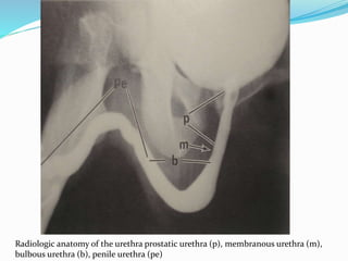
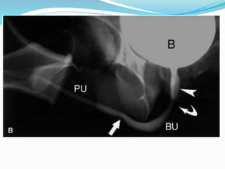
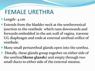
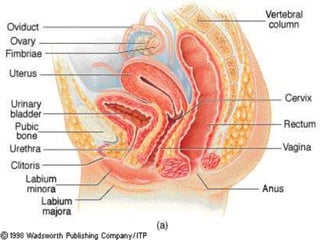
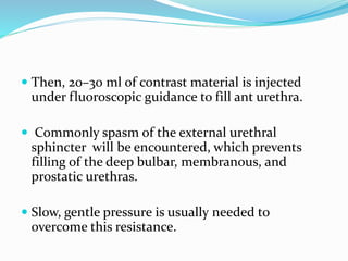
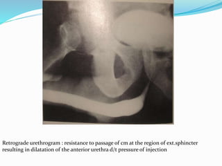
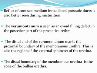
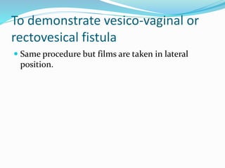
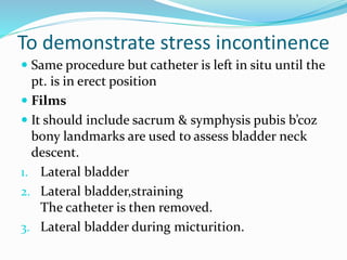
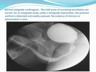
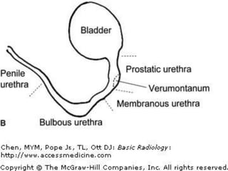
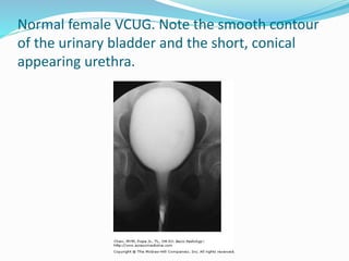
SlideShare a Scribd company logo
AMLENDU KUMAR
1st year resident
INTRODUCTION
 Retrograde urethrography and voiding cystourethrography -
modalities of choice for imaging the urethra.
 RGU-Primary imaging modality for evaluating traumatic
injuries, inflammatory and stricture diseases of male urethra.
VCUG frequently used to evaluate urethral diverticula in women
 USG, MRI and CT-useful for evaluating periurethral structures.
 MR imaging is also accurate in the local staging of urethral
tumors.
MALE URETHRA
 Length-17.5 to 20 cm.
 Consists of
-Anterior portion
-Posterior portion
 Each portion is
subdivided in two parts.
ANTERIOR URETHRA
 Anterior urethra - from external urethral
meatus to inferior edge of the urogenital
diaphragm, coursing through the corpus
spongiosum.
 The anterior urethra is conventionally divided
into
- Penile (or pendulous)
- Bulbous parts(at the penoscrotal junction)
 The penile portion terminates in the glans
penis to form the fossa navicularis, which is
1–1.5 cm long.
 The proximal portion of the bulbous
urethra is dilated called “sump” .
 Just proximal to the sump, the bulbous
urethra assumes a conical shape at the
bulbomembranous junction called “cone.”
POSTERIOR URETHRA
 Divided into
-Prostatic urethra
-Membranous urethra
PROSTATIC URETHRA
 Approx. 3.5 cm long.
 Passes through the prostate slightly anterior to the
midline.
 Urethral crest-longitudinal ridge of smooth muscle
that extends from bladder neck to membranous urethra
on posterior wall .
 Prostatic utricle- small saccular depression which is
remnant of mullerian duct opens over urethral crest at
the centre of Verumontanum.
 Just distal & lateral to utricle are the orifices of the
paired ejaculatory ducts.
MEMBRANOUS URETHRA
 1-1.5 cm long
 perforate UG diaphragm
 Surrounded by muscles fibers (sphincter urethrae) of
UG diaphragm( ext. sphincter)
GLANDS & DUCTS
 Periurethral Littre´ glands –in ant. Urethra & are more
numerous at the dorsal aspect.
 Cowper glands - lie within the urogenital diaphragm
on either side of the membranous urethra. The ducts of
the Cowper gland empty into the bulbous urethral sump.
 Ejaculatory duct-on either side of orifice of prostatic
utricle.
 Prostatic glands-opens directly in prostatic urethra via
multiple small openings aound the verumontanum.
Radiologic anatomy of the urethra prostatic urethra (p), membranous urethra (m),
bulbous urethra (b), penile urethra (pe)
FEMALE URETHRA
 Length- 4 cm
 Extends from the bladder neck at the urethrovesical
junction to the vestibule which runs downwards and
forwards embedded in the ant.wall of vagina, traverse
UG diaphragm and ends at external urethral orifice of
vestibule.
 Many small periurethral glands open into the urethra.
 Distally, these glands group together on either side of
the urethra(Skene glands) and empty through two
small ducts to either side of the external meatus.
Retrograde urethrogram
 Retrograde urethrography -Best initial study for
urethral and periurethral imaging in men and is
indicated in the evaluation of urethral injuries,
strictures and fistulas.
 Straight forward, readily available, cost-effective
examination
Indications
Strictures
Urethral tears
 congenital abnormalities
Periurethral or prostatic abscess
 fistula
Contraindications:
 Acute urethritis and balanitis
 Recent instrumentation
Contrast media
 HOCM or LOCM 200-300, 2w0 ml
 Pre-warming the contrast media will help
reduce the incidence of spasm of the external
sphincter.
Equipment
 Tilting radiography table with flouroscopy
unit & spot film device.
 Foley’s catheter 8F.
Patient preparation
 Empty urinary bladder
 Allergic to x-ray contrast material
 Consent
Preliminary film
 Coned supine PA view of bladder base &
urethra
Technique
 The pt. lies supine on x-ray table.
 Using aseptic technique the tip of the catheter is
inserted so that the balloon lies in the fossa
navicularis. Lubrication is not recommended.
 The patient should be reassured about the
discomfort that is experienced during balloon
inflation.
 Balloon is inflated with 1-2 ml of saline.
 The patient is placed in a supine oblique position.
 The penis should be placed laterally over the
proximal thigh with moderate traction.
 Then, 20–30 ml of contrast material is injected
under fluoroscopic guidance to fill ant urethra.
 Commonly spasm of the external urethral
sphincter will be encountered, which prevents
filling of the deep bulbar, membranous, and
prostatic urethras.
 Slow, gentle pressure is usually needed to
overcome this resistance.
Retrograde urethrogram : resistance to passage of cm at the region of ext.sphincter
resulting in dilatation of the anterior urethra d/t pressure of injection
Films
1) 30 degrees LAO, with right leg abducted &
knee flexed.
2) Supine PA
3) 30 degrees RAO, with left leg abducted &
knee flexed.
 Retrograde urethrography should be followed
by micturating cystourethrography to
demonstrate the proximal urethra .
 Reflux of contrast medium into dilated prostatic ducts is
also better seen during micturition.
 The verumontanum is seen as an ovoid filling defect in
the posterior part of the prostatic urethra.
 The distal end of the verumontanum marks the
proximal boundary of the membranous urethra. This is
also the region of the external sphincter of the urethra.
 The distal boundary of the membranous urethra is the
cone of the bulbar urethra.
Identification of bulbomembranous jn.
 The identification of bulbomembranous junction on RGU is very
important for assessing patients with urethral disease and for
planning urologic procedures.
 When the posterior urethra is optimally opacified and the
verumontanum visible, the bulbomembranous junction can be
identified 1–1.5 cm distal to the inferior margin of the
verumontanum.
 When the posterior urethra is suboptimally opacified, the
bulbomembranous junction can be arbitrarily localized where an
imaginary line connecting the inferior margins of the obturator
foramina intersects the urethra.
 The anterior urethra extends from its origin at the end of the
membranous urethra to the urethral meatus.
 There is usually mild angulation of the urethra where the
pedulous & bulbar segments join at the penoscrotal junction.
 Contraction or spasm of the constrictor nudae muscle, a deep
musculotendinous sling of the bulbocavernous muscle, may cause
circumferential indentation of the proximal bulbous urethra. It
should not be confused with urethral stricture
 The membranous urethra should not be confused with stricture.
 Narrowing elsewhere in the urethra will be clearly defined as
separate from the membranous urethra and, therefore,
representative of a pathologic stricture.
 If the patient is not positioned sufficiently oblique,
the bulbous urethra will appear foreshortened and
will therefore not be adequately evaluated .
 Filling of the Cowper ducts should not be
misinterpreted as extravasation .
 Opacification of the prostatic ducts, Cowper
ducts, and periurethral Littre´glands is often, but
not necessarily, associated with urethral
inflammatory and stricture disease.
 If the integrity of the urethral mucosal lining is
disrupted by increased pressure during contrast
material injection, intravasation of contrast
material with opacification of the corpora and
draining veins may occur.
After care
 None
Complications
Due to contrast medium
 Rare
Due to technique
 Acute UTI
 Urethral trauma
 Intravasation of contrast medium,esp. if excessive
presure is used to overcome stricture.
Antegrade Urethrogram
Definition: Filling the bladder with contrast
media through urethral catheter or by
percutaneous needling of bladder
suprapubically for examination of bladder and
the urethra( during voiding)- Voiding
cystourethrography (VCUG) or micturating
cystourethrography (MCU).
 Excretory micturition cystourethrography
(EMCU): variation of antegrade method,
the urethra is studied after opacification of
bladder by I.V urography.
 Often inadequate for study of the urethra
because of insufficient radiodensity of
bladder urine after IVU; however result can
be improved by having the patient void
against resistance e.g compress penis
between fingers during voiding.
Indications
 Vesicoureteric reflux
 Study of urethra during micturition
 Abnormalities of bladder
 Stress incontinence
Contraindication
 Acute UTI
 Hypersensitivity to contrast media
 Fever within the past 24 hours
Contrast medium
 HOCM or LOCM
 Water soluble contrast media (150 mg/ml iodine) are
used, which are diluted with normal saline in 1: 3 ratio.
Equipment
 Flouroscopy unit with spot film device & tilting table.
 Video recorder
 foley catheter
 In infants 5-7 F feeding tube is adequate.
Patient preparation:
Pt. micturates prior to the examination.
Preliminary films
 Coned view of the bladder.
Technique
To demonstrate vesico-ureteric reflux
 Indicated almost exclusively in children
 Pt. lies supine on x-ray table.
 Using aseptic technique ,a catheter lubricated with
sterile gel containing LA & antiseptic is
introduced in bladder.
 Residual urine is drained.
 Contrast material is slowly dripped in & bladder
filling is observed by intermittent flouroscopy
 Initial filling should be monitered by flouroscopy
as catheter may be in ureter(mimick vesico-
ureteric reflux) or vagina.
 Any reflux is recorded on spot films.
 The catheter should not be removed until the
radiologist is convinced that the patient will
micturate or until no more contrast media will
drip into the bladder.
 Older children & adults are given urine
receiver while small children are allowed to
micturate onto absorbent pads on which
they lie.
 Children can lie on table but adults will find
it easier to micturate while standing erect.
 In pt. of neuropathic bladder ,micturition can be
accomplished by surapubic pressure.
 Spot films are taken during micturition & any reflux
recorded.
 Lower ureter is best seen in anterior oblique position of
that side.
 Finally a full length view of the abdomen is taken to
demonstrate any reflux of contrast medium that might
have occurred unnoticed into the kidneys & to record
post micturition volume.
To demonstrate vesico-vaginal or
rectovesical fistula
 Same procedure but films are taken in lateral
position.
To demonstrate stress incontinence
 Same procedure but catheter is left in situ until the
pt. is in erect position
 Films
 It should include sacrum & symphysis pubis b’coz
bony landmarks are used to assess bladder neck
descent.
1. Lateral bladder
2. Lateral bladder,straining
The catheter is then removed.
3. Lateral bladder during micturition.
Normal antegrade urethrogram . The mild areas of narrowing and dilation are
normal. On an antegrade study, unlike a retrograde examination, the proximal
urethral is distended and readily assessed. No evidence of stricture or
extravasation is seen.
Normal female VCUG. Note the smooth contour
of the urinary bladder and the short, conical
appearing urethra.
Aftercare
 Pt. & parents of children should be warned that
dysuria, possibly leading to retention of urine
may rarely occur. In such cases analgesic should
be given & children may be helped by allowing
them to micturite in warm bath.
 If reflux is present, antibiotics should be
prescribed.
Complications
Due to contrast medium
 Contrast medium induced cystitis.
Due to the technique
 Acute UTI
 Catheter trauma-dysuria, increased frequency of
micturation, hematuria & urinary retention.
 Complication of bladder filling-perforation from
overdistention, prevented by using non-retaining catheter
eg. Jaques
 Retention of a foley cathter.
THANK YOU!
 Best investigation for Vesico ureteric reflux ?
a. IVU
b. MCU
c. retrograde pyelogram
d. RGU
 Posterior urethra is best diagnosed by ?
a. CT cystogram
b. voiding cystogram
c. retrograde urethrogram
d. static cystogram
Identify the defect in MCUG study, marked
by arrow…
 Narrowest and least distensible part of the
urethra is ?
a. prostratic urethra
b. membranous urethra
c. bulbous urethra
d. pendulous urethra
 which of the following are incorrect regarding ducts
that open into urethra;
A. Glands of littre: penile urethra
B. Ejaculatory duct: prostatic urethra
C. Skene glands: female urethra
D. Cowper’s duct: membranous urethra

More Related Content

What's hot

Anterograde/Retrograde urethrography (RGU/MCU)
Anterograde/Retrograde urethrography (RGU/MCU)Anterograde/Retrograde urethrography (RGU/MCU)
Anterograde/Retrograde urethrography (RGU/MCU)
Shubham Singhal
 
Loopogram
LoopogramLoopogram
Loopogram
Sam Shaikh
 
T-tube Cholangiogram
T-tube CholangiogramT-tube Cholangiogram
T-tube Cholangiogram
ricksw78
 
Magnetic resonance cholangiopancreatography ppt
Magnetic resonance cholangiopancreatography pptMagnetic resonance cholangiopancreatography ppt
Magnetic resonance cholangiopancreatography ppt
Anjan Dangal
 
Methods of imaging of the urinary tract using contrast
Methods of imaging of the urinary tract using contrastMethods of imaging of the urinary tract using contrast
Methods of imaging of the urinary tract using contrast
Dr Abdalla M. Gamal
 
Barium meal follow through
Barium meal follow throughBarium meal follow through
Barium meal follow through
Shiva Prakash
 
Procedure of ercp and t tube cholangiography
Procedure of ercp and t tube cholangiographyProcedure of ercp and t tube cholangiography
Procedure of ercp and t tube cholangiography
Yashawant Yadav
 
Barium swallow,,
Barium swallow,,Barium swallow,,
Barium swallow,,
Varsha Pathkala
 
Imaging of Obstructive jaundice
Imaging of Obstructive jaundiceImaging of Obstructive jaundice
Imaging of Obstructive jaundice
Dr. Yash Kumar Achantani
 
Sinogram and fistulogram
Sinogram and fistulogramSinogram and fistulogram
Sinogram and fistulogram
InosRagan
 
Enteroclysis
EnteroclysisEnteroclysis
PTBD (Percutaneus trans-hepatic biliary drainage) PPT,PDF PK
PTBD (Percutaneus trans-hepatic biliary drainage) PPT,PDF PKPTBD (Percutaneus trans-hepatic biliary drainage) PPT,PDF PK
PTBD (Percutaneus trans-hepatic biliary drainage) PPT,PDF PK
Dr pradeep Kumar
 
Retrograde Pyelography
Retrograde PyelographyRetrograde Pyelography
Retrograde Pyelography
Dr. Yash Kumar Achantani
 
Learn Barium Meal & Follow Through
Learn Barium Meal & Follow ThroughLearn Barium Meal & Follow Through
Learn Barium Meal & Follow Through
Dr.Santosh Atreya
 
Abdominal X ray
Abdominal X rayAbdominal X ray
Abdominal X ray
Richin Koshy
 
RGU (Retrograde urethrogram), MCU (Micturating cystourethrogram) and its inte...
RGU (Retrograde urethrogram), MCU (Micturating cystourethrogram) and its inte...RGU (Retrograde urethrogram), MCU (Micturating cystourethrogram) and its inte...
RGU (Retrograde urethrogram), MCU (Micturating cystourethrogram) and its inte...
Milan Silwal
 
Barium studies in git
Barium studies in gitBarium studies in git
Barium studies in git
Dev Lakhera
 
PERCITANEOUS NEPHROSTOMY and HYSTEROSALPIONGOGRAPHY
PERCITANEOUS NEPHROSTOMY and HYSTEROSALPIONGOGRAPHYPERCITANEOUS NEPHROSTOMY and HYSTEROSALPIONGOGRAPHY
PERCITANEOUS NEPHROSTOMY and HYSTEROSALPIONGOGRAPHY
SharmaRajan4
 
CT urography
CT urography CT urography
CT urography
SabitaMandal1
 
Barium swallow
Barium swallowBarium swallow
Barium swallow
Athul Nampoothiri
 

What's hot (20)

Anterograde/Retrograde urethrography (RGU/MCU)
Anterograde/Retrograde urethrography (RGU/MCU)Anterograde/Retrograde urethrography (RGU/MCU)
Anterograde/Retrograde urethrography (RGU/MCU)
 
Loopogram
LoopogramLoopogram
Loopogram
 
T-tube Cholangiogram
T-tube CholangiogramT-tube Cholangiogram
T-tube Cholangiogram
 
Magnetic resonance cholangiopancreatography ppt
Magnetic resonance cholangiopancreatography pptMagnetic resonance cholangiopancreatography ppt
Magnetic resonance cholangiopancreatography ppt
 
Methods of imaging of the urinary tract using contrast
Methods of imaging of the urinary tract using contrastMethods of imaging of the urinary tract using contrast
Methods of imaging of the urinary tract using contrast
 
Barium meal follow through
Barium meal follow throughBarium meal follow through
Barium meal follow through
 
Procedure of ercp and t tube cholangiography
Procedure of ercp and t tube cholangiographyProcedure of ercp and t tube cholangiography
Procedure of ercp and t tube cholangiography
 
Barium swallow,,
Barium swallow,,Barium swallow,,
Barium swallow,,
 
Imaging of Obstructive jaundice
Imaging of Obstructive jaundiceImaging of Obstructive jaundice
Imaging of Obstructive jaundice
 
Sinogram and fistulogram
Sinogram and fistulogramSinogram and fistulogram
Sinogram and fistulogram
 
Enteroclysis
EnteroclysisEnteroclysis
Enteroclysis
 
PTBD (Percutaneus trans-hepatic biliary drainage) PPT,PDF PK
PTBD (Percutaneus trans-hepatic biliary drainage) PPT,PDF PKPTBD (Percutaneus trans-hepatic biliary drainage) PPT,PDF PK
PTBD (Percutaneus trans-hepatic biliary drainage) PPT,PDF PK
 
Retrograde Pyelography
Retrograde PyelographyRetrograde Pyelography
Retrograde Pyelography
 
Learn Barium Meal & Follow Through
Learn Barium Meal & Follow ThroughLearn Barium Meal & Follow Through
Learn Barium Meal & Follow Through
 
Abdominal X ray
Abdominal X rayAbdominal X ray
Abdominal X ray
 
RGU (Retrograde urethrogram), MCU (Micturating cystourethrogram) and its inte...
RGU (Retrograde urethrogram), MCU (Micturating cystourethrogram) and its inte...RGU (Retrograde urethrogram), MCU (Micturating cystourethrogram) and its inte...
RGU (Retrograde urethrogram), MCU (Micturating cystourethrogram) and its inte...
 
Barium studies in git
Barium studies in gitBarium studies in git
Barium studies in git
 
PERCITANEOUS NEPHROSTOMY and HYSTEROSALPIONGOGRAPHY
PERCITANEOUS NEPHROSTOMY and HYSTEROSALPIONGOGRAPHYPERCITANEOUS NEPHROSTOMY and HYSTEROSALPIONGOGRAPHY
PERCITANEOUS NEPHROSTOMY and HYSTEROSALPIONGOGRAPHY
 
CT urography
CT urography CT urography
CT urography
 
Barium swallow
Barium swallowBarium swallow
Barium swallow
 

Similar to Antegrade & retrograde urethrogram

urethrogram.pptx
urethrogram.pptxurethrogram.pptx
urethrogram.pptx
AliyaIshak1
 
Imaging of urethral pathologies
Imaging of urethral pathologiesImaging of urethral pathologies
Imaging of urethral pathologies
Sunil Kumar
 
RGU and MCU by capt alauddin, MD phase A.pptx
RGU and MCU by capt alauddin, MD phase A.pptxRGU and MCU by capt alauddin, MD phase A.pptx
RGU and MCU by capt alauddin, MD phase A.pptx
Alauddin Md
 
anorectal malformation
anorectal malformationanorectal malformation
anorectal malformation
Pushpa Latha
 
Anorectal malformations.pptx
Anorectal malformations.pptxAnorectal malformations.pptx
Anorectal malformations.pptx
Pushpa Lal Bhadel
 
Vcu ppt
Vcu pptVcu ppt
Vcu ppt
dypradio
 
MCU AND RGU
MCU AND RGUMCU AND RGU
MCU AND RGU
dypradio
 
Radiological anatomy of the male urethra and techniques of imaging
Radiological  anatomy of the male urethra and techniques of imagingRadiological  anatomy of the male urethra and techniques of imaging
Radiological anatomy of the male urethra and techniques of imaging
HarrietAimua
 
Anorectal malformations
Anorectal malformationsAnorectal malformations
Anorectal malformations
rahulverma1194
 
Surgical Anatomy of Urinary bladder.pptx
Surgical Anatomy of Urinary bladder.pptxSurgical Anatomy of Urinary bladder.pptx
Surgical Anatomy of Urinary bladder.pptx
DurgaPrasadM10
 
Mcu
McuMcu
OPPOSING URETHROGRAM (OUG)
OPPOSING URETHROGRAM (OUG)OPPOSING URETHROGRAM (OUG)
OPPOSING URETHROGRAM (OUG)
Jai Kumar
 
RGU MCU and its interpretation in pathology of Urinary Bladder & Urethra
RGU MCU and its interpretation in pathology of Urinary Bladder & UrethraRGU MCU and its interpretation in pathology of Urinary Bladder & Urethra
RGU MCU and its interpretation in pathology of Urinary Bladder & Urethradbc9427
 
Anorectal malformation seminar
Anorectal malformation seminarAnorectal malformation seminar
Anorectal malformation seminar
Dr. Dixit
 
Uro procedure
Uro procedureUro procedure
Uro procedure
Dr-Girish Gunari
 
MCU and AUG - CYSTOURETHEROGRAM Urology.pptx
MCU and AUG - CYSTOURETHEROGRAM Urology.pptxMCU and AUG - CYSTOURETHEROGRAM Urology.pptx
MCU and AUG - CYSTOURETHEROGRAM Urology.pptx
Kuppan Thenappan
 
Anorectal malformations in children
Anorectal malformations in childrenAnorectal malformations in children
Anorectal malformations in children
dhanyav54
 
Causes and management of long ureteral defect
Causes and management of long ureteral defectCauses and management of long ureteral defect
Causes and management of long ureteral defect
د. نادر عبد الستار
 
Vaginal approach for Stress Urinary Incontinence surgery
Vaginal approach for Stress Urinary Incontinence surgeryVaginal approach for Stress Urinary Incontinence surgery
Vaginal approach for Stress Urinary Incontinence surgery
Rohan Sharma
 
URETHRAL STRICTURES.pdf
URETHRAL STRICTURES.pdfURETHRAL STRICTURES.pdf
URETHRAL STRICTURES.pdf
Shapi. MD
 

Similar to Antegrade & retrograde urethrogram (20)

urethrogram.pptx
urethrogram.pptxurethrogram.pptx
urethrogram.pptx
 
Imaging of urethral pathologies
Imaging of urethral pathologiesImaging of urethral pathologies
Imaging of urethral pathologies
 
RGU and MCU by capt alauddin, MD phase A.pptx
RGU and MCU by capt alauddin, MD phase A.pptxRGU and MCU by capt alauddin, MD phase A.pptx
RGU and MCU by capt alauddin, MD phase A.pptx
 
anorectal malformation
anorectal malformationanorectal malformation
anorectal malformation
 
Anorectal malformations.pptx
Anorectal malformations.pptxAnorectal malformations.pptx
Anorectal malformations.pptx
 
Vcu ppt
Vcu pptVcu ppt
Vcu ppt
 
MCU AND RGU
MCU AND RGUMCU AND RGU
MCU AND RGU
 
Radiological anatomy of the male urethra and techniques of imaging
Radiological  anatomy of the male urethra and techniques of imagingRadiological  anatomy of the male urethra and techniques of imaging
Radiological anatomy of the male urethra and techniques of imaging
 
Anorectal malformations
Anorectal malformationsAnorectal malformations
Anorectal malformations
 
Surgical Anatomy of Urinary bladder.pptx
Surgical Anatomy of Urinary bladder.pptxSurgical Anatomy of Urinary bladder.pptx
Surgical Anatomy of Urinary bladder.pptx
 
Mcu
McuMcu
Mcu
 
OPPOSING URETHROGRAM (OUG)
OPPOSING URETHROGRAM (OUG)OPPOSING URETHROGRAM (OUG)
OPPOSING URETHROGRAM (OUG)
 
RGU MCU and its interpretation in pathology of Urinary Bladder & Urethra
RGU MCU and its interpretation in pathology of Urinary Bladder & UrethraRGU MCU and its interpretation in pathology of Urinary Bladder & Urethra
RGU MCU and its interpretation in pathology of Urinary Bladder & Urethra
 
Anorectal malformation seminar
Anorectal malformation seminarAnorectal malformation seminar
Anorectal malformation seminar
 
Uro procedure
Uro procedureUro procedure
Uro procedure
 
MCU and AUG - CYSTOURETHEROGRAM Urology.pptx
MCU and AUG - CYSTOURETHEROGRAM Urology.pptxMCU and AUG - CYSTOURETHEROGRAM Urology.pptx
MCU and AUG - CYSTOURETHEROGRAM Urology.pptx
 
Anorectal malformations in children
Anorectal malformations in childrenAnorectal malformations in children
Anorectal malformations in children
 
Causes and management of long ureteral defect
Causes and management of long ureteral defectCauses and management of long ureteral defect
Causes and management of long ureteral defect
 
Vaginal approach for Stress Urinary Incontinence surgery
Vaginal approach for Stress Urinary Incontinence surgeryVaginal approach for Stress Urinary Incontinence surgery
Vaginal approach for Stress Urinary Incontinence surgery
 
URETHRAL STRICTURES.pdf
URETHRAL STRICTURES.pdfURETHRAL STRICTURES.pdf
URETHRAL STRICTURES.pdf
 

Recently uploaded

Cambridge International AS A Level Biology Coursebook - EBook (MaryFosbery J...
Cambridge International AS  A Level Biology Coursebook - EBook (MaryFosbery J...Cambridge International AS  A Level Biology Coursebook - EBook (MaryFosbery J...
Cambridge International AS A Level Biology Coursebook - EBook (MaryFosbery J...
AzmatAli747758
 
The French Revolution Class 9 Study Material pdf free download
The French Revolution Class 9 Study Material pdf free downloadThe French Revolution Class 9 Study Material pdf free download
The French Revolution Class 9 Study Material pdf free download
Vivekanand Anglo Vedic Academy
 
ESC Beyond Borders _From EU to You_ InfoPack general.pdf
ESC Beyond Borders _From EU to You_ InfoPack general.pdfESC Beyond Borders _From EU to You_ InfoPack general.pdf
ESC Beyond Borders _From EU to You_ InfoPack general.pdf
Fundacja Rozwoju Społeczeństwa Przedsiębiorczego
 
Polish students' mobility in the Czech Republic
Polish students' mobility in the Czech RepublicPolish students' mobility in the Czech Republic
Polish students' mobility in the Czech Republic
Anna Sz.
 
How to Break the cycle of negative Thoughts
How to Break the cycle of negative ThoughtsHow to Break the cycle of negative Thoughts
How to Break the cycle of negative Thoughts
Col Mukteshwar Prasad
 
Palestine last event orientationfvgnh .pptx
Palestine last event orientationfvgnh .pptxPalestine last event orientationfvgnh .pptx
Palestine last event orientationfvgnh .pptx
RaedMohamed3
 
Introduction to Quality Improvement Essentials
Introduction to Quality Improvement EssentialsIntroduction to Quality Improvement Essentials
Introduction to Quality Improvement Essentials
Excellence Foundation for South Sudan
 
GIÁO ÁN DẠY THÊM (KẾ HOẠCH BÀI BUỔI 2) - TIẾNG ANH 8 GLOBAL SUCCESS (2 CỘT) N...
GIÁO ÁN DẠY THÊM (KẾ HOẠCH BÀI BUỔI 2) - TIẾNG ANH 8 GLOBAL SUCCESS (2 CỘT) N...GIÁO ÁN DẠY THÊM (KẾ HOẠCH BÀI BUỔI 2) - TIẾNG ANH 8 GLOBAL SUCCESS (2 CỘT) N...
GIÁO ÁN DẠY THÊM (KẾ HOẠCH BÀI BUỔI 2) - TIẾNG ANH 8 GLOBAL SUCCESS (2 CỘT) N...
Nguyen Thanh Tu Collection
 
1.4 modern child centered education - mahatma gandhi-2.pptx
1.4 modern child centered education - mahatma gandhi-2.pptx1.4 modern child centered education - mahatma gandhi-2.pptx
1.4 modern child centered education - mahatma gandhi-2.pptx
JosvitaDsouza2
 
CLASS 11 CBSE B.St Project AIDS TO TRADE - INSURANCE
CLASS 11 CBSE B.St Project AIDS TO TRADE - INSURANCECLASS 11 CBSE B.St Project AIDS TO TRADE - INSURANCE
CLASS 11 CBSE B.St Project AIDS TO TRADE - INSURANCE
BhavyaRajput3
 
Model Attribute Check Company Auto Property
Model Attribute  Check Company Auto PropertyModel Attribute  Check Company Auto Property
Model Attribute Check Company Auto Property
Celine George
 
Thesis Statement for students diagnonsed withADHD.ppt
Thesis Statement for students diagnonsed withADHD.pptThesis Statement for students diagnonsed withADHD.ppt
Thesis Statement for students diagnonsed withADHD.ppt
EverAndrsGuerraGuerr
 
Instructions for Submissions thorugh G- Classroom.pptx
Instructions for Submissions thorugh G- Classroom.pptxInstructions for Submissions thorugh G- Classroom.pptx
Instructions for Submissions thorugh G- Classroom.pptx
Jheel Barad
 
The Roman Empire A Historical Colossus.pdf
The Roman Empire A Historical Colossus.pdfThe Roman Empire A Historical Colossus.pdf
The Roman Empire A Historical Colossus.pdf
kaushalkr1407
 
aaaaaaaaaaaaaaaaaaaaaaaaaaaaaaaaaaaaaaaaa
aaaaaaaaaaaaaaaaaaaaaaaaaaaaaaaaaaaaaaaaaaaaaaaaaaaaaaaaaaaaaaaaaaaaaaaaaaaaaaaaaa
aaaaaaaaaaaaaaaaaaaaaaaaaaaaaaaaaaaaaaaaa
siemaillard
 
aaaaaaaaaaaaaaaaaaaaaaaaaaaaaaaaaaaaaaaaaaaaaaaaaaaaaaa
aaaaaaaaaaaaaaaaaaaaaaaaaaaaaaaaaaaaaaaaaaaaaaaaaaaaaaaaaaaaaaaaaaaaaaaaaaaaaaaaaaaaaaaaaaaaaaaaaaaaaaaaaaaaaa
aaaaaaaaaaaaaaaaaaaaaaaaaaaaaaaaaaaaaaaaaaaaaaaaaaaaaaa
siemaillard
 
Synthetic Fiber Construction in lab .pptx
Synthetic Fiber Construction in lab .pptxSynthetic Fiber Construction in lab .pptx
Synthetic Fiber Construction in lab .pptx
Pavel ( NSTU)
 
Digital Tools and AI for Teaching Learning and Research
Digital Tools and AI for Teaching Learning and ResearchDigital Tools and AI for Teaching Learning and Research
Digital Tools and AI for Teaching Learning and Research
Vikramjit Singh
 
Basic phrases for greeting and assisting costumers
Basic phrases for greeting and assisting costumersBasic phrases for greeting and assisting costumers
Basic phrases for greeting and assisting costumers
PedroFerreira53928
 
Chapter 3 - Islamic Banking Products and Services.pptx
Chapter 3 - Islamic Banking Products and Services.pptxChapter 3 - Islamic Banking Products and Services.pptx
Chapter 3 - Islamic Banking Products and Services.pptx
Mohd Adib Abd Muin, Senior Lecturer at Universiti Utara Malaysia
 

Recently uploaded (20)

Cambridge International AS A Level Biology Coursebook - EBook (MaryFosbery J...
Cambridge International AS  A Level Biology Coursebook - EBook (MaryFosbery J...Cambridge International AS  A Level Biology Coursebook - EBook (MaryFosbery J...
Cambridge International AS A Level Biology Coursebook - EBook (MaryFosbery J...
 
The French Revolution Class 9 Study Material pdf free download
The French Revolution Class 9 Study Material pdf free downloadThe French Revolution Class 9 Study Material pdf free download
The French Revolution Class 9 Study Material pdf free download
 
ESC Beyond Borders _From EU to You_ InfoPack general.pdf
ESC Beyond Borders _From EU to You_ InfoPack general.pdfESC Beyond Borders _From EU to You_ InfoPack general.pdf
ESC Beyond Borders _From EU to You_ InfoPack general.pdf
 
Polish students' mobility in the Czech Republic
Polish students' mobility in the Czech RepublicPolish students' mobility in the Czech Republic
Polish students' mobility in the Czech Republic
 
How to Break the cycle of negative Thoughts
How to Break the cycle of negative ThoughtsHow to Break the cycle of negative Thoughts
How to Break the cycle of negative Thoughts
 
Palestine last event orientationfvgnh .pptx
Palestine last event orientationfvgnh .pptxPalestine last event orientationfvgnh .pptx
Palestine last event orientationfvgnh .pptx
 
Introduction to Quality Improvement Essentials
Introduction to Quality Improvement EssentialsIntroduction to Quality Improvement Essentials
Introduction to Quality Improvement Essentials
 
GIÁO ÁN DẠY THÊM (KẾ HOẠCH BÀI BUỔI 2) - TIẾNG ANH 8 GLOBAL SUCCESS (2 CỘT) N...
GIÁO ÁN DẠY THÊM (KẾ HOẠCH BÀI BUỔI 2) - TIẾNG ANH 8 GLOBAL SUCCESS (2 CỘT) N...GIÁO ÁN DẠY THÊM (KẾ HOẠCH BÀI BUỔI 2) - TIẾNG ANH 8 GLOBAL SUCCESS (2 CỘT) N...
GIÁO ÁN DẠY THÊM (KẾ HOẠCH BÀI BUỔI 2) - TIẾNG ANH 8 GLOBAL SUCCESS (2 CỘT) N...
 
1.4 modern child centered education - mahatma gandhi-2.pptx
1.4 modern child centered education - mahatma gandhi-2.pptx1.4 modern child centered education - mahatma gandhi-2.pptx
1.4 modern child centered education - mahatma gandhi-2.pptx
 
CLASS 11 CBSE B.St Project AIDS TO TRADE - INSURANCE
CLASS 11 CBSE B.St Project AIDS TO TRADE - INSURANCECLASS 11 CBSE B.St Project AIDS TO TRADE - INSURANCE
CLASS 11 CBSE B.St Project AIDS TO TRADE - INSURANCE
 
Model Attribute Check Company Auto Property
Model Attribute  Check Company Auto PropertyModel Attribute  Check Company Auto Property
Model Attribute Check Company Auto Property
 
Thesis Statement for students diagnonsed withADHD.ppt
Thesis Statement for students diagnonsed withADHD.pptThesis Statement for students diagnonsed withADHD.ppt
Thesis Statement for students diagnonsed withADHD.ppt
 
Instructions for Submissions thorugh G- Classroom.pptx
Instructions for Submissions thorugh G- Classroom.pptxInstructions for Submissions thorugh G- Classroom.pptx
Instructions for Submissions thorugh G- Classroom.pptx
 
The Roman Empire A Historical Colossus.pdf
The Roman Empire A Historical Colossus.pdfThe Roman Empire A Historical Colossus.pdf
The Roman Empire A Historical Colossus.pdf
 
aaaaaaaaaaaaaaaaaaaaaaaaaaaaaaaaaaaaaaaaa
aaaaaaaaaaaaaaaaaaaaaaaaaaaaaaaaaaaaaaaaaaaaaaaaaaaaaaaaaaaaaaaaaaaaaaaaaaaaaaaaaa
aaaaaaaaaaaaaaaaaaaaaaaaaaaaaaaaaaaaaaaaa
 
aaaaaaaaaaaaaaaaaaaaaaaaaaaaaaaaaaaaaaaaaaaaaaaaaaaaaaa
aaaaaaaaaaaaaaaaaaaaaaaaaaaaaaaaaaaaaaaaaaaaaaaaaaaaaaaaaaaaaaaaaaaaaaaaaaaaaaaaaaaaaaaaaaaaaaaaaaaaaaaaaaaaaa
aaaaaaaaaaaaaaaaaaaaaaaaaaaaaaaaaaaaaaaaaaaaaaaaaaaaaaa
 
Synthetic Fiber Construction in lab .pptx
Synthetic Fiber Construction in lab .pptxSynthetic Fiber Construction in lab .pptx
Synthetic Fiber Construction in lab .pptx
 
Digital Tools and AI for Teaching Learning and Research
Digital Tools and AI for Teaching Learning and ResearchDigital Tools and AI for Teaching Learning and Research
Digital Tools and AI for Teaching Learning and Research
 
Basic phrases for greeting and assisting costumers
Basic phrases for greeting and assisting costumersBasic phrases for greeting and assisting costumers
Basic phrases for greeting and assisting costumers
 
Chapter 3 - Islamic Banking Products and Services.pptx
Chapter 3 - Islamic Banking Products and Services.pptxChapter 3 - Islamic Banking Products and Services.pptx
Chapter 3 - Islamic Banking Products and Services.pptx
 

Antegrade & retrograde urethrogram

  • 2. INTRODUCTION  Retrograde urethrography and voiding cystourethrography - modalities of choice for imaging the urethra.  RGU-Primary imaging modality for evaluating traumatic injuries, inflammatory and stricture diseases of male urethra. VCUG frequently used to evaluate urethral diverticula in women  USG, MRI and CT-useful for evaluating periurethral structures.  MR imaging is also accurate in the local staging of urethral tumors.
  • 3. MALE URETHRA  Length-17.5 to 20 cm.  Consists of -Anterior portion -Posterior portion  Each portion is subdivided in two parts.
  • 4. ANTERIOR URETHRA  Anterior urethra - from external urethral meatus to inferior edge of the urogenital diaphragm, coursing through the corpus spongiosum.  The anterior urethra is conventionally divided into - Penile (or pendulous) - Bulbous parts(at the penoscrotal junction)
  • 5.  The penile portion terminates in the glans penis to form the fossa navicularis, which is 1–1.5 cm long.  The proximal portion of the bulbous urethra is dilated called “sump” .  Just proximal to the sump, the bulbous urethra assumes a conical shape at the bulbomembranous junction called “cone.”
  • 6. POSTERIOR URETHRA  Divided into -Prostatic urethra -Membranous urethra PROSTATIC URETHRA  Approx. 3.5 cm long.  Passes through the prostate slightly anterior to the midline.  Urethral crest-longitudinal ridge of smooth muscle that extends from bladder neck to membranous urethra on posterior wall .
  • 7.  Prostatic utricle- small saccular depression which is remnant of mullerian duct opens over urethral crest at the centre of Verumontanum.  Just distal & lateral to utricle are the orifices of the paired ejaculatory ducts. MEMBRANOUS URETHRA  1-1.5 cm long  perforate UG diaphragm  Surrounded by muscles fibers (sphincter urethrae) of UG diaphragm( ext. sphincter)
  • 8. GLANDS & DUCTS  Periurethral Littre´ glands –in ant. Urethra & are more numerous at the dorsal aspect.  Cowper glands - lie within the urogenital diaphragm on either side of the membranous urethra. The ducts of the Cowper gland empty into the bulbous urethral sump.  Ejaculatory duct-on either side of orifice of prostatic utricle.  Prostatic glands-opens directly in prostatic urethra via multiple small openings aound the verumontanum.
  • 9.
  • 10. Radiologic anatomy of the urethra prostatic urethra (p), membranous urethra (m), bulbous urethra (b), penile urethra (pe)
  • 11.
  • 12. FEMALE URETHRA  Length- 4 cm  Extends from the bladder neck at the urethrovesical junction to the vestibule which runs downwards and forwards embedded in the ant.wall of vagina, traverse UG diaphragm and ends at external urethral orifice of vestibule.  Many small periurethral glands open into the urethra.  Distally, these glands group together on either side of the urethra(Skene glands) and empty through two small ducts to either side of the external meatus.
  • 13.
  • 14. Retrograde urethrogram  Retrograde urethrography -Best initial study for urethral and periurethral imaging in men and is indicated in the evaluation of urethral injuries, strictures and fistulas.  Straight forward, readily available, cost-effective examination
  • 15. Indications Strictures Urethral tears  congenital abnormalities Periurethral or prostatic abscess  fistula Contraindications:  Acute urethritis and balanitis  Recent instrumentation
  • 16. Contrast media  HOCM or LOCM 200-300, 2w0 ml  Pre-warming the contrast media will help reduce the incidence of spasm of the external sphincter. Equipment  Tilting radiography table with flouroscopy unit & spot film device.  Foley’s catheter 8F.
  • 17. Patient preparation  Empty urinary bladder  Allergic to x-ray contrast material  Consent Preliminary film  Coned supine PA view of bladder base & urethra
  • 18. Technique  The pt. lies supine on x-ray table.  Using aseptic technique the tip of the catheter is inserted so that the balloon lies in the fossa navicularis. Lubrication is not recommended.  The patient should be reassured about the discomfort that is experienced during balloon inflation.  Balloon is inflated with 1-2 ml of saline.  The patient is placed in a supine oblique position.  The penis should be placed laterally over the proximal thigh with moderate traction.
  • 19.  Then, 20–30 ml of contrast material is injected under fluoroscopic guidance to fill ant urethra.  Commonly spasm of the external urethral sphincter will be encountered, which prevents filling of the deep bulbar, membranous, and prostatic urethras.  Slow, gentle pressure is usually needed to overcome this resistance.
  • 20. Retrograde urethrogram : resistance to passage of cm at the region of ext.sphincter resulting in dilatation of the anterior urethra d/t pressure of injection
  • 21. Films 1) 30 degrees LAO, with right leg abducted & knee flexed. 2) Supine PA 3) 30 degrees RAO, with left leg abducted & knee flexed.  Retrograde urethrography should be followed by micturating cystourethrography to demonstrate the proximal urethra .
  • 22.  Reflux of contrast medium into dilated prostatic ducts is also better seen during micturition.  The verumontanum is seen as an ovoid filling defect in the posterior part of the prostatic urethra.  The distal end of the verumontanum marks the proximal boundary of the membranous urethra. This is also the region of the external sphincter of the urethra.  The distal boundary of the membranous urethra is the cone of the bulbar urethra.
  • 23. Identification of bulbomembranous jn.  The identification of bulbomembranous junction on RGU is very important for assessing patients with urethral disease and for planning urologic procedures.  When the posterior urethra is optimally opacified and the verumontanum visible, the bulbomembranous junction can be identified 1–1.5 cm distal to the inferior margin of the verumontanum.  When the posterior urethra is suboptimally opacified, the bulbomembranous junction can be arbitrarily localized where an imaginary line connecting the inferior margins of the obturator foramina intersects the urethra.
  • 24.  The anterior urethra extends from its origin at the end of the membranous urethra to the urethral meatus.  There is usually mild angulation of the urethra where the pedulous & bulbar segments join at the penoscrotal junction.  Contraction or spasm of the constrictor nudae muscle, a deep musculotendinous sling of the bulbocavernous muscle, may cause circumferential indentation of the proximal bulbous urethra. It should not be confused with urethral stricture  The membranous urethra should not be confused with stricture.  Narrowing elsewhere in the urethra will be clearly defined as separate from the membranous urethra and, therefore, representative of a pathologic stricture.
  • 25.
  • 26.  If the patient is not positioned sufficiently oblique, the bulbous urethra will appear foreshortened and will therefore not be adequately evaluated .
  • 27.
  • 28.  Filling of the Cowper ducts should not be misinterpreted as extravasation .  Opacification of the prostatic ducts, Cowper ducts, and periurethral Littre´glands is often, but not necessarily, associated with urethral inflammatory and stricture disease.  If the integrity of the urethral mucosal lining is disrupted by increased pressure during contrast material injection, intravasation of contrast material with opacification of the corpora and draining veins may occur.
  • 29. After care  None Complications Due to contrast medium  Rare Due to technique  Acute UTI  Urethral trauma  Intravasation of contrast medium,esp. if excessive presure is used to overcome stricture.
  • 30. Antegrade Urethrogram Definition: Filling the bladder with contrast media through urethral catheter or by percutaneous needling of bladder suprapubically for examination of bladder and the urethra( during voiding)- Voiding cystourethrography (VCUG) or micturating cystourethrography (MCU).
  • 31.  Excretory micturition cystourethrography (EMCU): variation of antegrade method, the urethra is studied after opacification of bladder by I.V urography.  Often inadequate for study of the urethra because of insufficient radiodensity of bladder urine after IVU; however result can be improved by having the patient void against resistance e.g compress penis between fingers during voiding.
  • 32. Indications  Vesicoureteric reflux  Study of urethra during micturition  Abnormalities of bladder  Stress incontinence Contraindication  Acute UTI  Hypersensitivity to contrast media  Fever within the past 24 hours
  • 33. Contrast medium  HOCM or LOCM  Water soluble contrast media (150 mg/ml iodine) are used, which are diluted with normal saline in 1: 3 ratio. Equipment  Flouroscopy unit with spot film device & tilting table.  Video recorder  foley catheter  In infants 5-7 F feeding tube is adequate. Patient preparation: Pt. micturates prior to the examination. Preliminary films  Coned view of the bladder.
  • 34. Technique To demonstrate vesico-ureteric reflux  Indicated almost exclusively in children  Pt. lies supine on x-ray table.  Using aseptic technique ,a catheter lubricated with sterile gel containing LA & antiseptic is introduced in bladder.  Residual urine is drained.  Contrast material is slowly dripped in & bladder filling is observed by intermittent flouroscopy
  • 35.  Initial filling should be monitered by flouroscopy as catheter may be in ureter(mimick vesico- ureteric reflux) or vagina.  Any reflux is recorded on spot films.  The catheter should not be removed until the radiologist is convinced that the patient will micturate or until no more contrast media will drip into the bladder.
  • 36.  Older children & adults are given urine receiver while small children are allowed to micturate onto absorbent pads on which they lie.  Children can lie on table but adults will find it easier to micturate while standing erect.
  • 37.  In pt. of neuropathic bladder ,micturition can be accomplished by surapubic pressure.  Spot films are taken during micturition & any reflux recorded.  Lower ureter is best seen in anterior oblique position of that side.  Finally a full length view of the abdomen is taken to demonstrate any reflux of contrast medium that might have occurred unnoticed into the kidneys & to record post micturition volume.
  • 38. To demonstrate vesico-vaginal or rectovesical fistula  Same procedure but films are taken in lateral position.
  • 39. To demonstrate stress incontinence  Same procedure but catheter is left in situ until the pt. is in erect position  Films  It should include sacrum & symphysis pubis b’coz bony landmarks are used to assess bladder neck descent. 1. Lateral bladder 2. Lateral bladder,straining The catheter is then removed. 3. Lateral bladder during micturition.
  • 40. Normal antegrade urethrogram . The mild areas of narrowing and dilation are normal. On an antegrade study, unlike a retrograde examination, the proximal urethral is distended and readily assessed. No evidence of stricture or extravasation is seen.
  • 41.
  • 42. Normal female VCUG. Note the smooth contour of the urinary bladder and the short, conical appearing urethra.
  • 43. Aftercare  Pt. & parents of children should be warned that dysuria, possibly leading to retention of urine may rarely occur. In such cases analgesic should be given & children may be helped by allowing them to micturite in warm bath.  If reflux is present, antibiotics should be prescribed.
  • 44. Complications Due to contrast medium  Contrast medium induced cystitis. Due to the technique  Acute UTI  Catheter trauma-dysuria, increased frequency of micturation, hematuria & urinary retention.  Complication of bladder filling-perforation from overdistention, prevented by using non-retaining catheter eg. Jaques  Retention of a foley cathter.
  • 46.  Best investigation for Vesico ureteric reflux ? a. IVU b. MCU c. retrograde pyelogram d. RGU
  • 47.  Posterior urethra is best diagnosed by ? a. CT cystogram b. voiding cystogram c. retrograde urethrogram d. static cystogram
  • 48. Identify the defect in MCUG study, marked by arrow…
  • 49.
  • 50.  Narrowest and least distensible part of the urethra is ? a. prostratic urethra b. membranous urethra c. bulbous urethra d. pendulous urethra
  • 51.  which of the following are incorrect regarding ducts that open into urethra; A. Glands of littre: penile urethra B. Ejaculatory duct: prostatic urethra C. Skene glands: female urethra D. Cowper’s duct: membranous urethra