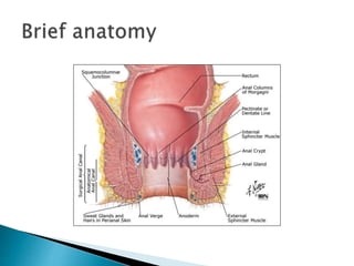
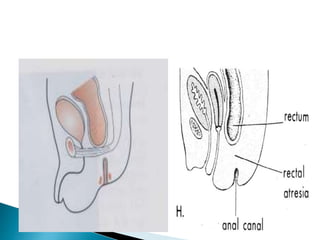
This document provides an overview of anorectal malformations. It begins with an anatomical description of anal canal development. It then discusses the epidemiology and etiology of anorectal malformations. The document classifies anorectal malformations and describes associated comorbidities. It outlines approaches for clinical evaluation, diagnosis and surgical management. Surgical correction depends on whether the malformation is high or low. The newest surgical technique described is posterior sagittal anorectoplasty.
Hirschsprung Disease - Approach & ManagementVikas V
Hirschsprung Disease. - A developmental Disorder of Intrinsic Component of Enteric Nervous System.
Also known Congenital Megacolon.
This Presentation deals with The eitology, presentation, diagnosis, medical and surgical management & complications of the same.
POSTERIOR URETHRAL VALVES- Pediatric Surgery
• Dear viewers,
• Greetings from “ Surgical Educator”
• Today I have uploaded one more video in Pediatric Surgery/Pediatric Urology- “ Posterior Urethral Valves”
• Posterior Urethral Valves is the congenital cause for Bladder Outlet Obstruction, resulting in abnormal development of the kidneys as well as the bladder.
• In this video, I talked about the learning outcomes, introduction, etiopathogenesis, clinical features, investigations, differential diagnosis, treatment, follow-up and prognosis of “ Posterior Urethral Valves”
• I hope you will enjoy the video for its educational value.
• You can watch all my teaching videos in the following links
• surgicaleducator.blogspot.com youtube.com/c/surgicaleducator
• Thank you for watching the video.
Title: Sense of Taste
Presenter: Dr. Faiza, Assistant Professor of Physiology
Qualifications:
MBBS (Best Graduate, AIMC Lahore)
FCPS Physiology
ICMT, CHPE, DHPE (STMU)
MPH (GC University, Faisalabad)
MBA (Virtual University of Pakistan)
Learning Objectives:
Describe the structure and function of taste buds.
Describe the relationship between the taste threshold and taste index of common substances.
Explain the chemical basis and signal transduction of taste perception for each type of primary taste sensation.
Recognize different abnormalities of taste perception and their causes.
Key Topics:
Significance of Taste Sensation:
Differentiation between pleasant and harmful food
Influence on behavior
Selection of food based on metabolic needs
Receptors of Taste:
Taste buds on the tongue
Influence of sense of smell, texture of food, and pain stimulation (e.g., by pepper)
Primary and Secondary Taste Sensations:
Primary taste sensations: Sweet, Sour, Salty, Bitter, Umami
Chemical basis and signal transduction mechanisms for each taste
Taste Threshold and Index:
Taste threshold values for Sweet (sucrose), Salty (NaCl), Sour (HCl), and Bitter (Quinine)
Taste index relationship: Inversely proportional to taste threshold
Taste Blindness:
Inability to taste certain substances, particularly thiourea compounds
Example: Phenylthiocarbamide
Structure and Function of Taste Buds:
Composition: Epithelial cells, Sustentacular/Supporting cells, Taste cells, Basal cells
Features: Taste pores, Taste hairs/microvilli, and Taste nerve fibers
Location of Taste Buds:
Found in papillae of the tongue (Fungiform, Circumvallate, Foliate)
Also present on the palate, tonsillar pillars, epiglottis, and proximal esophagus
Mechanism of Taste Stimulation:
Interaction of taste substances with receptors on microvilli
Signal transduction pathways for Umami, Sweet, Bitter, Sour, and Salty tastes
Taste Sensitivity and Adaptation:
Decrease in sensitivity with age
Rapid adaptation of taste sensation
Role of Saliva in Taste:
Dissolution of tastants to reach receptors
Washing away the stimulus
Taste Preferences and Aversions:
Mechanisms behind taste preference and aversion
Influence of receptors and neural pathways
Impact of Sensory Nerve Damage:
Degeneration of taste buds if the sensory nerve fiber is cut
Abnormalities of Taste Detection:
Conditions: Ageusia, Hypogeusia, Dysgeusia (parageusia)
Causes: Nerve damage, neurological disorders, infections, poor oral hygiene, adverse drug effects, deficiencies, aging, tobacco use, altered neurotransmitter levels
Neurotransmitters and Taste Threshold:
Effects of serotonin (5-HT) and norepinephrine (NE) on taste sensitivity
Supertasters:
25% of the population with heightened sensitivity to taste, especially bitterness
Increased number of fungiform papillae
micro teaching on communication m.sc nursing.pdfAnurag Sharma
Microteaching is a unique model of practice teaching. It is a viable instrument for the. desired change in the teaching behavior or the behavior potential which, in specified types of real. classroom situations, tends to facilitate the achievement of specified types of objectives.
Adv. biopharm. APPLICATION OF PHARMACOKINETICS : TARGETED DRUG DELIVERY SYSTEMSAkankshaAshtankar
MIP 201T & MPH 202T
ADVANCED BIOPHARMACEUTICS & PHARMACOKINETICS : UNIT 5
APPLICATION OF PHARMACOKINETICS : TARGETED DRUG DELIVERY SYSTEMS By - AKANKSHA ASHTANKAR
These simplified slides by Dr. Sidra Arshad present an overview of the non-respiratory functions of the respiratory tract.
Learning objectives:
1. Enlist the non-respiratory functions of the respiratory tract
2. Briefly explain how these functions are carried out
3. Discuss the significance of dead space
4. Differentiate between minute ventilation and alveolar ventilation
5. Describe the cough and sneeze reflexes
Study Resources:
1. Chapter 39, Guyton and Hall Textbook of Medical Physiology, 14th edition
2. Chapter 34, Ganong’s Review of Medical Physiology, 26th edition
3. Chapter 17, Human Physiology by Lauralee Sherwood, 9th edition
4. Non-respiratory functions of the lungs https://academic.oup.com/bjaed/article/13/3/98/278874
Rasamanikya is a excellent preparation in the field of Rasashastra, it is used in various Kushtha Roga, Shwasa, Vicharchika, Bhagandara, Vatarakta, and Phiranga Roga. In this article Preparation& Comparative analytical profile for both Formulationon i.e Rasamanikya prepared by Kushmanda swarasa & Churnodhaka Shodita Haratala. The study aims to provide insights into the comparative efficacy and analytical aspects of these formulations for enhanced therapeutic outcomes.
Local Advanced Lung Cancer: Artificial Intelligence, Synergetics, Complex Sys...Oleg Kshivets
Overall life span (LS) was 1671.7±1721.6 days and cumulative 5YS reached 62.4%, 10 years – 50.4%, 20 years – 44.6%. 94 LCP lived more than 5 years without cancer (LS=2958.6±1723.6 days), 22 – more than 10 years (LS=5571±1841.8 days). 67 LCP died because of LC (LS=471.9±344 days). AT significantly improved 5YS (68% vs. 53.7%) (P=0.028 by log-rank test). Cox modeling displayed that 5YS of LCP significantly depended on: N0-N12, T3-4, blood cell circuit, cell ratio factors (ratio between cancer cells-CC and blood cells subpopulations), LC cell dynamics, recalcification time, heparin tolerance, prothrombin index, protein, AT, procedure type (P=0.000-0.031). Neural networks, genetic algorithm selection and bootstrap simulation revealed relationships between 5YS and N0-12 (rank=1), thrombocytes/CC (rank=2), segmented neutrophils/CC (3), eosinophils/CC (4), erythrocytes/CC (5), healthy cells/CC (6), lymphocytes/CC (7), stick neutrophils/CC (8), leucocytes/CC (9), monocytes/CC (10). Correct prediction of 5YS was 100% by neural networks computing (error=0.000; area under ROC curve=1.0).
NVBDCP.pptx Nation vector borne disease control programSapna Thakur
NVBDCP was launched in 2003-2004 . Vector-Borne Disease: Disease that results from an infection transmitted to humans and other animals by blood-feeding arthropods, such as mosquitoes, ticks, and fleas. Examples of vector-borne diseases include Dengue fever, West Nile Virus, Lyme disease, and malaria.
Here is the updated list of Top Best Ayurvedic medicine for Gas and Indigestion and those are Gas-O-Go Syp for Dyspepsia | Lavizyme Syrup for Acidity | Yumzyme Hepatoprotective Capsules etc
3. Anorectal malformations comprise a wide
spectrum of diseases, which can affect males
and female,and can involve the distal anus
and rectum as well as the urinary and genital
tracts.
4.
5. The rectum and the proximal half of the anus, which are
endodermal in origin, are developed from the dorsal pan of
the cloaca while the distal half of the anus, which is
Ectodermal in origin, arises from the anal pit or proctodeum.
The cloaca is the blind terminal portion of the hindgut into
Which the allantois opens. A urorectal septum arises from
the
angle between the opening of the allantois and the cloaca
and grows caudally to fuse with the blind end thus dividing
the dorsal portion, the rectum, from the ventral portion of
the urogenital sinus.
6. The rectal portion of the blind end of the
cloaca is the anal membrane separating the
proximal portion of the anus from the
ectodermal depression, the proctodeum,
which later becomes the distal portion of the
anus. The anal membrane lies at the level of
the anal valves and breaks down by the
eighth week to establish continuity of the
lumen.
7. They occur in approximately 1 in 5000 live
births.
males are at a greater risks than females
8. The etiology of such malformations remains
unclear and is likely multifactorial.
Genetics
Increased risk in trisomy 21 (Down's syndrome)
9. Anorectal anomalies can classified has either
High(40%) or,
Low(60%) .
This depends on whether the malformation is
above or below the level of the puborectalis
muscle.
Some has also included intermediate.
10. Termination or malformation of bowel below the
pelvic floor. continence is maintained in 90% of
the cases, easy to Diagnose
Simple to treat
Prognosis(outlook) is good
They include
Covered anus
Ectopic anus
Membranous stenosis/persistent anal memb
Anal stenosis
Anal agenesis
11. In this type, the anal canal and sphincters are
normally developed, but the anal orifice is
covered by skin arising from the excessive
fusion of the anal tubercles and lateral genital
folds.
12.
13. The anus opens in an ectopic site usually in
the vulva, vestibule, lower vagina or the
perineum. It is caused by failure of the anal
opening to "migrate" posteriorly to its proper
position. The sphincters are normally
developed. Ectopic anus is more common in
females.
15. The rectum and the anus are normally
developed but there is a complete or
incomplete diaphragm in the anal canal at the
level of the anal valves (pectinate line). Anus
is normally sited covered with a thin
membrane that bulges with retained
meconium
16.
17. It results from non-development of the anal
pit and excessive fusion of the anal tubercles.
19. Rare; blind rectal pouch lies just above the
pelvic floor
In ♂ anterior aspect attached to the bladder &
often there is a rectovesical fistula or
rectourethral fistula (manifested by passage
of gas or meconium in the urine) or perineal
fistula
In♀ fistula to the posterior fornix >>
rectovestibular (or rectovaginal)
low RVF easy to correct
post op. Function is good
high RVF difficult to correct
post op function is poor
20.
21. Rare too,
Anal canal is normal but ends blindly at the
level of pelvic floor & the rectum also ends
blindly above the pelvic floor without a
fistulas opening.
22.
23. Occurs only in ♀
Bowel, urinary & genital tracts all open into a
common wide cavity
Commonly associated with other
developmental abnormalities e.g.
tracheoesophagial fistula
24. GI anomalies
esophageal atresia, intestinal atresia
Skeletal abnormalities;
sacral agenesis(40%) of patients with anomaly
urinary abnormalities(30%); megaureter,
hydronephrosis
malformation of the kidneys can also occur in
about a third of pts
Genital anomalies; Congenital heart lesions
Nervous system anomalies( tethered cord,stenosis
of the dural sac, narrow spinal cord)
25. History >>> the systems involved
Examination >>> DDx
Investigation >>> Dx
Management
26. Failure to pass meconium within the 1st 24
hours of life.
Patients with anterior ectopic anus may
present in childhood or in adulthood. These
patients have lifelong histories of
constipation and painful defecation and
usually have required laxatives or enemas for
management
27. Careful examinations of the perineum,
vulva,vagaina,scrotum,and penis for
abnormal openings. Bulge of anal membrane
should be looked for, a flat perineum without
an anal dimple, suggests high anom, while an
anal dimple suggests otherwise.
The urine should be examine for
meconium.other congenital anomlies should
be looked for.
28. Some can also present with features of
obstruction; late vomiting, distended
abdomen,
29. Low
• Bucket handle defect
• Gas/meconium from perineum
• Perineal fistula if skin bridge
between vestibule and fistula.
• Anterior ectopic anus
i.e. normal sized anus
surrounded by normal muscle.
• Urine free of meconium
(NB average of 20 hrs for meconium to appear in urine).
High
•Flat bottom
•Short sacrum
•Little muscle contraction
•Meconium per urethra
•Rectovestibular fistula if within mucosal
margin of the vestibule.
•Sphincter located close to the scrotum
or bifid scrotum often associated
with a Prostatic Fistula.
•Cloacae are often missed if urethral
opening is not identified in female
with a “recto-vaginal” fistula.
•Furthermore, cloacae often have small
openings and may be a cause of
referral for ambiguous genitalia.
30. Very Important to:
1. Exclude other anomalies!!
2.Determine whether abnormality is high or
low!!
32. 1) X-ray (Invertogram)
with a metal button or a coin strapped to the site
of the anus or a metal bougie inserted into the
blind anal canal
Infant is held upside down for 3-4 minutes
Then radiograph in the inverted lateral position (
both the greater trochanters should be on the
same line)
The gas in the rectum will rise to the top indicates
the distance between the site of the metal indicator
and the blind end of the rectum >>> if the
distance > 2.5 cm, the abnormality is high!!
33. if the rectum ends above the PC line
(pubococcygeal line = from the symphysis pubis to
the last vertebra), the abnormality is Low & vise
versa or according to ischeal line (between ant.-
sup. Iliac spines).
When to be done?
Although it is a useful method, sometimes vitiated
by a plug of meconium in the rectum causing an
apparent gap far in excess of that actually present.
So, it may be necessary to wait until the baby is 24
hrs old before rectal gas appears
35. IVU and voiding cystourethrogram to find out
urinary abnormalities
USS, CT, MRI. Gives a clear definition of the
end of the bowel and any associated
anomalies
Also, a urine culture may be done (presence
of proteus or psuedomonas may suggest
meconium
36. Low Anomalies
The aim of treatment is to restore the anus
to its normal anatomical position within the
external sphincter. The defects are treated at
birth.
37. 1)Covered anus:
The track should be opened by scissors
Followed by routine dilatation of the anus (for
about 3 months)
2)Ectopic anus:
Plastic “cut-back”/midline operation and
subsequent dilatation
3)Stenosed anus:
Regular dilatation
4) Anal Membrane– incision and dilatation
38. Whether there is obstruction or not, a
sigmoid colostomy is done first. At 3-6
months or when the baby is 9kg, the
definitive operation of posterior sagittal
anorectoplasty is PSARP
done.
“Pull -through” operation
39. 1. Incontinence especially in high anomalies
is a problem.
2. Stenosis or stricture at the muco-
cutaneous junction
after abdominal pulbthrough may occur.
Dilatation is performed.
3. Prolapse of mucosa may occur, and must
be excised.
40. In high % of cases, imperforate anus is
associated with other congenital
abnormalities especially of the urinary organs
& nearly 50% of deaths in cases of
imperforate anus are due to other
malformations
41. Lower bowel is mobilized
New passage is created through the pelvic
floor by passing a pair of curved forceps &
through it, keeping close to the urethra, to
the site of the future anus.
This is dilated by Hegar’s dilator so that the
bowel can be pulled down and its mucosa
stitched to the skin of the newly formed
anus.
Daily dilatation will be required for at least
3 mo. after the operation and it may be
necessary for years.
42. Published by Dr. Albeto Peña >> It is assumed that the
innervation to the sphincter mechanism is paired and meets
in the midline. The PSARP dissection is along the midline
providing excellent exposure with minimal trauma to nerves
and musculature.
It involves:
• Stimulation of muscles to demonstrate the midline and
sphincter
• Posterior sagittal incision - length depends on severity of
abnormality and required extent of dissection
• Rectum identified. Abdominal approach may be required in
addition in 10% of males and 40% of cloacae
• Rectum dissected
• Separation from genitourinary tract
• Repositioning the neoanus within the sphincteric mechanism
43.
44.
45.
46. 1 opening >>> Cloaca
2 openings >>> anorectal agenesis with
rectovaginal fistula
3 openings >>> ectopic anus, stenosed
anus, membranous anus, rectal atresia or
even normal anus!!!
The most important investigation is the
invertogram
It is very important to rule out other
anomalies
The best & the newest operation is PSARP