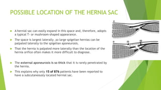
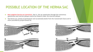
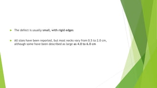
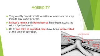
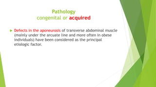
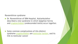
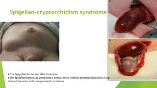
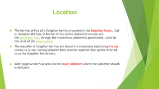
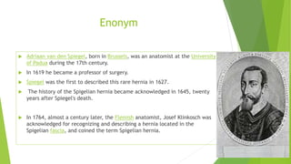
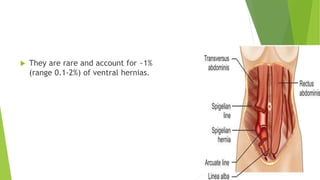
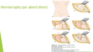
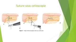
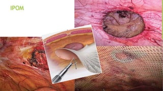
Spigelean hernia
Georges KHALIFEH FFI
GHPSO
Chirurgie digestive et visceral
Enonym
 Adriaan van den Spiegel, born in Brussels, was an anatomist at the University
of Padua during the 17th century.
 In 1619 he became a professor of surgery.
 Spiegel was the first to described this rare hernia in 1627.
 The history of the Spigelian hernia became acknowledged in 1645, twenty
years after Spiegel's death.
 In 1764, almost a century later, the Flemish anatomist, Josef Klinkosch was
acknowledged for recognizing and describing a hernia located in the
Spigelian fascia, and coined the term Spigelian hernia.
 They are rare and account for ~1%
(range 0.1-2%) of ventral hernias.
Epidemiology
Review of the literature (876 patients)
 Female-to-male ratio was 1.4:1
 Right-to-left-side ratio was 1.18:1.
 Twenty-nine instances of bilateral hernias were reported.
 In 6 patients, there was more than 1 hernia on the same side.
 29 of the hernias were located above the umbilicus.
 Most spigelian hernias have been diagnosed in patients between 40 and 70 years of
age.
 Twenty eight children, 17 boys and 11 girls, younger than 16 years of age were
operated on for spigelian hernia.
 Incarceration at the time of operation was seen in t01 of 419 reported hernias
(24.1%).
ANATOMY
Linea semilunaris (Spigelii): This is the line forming and
marking the transition from muscle to aponeurosis in the
transversus abdominis muscle
The part of the aponeurosis that lies lateral to the rectus
abdominis muscle is usually called the spigelian fascia.
Spigelian hernia belt
A hernia may occur throughout the length of the spigelian
aponeurosis.
The term spigelian hernia usually refers to hernias
located above the inferior epigastric vessels.
About 9 of 10 of these hernias occur within a transverse
belt lying 0-6 cm cranial to the interspinal plane
Above the umbilicus
29876
 Above the umbilicus the fibers of the transversus abdominis and internal
oblique muscles cross one another at angles, making herniation more
unlikely than if the fibers were to run parallel, as they do below Nthe
umbilicus.
low spigelian hernias
 caudal and medial to the
inferior epigastric artery
within the Hesselbach
triangle
POSSIBLE LOCATION OF THE HERNIA SAC
 A hernial sac can easily expand in this space and, therefore, adopts
a typical T- or mushroom-shaped appearance.
 The space is largest laterally ,so large spigelian hernias can be
palpated laterally to the spigelian aponeurosis.
 That the hernia is palpated more laterally than the location of the
hernia orifice often makes it more difficult to diagnose.
 The external aponeurosis is so thick that it is rarely penetrated by
the hernia.
 This explains why only 15 of 876 patients have been reported to
have a subcutaneously located hernial sac.
POSSIBLE LOCATION OF THE HERNIA SAC
 Most spigelian hernias are interstitial, that is, the sac penetrates through the transversus
aponeurosis and internal oblique muscle but dissects under the external oblique.
 The hernia sac consists of peritoneum and occasionally bands from the transversalis fascia and is
often preceded by preperitoneal fat.
 The defect is usually small, with rigid edges
 All sizes have been reported, but most necks vary from 0.5 to 2.0 cm,
although some have been described as large as 4.0 to 6.0 cm
MORBIDITY
 They usually contain small intestine or omentum but may
include any viscus or organ.
 Richter’s hernia and sliding hernias have been associated
with spigelian hernia
 Up to one third of reported cases have been incarcerated
at the time of operation.
Pathology
congenital or acquired
 Defects in the aponeurosis of transverse abdominal muscle
(mainly under the arcuate line and more often in obese
individuals) have been considered as the principal
etiologic factor.
Pathology
congenital or acquired
Pediatric cases, especially neonates and infants, are mostly congenital
A Spigelian hernia is associated with ipsilateral cryptorchidism among
75% male infants .
Two hypotheses have been proposed to explain the association, but the
exact mechanism is still in debate
 Spigelian-cryptorchidism syndrome (failure in the development of a
gubernaculum)
 Raveenthiran syndrome (ectopic testis from a potential hernia sac)
Raveenthiran syndrome
 Dr. Raveenthiran of SRM Hospital, Kattankulathur
described a new syndrome in which Spigelian hernia
and cryptorchidism (undescended testis) occur together.
 Some common complications of this distinct
syndrome cryptorchidism are testicular torsion, and its
link to testicular cancer
Spigelian-cryptorchidism syndrome
a The Spigelian hernia sac after dissection,
b the Spigelian hernia sac containing a normal testis without gubernaculum and a loop
of small intestine with compromised circulation
Location
 The hernial orifice of a Spigelian hernia is located in the Spigelian fascia, that
is, between the lateral border of the rectus abdominis muscle and
the semilunar line, through the transversus abdominis aponeurosis, close to
the level of the arcuate line.
 The majority of Spigelian hernias are found in a transverse band lying 0-6 cm
cranial to a line running between both anterior superior iliac spines referred
to as the Spigelian hernia belt.
 Most Spigelian hernias occur in the lower abdomen where the posterior sheath
is deficient
DIAGNOSIS
 The diagnosis of a Spigelian hernia at times presents greater challenge than
its treatment.
 The clinical presentation varies, depending on the contents of the hernial
sac and the degree and type of herniation.
 The pain, which is the most common symptom, varies, and there is no pain
typical to a Spigelian hernia.
 Findings to facilitate diagnosis are a palpable hernia and a palpable hernial
orifice.
 It should be stressed, though, that since the hernia lies deep to a muscle, it
commonly does not cause a noticeable bulge in the abdominal wall.
Radiographic features
 Ultrasound
Ultrasound can be recommended for verification of the diagnosis in both palpable
and nonpalpable Spigelian hernia.
 CT
The hernial orifice and sac can be well demonstrated by computed tomography,
which gives more detailed information on the contents of the sac than does
ultrasound scanning.
Treatment
 Anterior herniorraphy + mech
 Laparoscopic:
1. Primary closure
2. TEP totally extraperitoneal
3. TAPP transabdominal preperitoneal
4. IPOM intra abdominal ONLAY mesh
Herniorraphy par abord direct
Hernioplastie prosthetique par abord
direct
Suture sous celioscopie
IPOM
Conclusion
 Laparoscopic technique : morbidity , hospital
stay ,As outpatient
 Non complicated SH we recommend TEP
 If associated hernia TAP
 Anterior hernioplasty for complications or
emergency
How to best handle with recurrence
Which mesh and how should it be fixed?
TAPP and IPOM
 Easy
 Precise location of the defect
 Access to hernia contents
 Concomitant treatment of any other pathology( hernia )
 Efficient and safe in emergencies
Which mesh and how should it be fixed?
 Standard polypropylene mesh (TEP preserved peritoneum TAPP))
 Composite mesh
How to best handle with recurrence?
Recurrence rate 5-14%
Larson (70 patients): repaired using simple suture (recurrence rate 4.3%)
Bames (26 patients): zero recurrences after laparoscopic surgery (F/U 4 year)
Some authors mesh for defect >2 cm or weak local tissue
Open and laparoscopic both safe similar results
Patient settings (SH is unique )
Intra abdominal laparoscopic approach is most
advisable for recurrent cases
 THANK YOU

Spigelean hernia

  • 1.
    Spigelean hernia Georges KHALIFEHFFI GHPSO Chirurgie digestive et visceral
  • 2.
    Enonym  Adriaan vanden Spiegel, born in Brussels, was an anatomist at the University of Padua during the 17th century.  In 1619 he became a professor of surgery.  Spiegel was the first to described this rare hernia in 1627.  The history of the Spigelian hernia became acknowledged in 1645, twenty years after Spiegel's death.  In 1764, almost a century later, the Flemish anatomist, Josef Klinkosch was acknowledged for recognizing and describing a hernia located in the Spigelian fascia, and coined the term Spigelian hernia.
  • 3.
     They arerare and account for ~1% (range 0.1-2%) of ventral hernias.
  • 4.
    Epidemiology Review of theliterature (876 patients)  Female-to-male ratio was 1.4:1  Right-to-left-side ratio was 1.18:1.  Twenty-nine instances of bilateral hernias were reported.  In 6 patients, there was more than 1 hernia on the same side.  29 of the hernias were located above the umbilicus.  Most spigelian hernias have been diagnosed in patients between 40 and 70 years of age.  Twenty eight children, 17 boys and 11 girls, younger than 16 years of age were operated on for spigelian hernia.  Incarceration at the time of operation was seen in t01 of 419 reported hernias (24.1%).
  • 5.
    ANATOMY Linea semilunaris (Spigelii):This is the line forming and marking the transition from muscle to aponeurosis in the transversus abdominis muscle The part of the aponeurosis that lies lateral to the rectus abdominis muscle is usually called the spigelian fascia.
  • 6.
    Spigelian hernia belt Ahernia may occur throughout the length of the spigelian aponeurosis. The term spigelian hernia usually refers to hernias located above the inferior epigastric vessels. About 9 of 10 of these hernias occur within a transverse belt lying 0-6 cm cranial to the interspinal plane
  • 7.
    Above the umbilicus 29876 Above the umbilicus the fibers of the transversus abdominis and internal oblique muscles cross one another at angles, making herniation more unlikely than if the fibers were to run parallel, as they do below Nthe umbilicus.
  • 8.
    low spigelian hernias caudal and medial to the inferior epigastric artery within the Hesselbach triangle
  • 9.
    POSSIBLE LOCATION OFTHE HERNIA SAC  A hernial sac can easily expand in this space and, therefore, adopts a typical T- or mushroom-shaped appearance.  The space is largest laterally ,so large spigelian hernias can be palpated laterally to the spigelian aponeurosis.  That the hernia is palpated more laterally than the location of the hernia orifice often makes it more difficult to diagnose.  The external aponeurosis is so thick that it is rarely penetrated by the hernia.  This explains why only 15 of 876 patients have been reported to have a subcutaneously located hernial sac.
  • 10.
    POSSIBLE LOCATION OFTHE HERNIA SAC  Most spigelian hernias are interstitial, that is, the sac penetrates through the transversus aponeurosis and internal oblique muscle but dissects under the external oblique.  The hernia sac consists of peritoneum and occasionally bands from the transversalis fascia and is often preceded by preperitoneal fat.
  • 11.
     The defectis usually small, with rigid edges  All sizes have been reported, but most necks vary from 0.5 to 2.0 cm, although some have been described as large as 4.0 to 6.0 cm
  • 12.
    MORBIDITY  They usuallycontain small intestine or omentum but may include any viscus or organ.  Richter’s hernia and sliding hernias have been associated with spigelian hernia  Up to one third of reported cases have been incarcerated at the time of operation.
  • 13.
    Pathology congenital or acquired Defects in the aponeurosis of transverse abdominal muscle (mainly under the arcuate line and more often in obese individuals) have been considered as the principal etiologic factor.
  • 14.
    Pathology congenital or acquired Pediatriccases, especially neonates and infants, are mostly congenital A Spigelian hernia is associated with ipsilateral cryptorchidism among 75% male infants . Two hypotheses have been proposed to explain the association, but the exact mechanism is still in debate  Spigelian-cryptorchidism syndrome (failure in the development of a gubernaculum)  Raveenthiran syndrome (ectopic testis from a potential hernia sac)
  • 15.
    Raveenthiran syndrome  Dr.Raveenthiran of SRM Hospital, Kattankulathur described a new syndrome in which Spigelian hernia and cryptorchidism (undescended testis) occur together.  Some common complications of this distinct syndrome cryptorchidism are testicular torsion, and its link to testicular cancer
  • 16.
    Spigelian-cryptorchidism syndrome a TheSpigelian hernia sac after dissection, b the Spigelian hernia sac containing a normal testis without gubernaculum and a loop of small intestine with compromised circulation
  • 17.
    Location  The hernialorifice of a Spigelian hernia is located in the Spigelian fascia, that is, between the lateral border of the rectus abdominis muscle and the semilunar line, through the transversus abdominis aponeurosis, close to the level of the arcuate line.  The majority of Spigelian hernias are found in a transverse band lying 0-6 cm cranial to a line running between both anterior superior iliac spines referred to as the Spigelian hernia belt.  Most Spigelian hernias occur in the lower abdomen where the posterior sheath is deficient
  • 18.
    DIAGNOSIS  The diagnosisof a Spigelian hernia at times presents greater challenge than its treatment.  The clinical presentation varies, depending on the contents of the hernial sac and the degree and type of herniation.  The pain, which is the most common symptom, varies, and there is no pain typical to a Spigelian hernia.  Findings to facilitate diagnosis are a palpable hernia and a palpable hernial orifice.  It should be stressed, though, that since the hernia lies deep to a muscle, it commonly does not cause a noticeable bulge in the abdominal wall.
  • 19.
    Radiographic features  Ultrasound Ultrasoundcan be recommended for verification of the diagnosis in both palpable and nonpalpable Spigelian hernia.  CT The hernial orifice and sac can be well demonstrated by computed tomography, which gives more detailed information on the contents of the sac than does ultrasound scanning.
  • 20.
    Treatment  Anterior herniorraphy+ mech  Laparoscopic: 1. Primary closure 2. TEP totally extraperitoneal 3. TAPP transabdominal preperitoneal 4. IPOM intra abdominal ONLAY mesh
  • 21.
  • 22.
  • 23.
  • 24.
  • 29.
    Conclusion  Laparoscopic technique: morbidity , hospital stay ,As outpatient  Non complicated SH we recommend TEP  If associated hernia TAP  Anterior hernioplasty for complications or emergency
  • 30.
    How to besthandle with recurrence Which mesh and how should it be fixed?
  • 35.
    TAPP and IPOM Easy  Precise location of the defect  Access to hernia contents  Concomitant treatment of any other pathology( hernia )  Efficient and safe in emergencies
  • 36.
    Which mesh andhow should it be fixed?  Standard polypropylene mesh (TEP preserved peritoneum TAPP))  Composite mesh
  • 37.
    How to besthandle with recurrence? Recurrence rate 5-14% Larson (70 patients): repaired using simple suture (recurrence rate 4.3%) Bames (26 patients): zero recurrences after laparoscopic surgery (F/U 4 year) Some authors mesh for defect >2 cm or weak local tissue Open and laparoscopic both safe similar results Patient settings (SH is unique ) Intra abdominal laparoscopic approach is most advisable for recurrent cases
  • 41.

Editor's Notes

  • #17 Etiology Spigelian hernia occurs with the failure of the development of the gubernaculum, which in turn halts the development of the inguinal canal and further descent of the testis from its intra-abdominal position to the scrotum. The arrested testis induces a sort of "rescue canal" through a weak area in the abdominal wall in the absence of inguinal canal resulting Spigelian hernia