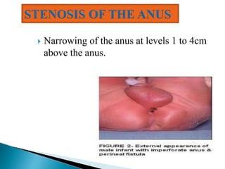
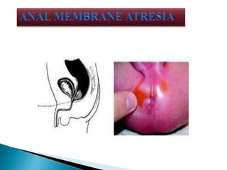
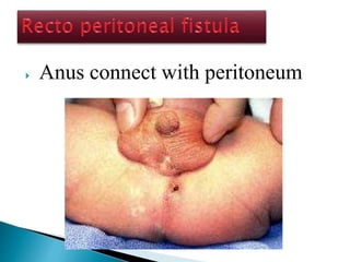
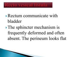
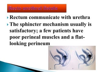
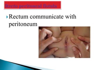
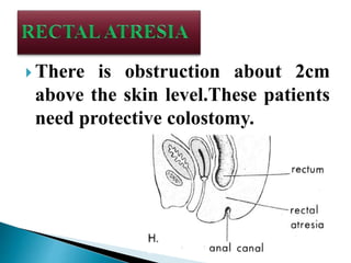
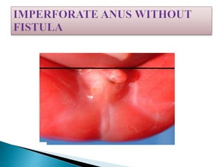
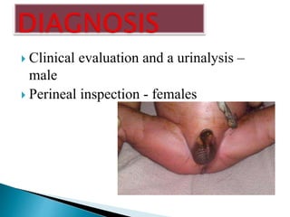
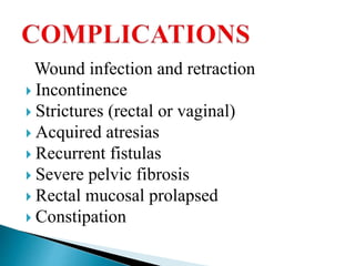
DHANYA VJ
ASSISTANT PROFESSOR
CHILD HEALTH NURSING
SUM NURSING COLLEGE
 Anorectal malformations are defined
as an arrest of rectal descent resulting
in absence of an anal opening and
occur during the 4th -16th week of
gestation.
 worldwide- 1 in 5000 live births
 genetic predisposition in certain families
 slight male preponderance
 common defect in females is recto vestibular
fistula
 Males- recto urethral fistula
 association with certain syndromes
 unknown
 associated with several other congenital
anomalies of the urinary tract, rectal
atresia and intestines.
 The hind gut forms and extends into the tail fold in the
2nd week of gestation
 At about 13 day, a ventral diverticulum, the allentois or
primitive bladder forms
 The junction of allentois and hindgut becomes the
cloaca into which the genital, urinary and intestinal
tubes empty
 This is covered by a cloacal membrane
 The urorectal septum descends to divide the common
channel into by forming the lateral ridges, which
grows in and fuse by 7th week.
 Opening of posterior portion of membrane occurs in
the 8th week
 Failures in any of these steps lead to anorectal
malformations.
 Stenosis of the anus
 Anal membrane atresia
 Anal agencies
 Rectal atresia
 Narrowing of the anus at levels 1 to 4cm
above the anus.
 Present with only anal dimple
 In female
 Imperforate anus type 3 with associated fistula
 Low rectovaginal fistula
 High rectovaginal fistula
 Recto peritoneal fistula
TYPES
Recto vesical
Recto urethral
Recto peritoneal
Anus connected with lower part of
the vagina
 Anus communicate with upper part
of the vagina
 Anus connect with peritoneum
Rectum communicate with
bladder
The sphincter mechanism is
frequently deformed and often
absent. The perineum looks flat
 Rectum communicate with urethra
 The sphincter mechanism usually is
satisfactory; a few patients have
poor perineal muscles and a flat-
looking perineum
Rectum communicate with
peritoneum
 Pouch like structure in anus and rectum
 Rare anorectal malformations
 The defect is frequently discovered while
rectal temperature is being taken.
 There is obstruction about 2cm
above the skin level.These patients
need protective colostomy.
 The rectum is completely blind and is
usually found approximately 2cm above
the perineal skin.
 The sacrum and the sphincteric
mechanism are usually well developed .
 A colostomy is indicated during the
newborn period.
 This defect is frequently associated with
down syndrome.
GENITOURINARY
 Vesicoureteric reflux
 Renal agenesis
 Ureteral duplication
 Cryptorchidism
 Hypospadiasis
 Bicornuate uterus
 Vaginal septums
 VERTEBRAL
 Presacral masses
 Meningocele
 Lipoma
 Dermoid
CARDIOVASCULAR
 Tetralogy of Fallot
 VSD
 TGA
 Hypoplastic left heart syndrome
Gastrointestinal
 Tracheoesophagel fistula
 Duodenal atresia
 Malrotation
 Hirschsprung disease
Low lesions
 Newborn examination reveals absence of an
anal orifice in the correct position
 Normal position of anus is halfway between
the coccyx and the scrotum or introitus
 Primary symptom is constipation
 Cases with anterior ectopic anus have no
symptoms
 If no anus or fistula present, it is called
“covered anus”
 After 24 hours meconium bulging may be
seen creating a blue or black appearance.
In male, the perineal fistula may track
anteriorly along the median raphe across the
scrotum and even down the penile shaft, this is
usually a thin track, with a normal rectum
often just a few millimeter from the skin
 In females, a low lesion enters the
vestibule or fourchette.
 In this case, the rectum has
descended through the sphincter
complex.
 In a boy with high imperforate anus, the
perineum appears flat
 There may be air or meconium passed via
the penis (urethra) when the fistula is
high
 In rectourethral fistula, the sphincter
mechanism is satisfactory, the sacrum
may be bifid and the anal dimple is near
the scrotum
 In retrovestibular fistulas, the sphincter
mechanism is poorly developed and
sacrum is hypoplastic or absent.
 In boys with trisomy 21, all the features
may be present, but there will be no
fistula, the sacrum and sphincter
mechanisms are usually well developed
and prognosis is good.
 In girls with high imperforate anus, there
may be the appearance of a rectovaginal
fistula.
 A true rectovaginal fistula is rare. Most
fistulas to the vestibule outside the
hymen orifice.
 These have a good prognosis with a
normal sacrum and anal dimple and intact
sphincter function
 Clinical evaluation and a urinalysis –
male
 Perineal inspection - females
 Invertogram
 Mainly required before redo surgery.
 Clearly shows the anatomy of sphincter
muscles, levator ani, muscle complex.
 Delineates the rectal pouch and fistula.
 Clearly shows the relationship between
intestine and surrounding muscles.
 Provides exquisite soft tissue imaging in
all three plane.
 Better than CT in soft tissue imaging and
no radiation hazard.
 Scan is expanded to include pelvis ,
kidneys and spinal cord.
 Clearly shows whether the pulled through
intestine is within levator ani sling or not
 Early decision-making
 IV line - fluids and antibiotics
 Insertion of nasogastric tube is inserted
 Meconium is usually not seen at the
perineum in a baby with a recto-perineal
fistula until at least 16–24 hours.
 Clinical inspection of the buttocks -
flat "bottom" or flat perineum
Operation can be performed during
newborn period, and after 1year
corrective surgical repair is
performed
 colostomy or an anoplasty
 Clinical evaluation and urine analysis
 X-ray, cross-table lateral film with
the baby in prone position
 colostomy
 Surgically created opening between the
colon and the abdominal wall to allow
fecal elimination.
 It may be temporary or permanent
diversion
 TPYES
 Transverse colostomy
 Descending colostomy
 DESCENDING COLOSTOMY
 made from the descending portion of the
colon located in the lower-left quadrant of
the abdomen, with separated stomas . The
proximal stoma is connected to the upper
gastrointestinal tract and drains stool. The
distal stoma is connected to the rectum and
will drain small amounts of mucus material.
 Performed at 1–2 months of age
 90% of male patients done anorectal repair,
 while 10% require laparotomy
Rectobladder neck fistula
 operation involves both a posterior sagittal
incision and an abdominal component, which
can be done with laparoscopy or laparotomy.
 Rectovestibular fistula is the most common defect in
girls and has an excellent functional prognosis.
 The diagnosis is based on clinical examination.
A meticulous inspection of the neonatal genitalia allows
the clinician to observe a normal urethral meatus and a
normal vagina, with a third hole in the vestibule, which
is the rectovestibular fistula .
 The laparoscopically-assisted anorectal
approach consists in mobilizing and bringing
the rectum through the pelvic floor sphincter
muscles through a minimal posterior
incision. Perianal dissection towards the
laparoscopic light source favours accurate
placement of a trocar to pull the rectum
through the external sphincter muscle
complex
o Thorough physical examination by the
attending nurse to identify anorectal
malformation
o kept nil per orally
o Colostomy care
o Start feeding after the child has recovered
from anaesthesia
 After the diagnosis of occlusion had been
made, gastric suction may be used.
 IV feeding and nasogastric suction help
prevent abdominal distension immediate
post operatively
 Keep the anal site clean and dry
 Diaper should not be used.
 The dressing must be changed frequently
Wound infection and retraction
 Incontinence
 Strictures (rectal or vaginal)
 Acquired atresias
 Recurrent fistulas
 Severe pelvic fibrosis
 Rectal mucosal prolapsed
 Constipation
Anorectal malformations in children

Anorectal malformations in children

  • 1.
    DHANYA VJ ASSISTANT PROFESSOR CHILDHEALTH NURSING SUM NURSING COLLEGE
  • 2.
     Anorectal malformationsare defined as an arrest of rectal descent resulting in absence of an anal opening and occur during the 4th -16th week of gestation.
  • 3.
     worldwide- 1in 5000 live births  genetic predisposition in certain families  slight male preponderance  common defect in females is recto vestibular fistula  Males- recto urethral fistula  association with certain syndromes
  • 4.
     unknown  associatedwith several other congenital anomalies of the urinary tract, rectal atresia and intestines.
  • 5.
     The hindgut forms and extends into the tail fold in the 2nd week of gestation  At about 13 day, a ventral diverticulum, the allentois or primitive bladder forms  The junction of allentois and hindgut becomes the cloaca into which the genital, urinary and intestinal tubes empty  This is covered by a cloacal membrane
  • 6.
     The urorectalseptum descends to divide the common channel into by forming the lateral ridges, which grows in and fuse by 7th week.  Opening of posterior portion of membrane occurs in the 8th week  Failures in any of these steps lead to anorectal malformations.
  • 7.
     Stenosis ofthe anus  Anal membrane atresia  Anal agencies  Rectal atresia
  • 8.
     Narrowing ofthe anus at levels 1 to 4cm above the anus.
  • 10.
     Present withonly anal dimple  In female  Imperforate anus type 3 with associated fistula  Low rectovaginal fistula  High rectovaginal fistula  Recto peritoneal fistula TYPES
  • 11.
  • 12.
    Anus connected withlower part of the vagina
  • 13.
     Anus communicatewith upper part of the vagina
  • 14.
     Anus connectwith peritoneum
  • 15.
    Rectum communicate with bladder Thesphincter mechanism is frequently deformed and often absent. The perineum looks flat
  • 16.
     Rectum communicatewith urethra  The sphincter mechanism usually is satisfactory; a few patients have poor perineal muscles and a flat- looking perineum
  • 17.
  • 18.
     Pouch likestructure in anus and rectum  Rare anorectal malformations  The defect is frequently discovered while rectal temperature is being taken.
  • 19.
     There isobstruction about 2cm above the skin level.These patients need protective colostomy.
  • 20.
     The rectumis completely blind and is usually found approximately 2cm above the perineal skin.  The sacrum and the sphincteric mechanism are usually well developed .  A colostomy is indicated during the newborn period.  This defect is frequently associated with down syndrome.
  • 22.
    GENITOURINARY  Vesicoureteric reflux Renal agenesis  Ureteral duplication  Cryptorchidism  Hypospadiasis  Bicornuate uterus  Vaginal septums
  • 23.
     VERTEBRAL  Presacralmasses  Meningocele  Lipoma  Dermoid
  • 24.
    CARDIOVASCULAR  Tetralogy ofFallot  VSD  TGA  Hypoplastic left heart syndrome
  • 25.
    Gastrointestinal  Tracheoesophagel fistula Duodenal atresia  Malrotation  Hirschsprung disease
  • 26.
    Low lesions  Newbornexamination reveals absence of an anal orifice in the correct position  Normal position of anus is halfway between the coccyx and the scrotum or introitus  Primary symptom is constipation  Cases with anterior ectopic anus have no symptoms
  • 27.
     If noanus or fistula present, it is called “covered anus”  After 24 hours meconium bulging may be seen creating a blue or black appearance. In male, the perineal fistula may track anteriorly along the median raphe across the scrotum and even down the penile shaft, this is usually a thin track, with a normal rectum often just a few millimeter from the skin
  • 28.
     In females,a low lesion enters the vestibule or fourchette.  In this case, the rectum has descended through the sphincter complex.
  • 29.
     In aboy with high imperforate anus, the perineum appears flat  There may be air or meconium passed via the penis (urethra) when the fistula is high  In rectourethral fistula, the sphincter mechanism is satisfactory, the sacrum may be bifid and the anal dimple is near the scrotum
  • 30.
     In retrovestibularfistulas, the sphincter mechanism is poorly developed and sacrum is hypoplastic or absent.  In boys with trisomy 21, all the features may be present, but there will be no fistula, the sacrum and sphincter mechanisms are usually well developed and prognosis is good.
  • 31.
     In girlswith high imperforate anus, there may be the appearance of a rectovaginal fistula.  A true rectovaginal fistula is rare. Most fistulas to the vestibule outside the hymen orifice.  These have a good prognosis with a normal sacrum and anal dimple and intact sphincter function
  • 32.
     Clinical evaluationand a urinalysis – male  Perineal inspection - females
  • 33.
  • 34.
     Mainly requiredbefore redo surgery.  Clearly shows the anatomy of sphincter muscles, levator ani, muscle complex.  Delineates the rectal pouch and fistula.  Clearly shows the relationship between intestine and surrounding muscles.
  • 35.
     Provides exquisitesoft tissue imaging in all three plane.  Better than CT in soft tissue imaging and no radiation hazard.  Scan is expanded to include pelvis , kidneys and spinal cord.  Clearly shows whether the pulled through intestine is within levator ani sling or not
  • 36.
     Early decision-making IV line - fluids and antibiotics  Insertion of nasogastric tube is inserted  Meconium is usually not seen at the perineum in a baby with a recto-perineal fistula until at least 16–24 hours.
  • 37.
     Clinical inspectionof the buttocks - flat "bottom" or flat perineum Operation can be performed during newborn period, and after 1year corrective surgical repair is performed  colostomy or an anoplasty
  • 38.
     Clinical evaluationand urine analysis  X-ray, cross-table lateral film with the baby in prone position  colostomy
  • 40.
     Surgically createdopening between the colon and the abdominal wall to allow fecal elimination.  It may be temporary or permanent diversion  TPYES  Transverse colostomy  Descending colostomy
  • 41.
     DESCENDING COLOSTOMY made from the descending portion of the colon located in the lower-left quadrant of the abdomen, with separated stomas . The proximal stoma is connected to the upper gastrointestinal tract and drains stool. The distal stoma is connected to the rectum and will drain small amounts of mucus material.
  • 43.
     Performed at1–2 months of age  90% of male patients done anorectal repair,  while 10% require laparotomy Rectobladder neck fistula  operation involves both a posterior sagittal incision and an abdominal component, which can be done with laparoscopy or laparotomy.
  • 44.
     Rectovestibular fistulais the most common defect in girls and has an excellent functional prognosis.  The diagnosis is based on clinical examination. A meticulous inspection of the neonatal genitalia allows the clinician to observe a normal urethral meatus and a normal vagina, with a third hole in the vestibule, which is the rectovestibular fistula .
  • 46.
     The laparoscopically-assistedanorectal approach consists in mobilizing and bringing the rectum through the pelvic floor sphincter muscles through a minimal posterior incision. Perianal dissection towards the laparoscopic light source favours accurate placement of a trocar to pull the rectum through the external sphincter muscle complex
  • 47.
    o Thorough physicalexamination by the attending nurse to identify anorectal malformation o kept nil per orally o Colostomy care o Start feeding after the child has recovered from anaesthesia
  • 48.
     After thediagnosis of occlusion had been made, gastric suction may be used.  IV feeding and nasogastric suction help prevent abdominal distension immediate post operatively  Keep the anal site clean and dry  Diaper should not be used.  The dressing must be changed frequently
  • 49.
    Wound infection andretraction  Incontinence  Strictures (rectal or vaginal)  Acquired atresias  Recurrent fistulas  Severe pelvic fibrosis  Rectal mucosal prolapsed  Constipation