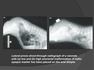
2. Introduction :
Early in embryonic life there is a common chamber (the
cloaca) into which, the hindgut and allantois open.
The cloaca becomes divided into two parts, dorsal
(rectum) and ventral (urogenital sinus) by the
downgrowth of a septum.
The dorsal part of the cloacal membrane known as the
anal membrane and is composed of an outer layer of
ectoderm and an inner layer of endoderm.
Resorption of this anal membrane by the 8th week of
embryonic life creates the anal canal.
3. It is due to imperfect fusion of the post-allantoic
gut with the proctodaeum.
Incidence is 1 in 4500 of live births.
The condition is divided into two main groups;
the high and the low, depending on whether the
termination of the bowel is above or below the
pelvic floor.
4. 1-Low abnormalities :
Below the level of pelvic floor (Puborectalis).
Easy to diagnose and treat with good outcome. they may be:
1. Covered anus : The anal canal is covered by a bar of skin.
2. Ectopic anus : The anus situated anteriorly in the
perineum in boys, or Anovestibular fistula in girls.
3. Anal stenosis : The anus is microscopic which respond to
regular dilatation.
4. Anal membrane
5. 2- High abnormalities :
They may be :
1. Anorectal agenesis : The blind rectal pouch lies just above
the pelvic floor with a fistula into the bladder, urethra, uterus,
vagina.
2. Rectal atresia : The anal canal is normal but ends blindly at
the level of the pelvic floor and the bowel also ends blindly
above the pelvic floor without a fistulous opening.
3. Cloaca : This occurs only in females, where the bowel,
urinary and genital tracts all open into a common wide cavity
8. Clinical assessment :
Newborn presents with :
• inability to pass meconium.
• abdominal distension.
• features of intestinal obstruction.
• improper anal dimple.
• sometimes with complaints of passing
meconium per urethra.
9. • It can be associated with VACTER anomaly.
• Most common ARM in boys is rectobulbar
fistula.
• Most common ARM in females is rectovestibular
fistula.
10. Radiological examination:
Sufficient air may collect in the large intestine, six
hours after birth, to give an x-ray shadow.
With a radio-opaque marker like coin strapped to
the site of the anus, the infant is held in upside
down for 3-4 minutes radiographed in the inverted
position (or lateral prone x-ray).
If the distance between the site of the marker and
the blind end of the rectum is more than 2.5 cm,
the abnormality is high.
11. Lateral prone shoot-through radiograph of a neonate
with (a) low and (b) high anorectal malformation. A radio-
opaque marker has been placed on the anal dimple.
12. If a high abnormality is suspected, obtain a
specimen of urine , the presence of proteus
usually indicates presence of fistula.
13. Treatment :
In low fistula, single stage reconstruction is
done under GA with very good results.
Anoplasty.
Anovestibuloplasty.
Anal dilatation.
Incision of anal membrane.
14. High abnormalities require :
Early colostomy, with definitive repair performed
several months later. This may involve posterior
sagittal anorectoplasty (PSARP) with or without
transabdominal mobilization of the left colon and
division of any communication (fistula) with the urinary
tract.
Laparotomy, division of fistula and transverse
colostomy. A rectal "pull-through" operation can be
done later.
Laparotomy, division of fistula and "pull-through"
operation in one stage.
Colostomy only (for the cloacal variety).