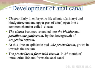
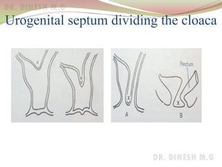
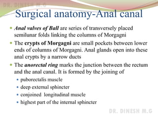
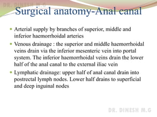
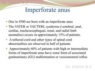
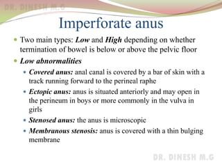
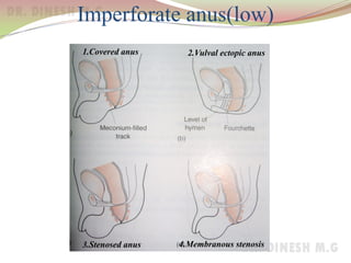
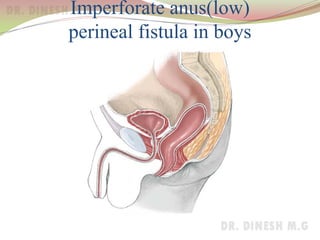
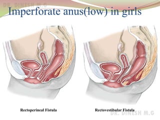
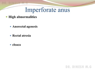
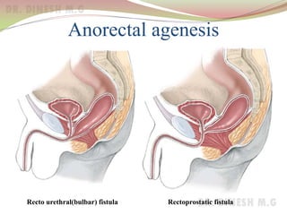
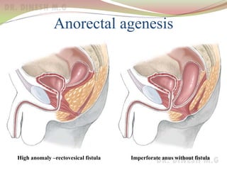
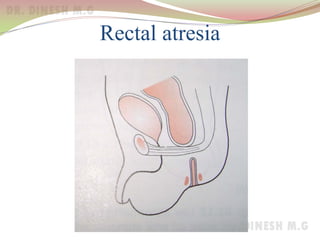
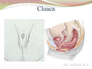
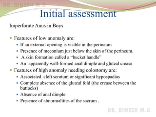
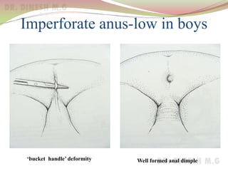
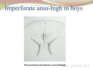
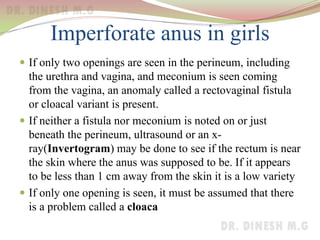
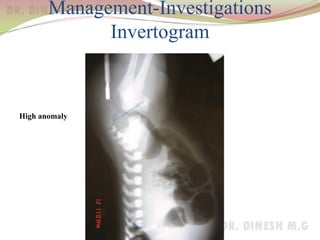
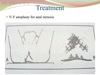
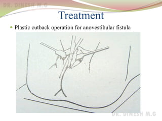
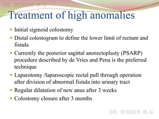
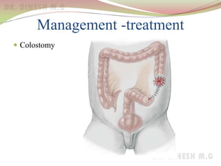
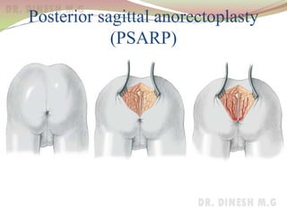
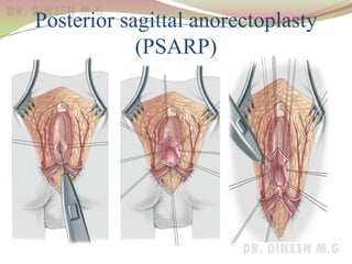
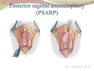
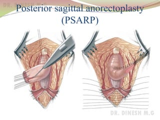
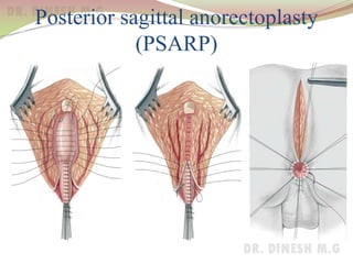
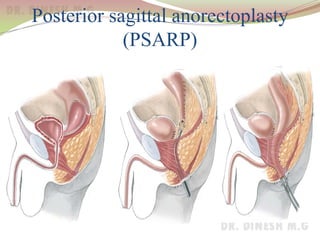
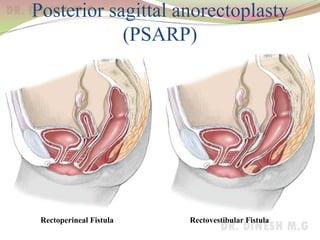
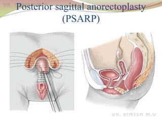
Imperforate anus
Dr. Dinesh. M.G
Professor of Surgery
J.J.M.M.C.
Davangere
Development of anal canal
 Cloaca: Early in embryonic life allantois(urinary) and
hindgut(rectum and upper part of anus) open into a
common chamber called cloaca
 The cloaca becomes separated into the bladder and
postallantoic gut(rectum) by the downgrowth of
urogenital septum.
 At this time an epiblastic bud , the proctodaeum, grows in
towards the rectum
 The proctodaeum fuses with rectum in 3rd month of
intrauterine life and forms the anal canal
Urogenital septum dividing the cloaca
Surgical anatomy-Anal canal
 Anal canal starts where rectum passes through the pelvic
diaphragm and ends at the anal verge
 The muscular junction between the rectum and anal canal
is felt as a anorectal bundle or ring
 The internal sphincter is a thickened continuation of
circular muscle coat of rectum. It is pearly white in colour
and involuntary muscle
 The longitudinal muscle is a continuation of longitudinal
muscle coat of rectum
 The external sphincter is pink in colour and composed of
voluntary muscle
Surgical anatomy-Anal canal
 Intersphincteric space exists between external and
internal sphincter
 The puborectalis maintains the anorectal angle and is
essential for preservation of continence
Surgical anatomy-Anal canal
 Pink columnar epithelium rectal mucosa changes into red
cubical epithelium in anal canal
 Mucosa is thrown into 8- 12 anal columns of Morgagni
which are joined at their lower ends by anal valves. This
wavy junction is called dentate line
 The dentate line separates
Above Below
• Cubical epithelium from squamous epithelium
• Autonomic nerve supply from somatic nerve supply
• Portal venous system from systemic venous system
Surgical anatomy-Anal canal
 Anal valves of Ball are series of transversely placed
semilunar folds linking the columns of Morgagni
 The crypts of Morgagni are small pockets between lower
ends of columns of Morgagni. Anal glands open into these
anal crypts by a narrow ducts
 The anorectal ring marks the junction between the rectum
and the anal canal. It is formed by the joining of
 puborectalis muscle
 deep external sphincter
 conjoined longitudinal muscle
 highest part of the internal sphincter
Surgical anatomy-Anal canal
 Arterial supply by branches of superior, middle and
inferior haemorrhoidal arteries
 Venous drainage : the superior and middle haemorrhoidal
veins drain via the inferior mesenteric vein into portal
system. The inferior haemorrhoidal veins drain the lower
half of the anal canal to the external iliac vein
 Lymphatic drainage: upper half of anal canal drain into
postrectal lymph nodes. Lower half drains to superficial
and deep inguinal nodes
Imperforate anus
 One in 4500 are born with an imperforate anus
 The VATER or VACTERL syndrome (vertebral, anal,
cardiac, tracheoesophageal, renal, and radial limb
anomalies) occurs in approximately 15% of patients.
 A tethered cord and other types of spinal cord
abnormalities are observed in half of patients
 Approximately 60% of patients with high or intermediate
forms of imperforate anus have some form of associated
genitourinary (GU) malformation or vesicoureteral reflux
Imperforate anus
 Two main types: Low and High depending on whether
termination of bowel is below or above the pelvic floor
 Low abnormalities
 Covered anus: anal canal is covered by a bar of skin with a
track running forward to the perineal raphe
 Ectopic anus: anus is situated anteriorly and may open in
the perineum in boys or more commonly in the vulva in
girls
 Stenosed anus: the anus is microscopic
 Membranous stenosis: anus is covered with a thin bulging
membrane
Imperforate anus(low)
1.Covered anus 2.Vulval ectopic anus
3.Stenosed anus 4.Membranous stenosis
Imperforate anus(low)
perineal fistula in boys
Imperforate anus(low) in girls
Rectoperineal Fistula Rectovestibular Fistula
Imperforate anus
 High abnormalities
 Anorectal agenesis
 Rectal atresia
 cloaca
Anorectal agenesis
Recto urethral(bulbar) fistula Rectoprostatic fistula
Anorectal agenesis
Imperforate anus without fistulaHigh anomaly –rectovesical fistula
Rectal atresia
Cloaca
Initial Management of the Newborn
 It is best to wait 24 hours prior to any surgery to allow
progression of gas or meconium through the bowel
 To decide whether temporary colostomy is needed
Initial assessment
Imperforate Anus in Boys
 Features of low anomaly are:
 If an external opening is visible in the perineum
 Presence of meconium just below the skin of the perineum.
 A skin formation called a “bucket handle”
 An apparently well-formed anal dimple and gluteal crease
 Features of high anomaly needing colostomy are:
 Associated cleft scrotum or significant hypospadias
 Complete absence of the gluteal fold (the crease between the
buttocks)
 Absence of anal dimple
 Presence of abnormalities of the sacrum .
Imperforate anus-low in boys
‘bucket handle’ deformity Well formed anal dimple
Imperforate anus-high in boys
Flat perineum and absence of anal dimple
Imperforate anus in girls
 If only two openings are seen in the perineum, including
the urethra and vagina, and meconium is seen coming
from the vagina, an anomaly called a rectovaginal fistula
or cloacal variant is present.
 If neither a fistula nor meconium is noted on or just
beneath the perineum, ultrasound or an x-
ray(Invertogram) may be done to see if the rectum is near
the skin where the anus was supposed to be. If it appears
to be less than 1 cm away from the skin it is a low variety
 If only one opening is seen, it must be assumed that there
is a problem called a cloaca
Management-Investigations
Invertogram
High anomaly
Treatment of low anomalies
 Initially by dilatations
After 3-4 weeks
 V-Y anoplasty for stenosed anus
 Anal transposition for ectopic stenosed anus
 Cutback procedure for ectopic stenosed anus
 Regular dilatation of anoplasty by the parents after 3
weeks to prevent stenosis
Treatment
 V-Y anoplasty for anal stenosis
Treatment
 Plastic cutback operation for anovestibular fistula
Treatment of high anomalies
 Initial sigmoid colostomy
 Distal colostogram to define the lower limit of rectum and
fistula
 Currently the posterior sagittal anorectoplasty (PSARP)
procedure described by de Vries and Pena is the preferred
technique
 Laparotomy /laparoscopic rectal pull through operation
after division of abnormal fistula into urinary tract
 Regular dilatation of new anus after 3 weeks
 Colostomy closure after 3 months
Management -treatment
 Colostomy
Anorectal agenesis
Imperforate anus without fistulaHigh anomaly –rectovesical fistula
Posterior sagittal anorectoplasty
(PSARP)
Posterior sagittal anorectoplasty
(PSARP)
Posterior sagittal anorectoplasty
(PSARP)
Posterior sagittal anorectoplasty
(PSARP)
Posterior sagittal anorectoplasty
(PSARP)
Posterior sagittal anorectoplasty
(PSARP)
Posterior sagittal anorectoplasty
(PSARP)
Rectoperineal Fistula Rectovestibular Fistula
Posterior sagittal anorectoplasty
(PSARP)
Conclusions
 Clinical results are different for each type of the
malformations.
 Faecal continence is good in low anomalies
 Constipation is a common sequelae seen after the repair of
an anorectal malformation.
 Urinary control can be expected in the overwhelming
majority of male patients after repair of imperforate anus
provided a good surgical technique was performed
Thank you

Imperforate Anus

  • 1.
    Imperforate anus Dr. Dinesh.M.G Professor of Surgery J.J.M.M.C. Davangere
  • 2.
    Development of analcanal  Cloaca: Early in embryonic life allantois(urinary) and hindgut(rectum and upper part of anus) open into a common chamber called cloaca  The cloaca becomes separated into the bladder and postallantoic gut(rectum) by the downgrowth of urogenital septum.  At this time an epiblastic bud , the proctodaeum, grows in towards the rectum  The proctodaeum fuses with rectum in 3rd month of intrauterine life and forms the anal canal
  • 3.
  • 4.
  • 5.
    Surgical anatomy-Anal canal Anal canal starts where rectum passes through the pelvic diaphragm and ends at the anal verge  The muscular junction between the rectum and anal canal is felt as a anorectal bundle or ring  The internal sphincter is a thickened continuation of circular muscle coat of rectum. It is pearly white in colour and involuntary muscle  The longitudinal muscle is a continuation of longitudinal muscle coat of rectum  The external sphincter is pink in colour and composed of voluntary muscle
  • 6.
    Surgical anatomy-Anal canal Intersphincteric space exists between external and internal sphincter  The puborectalis maintains the anorectal angle and is essential for preservation of continence
  • 7.
    Surgical anatomy-Anal canal Pink columnar epithelium rectal mucosa changes into red cubical epithelium in anal canal  Mucosa is thrown into 8- 12 anal columns of Morgagni which are joined at their lower ends by anal valves. This wavy junction is called dentate line  The dentate line separates Above Below • Cubical epithelium from squamous epithelium • Autonomic nerve supply from somatic nerve supply • Portal venous system from systemic venous system
  • 8.
    Surgical anatomy-Anal canal Anal valves of Ball are series of transversely placed semilunar folds linking the columns of Morgagni  The crypts of Morgagni are small pockets between lower ends of columns of Morgagni. Anal glands open into these anal crypts by a narrow ducts  The anorectal ring marks the junction between the rectum and the anal canal. It is formed by the joining of  puborectalis muscle  deep external sphincter  conjoined longitudinal muscle  highest part of the internal sphincter
  • 9.
    Surgical anatomy-Anal canal Arterial supply by branches of superior, middle and inferior haemorrhoidal arteries  Venous drainage : the superior and middle haemorrhoidal veins drain via the inferior mesenteric vein into portal system. The inferior haemorrhoidal veins drain the lower half of the anal canal to the external iliac vein  Lymphatic drainage: upper half of anal canal drain into postrectal lymph nodes. Lower half drains to superficial and deep inguinal nodes
  • 10.
    Imperforate anus  Onein 4500 are born with an imperforate anus  The VATER or VACTERL syndrome (vertebral, anal, cardiac, tracheoesophageal, renal, and radial limb anomalies) occurs in approximately 15% of patients.  A tethered cord and other types of spinal cord abnormalities are observed in half of patients  Approximately 60% of patients with high or intermediate forms of imperforate anus have some form of associated genitourinary (GU) malformation or vesicoureteral reflux
  • 11.
    Imperforate anus  Twomain types: Low and High depending on whether termination of bowel is below or above the pelvic floor  Low abnormalities  Covered anus: anal canal is covered by a bar of skin with a track running forward to the perineal raphe  Ectopic anus: anus is situated anteriorly and may open in the perineum in boys or more commonly in the vulva in girls  Stenosed anus: the anus is microscopic  Membranous stenosis: anus is covered with a thin bulging membrane
  • 12.
    Imperforate anus(low) 1.Covered anus2.Vulval ectopic anus 3.Stenosed anus 4.Membranous stenosis
  • 13.
  • 14.
    Imperforate anus(low) ingirls Rectoperineal Fistula Rectovestibular Fistula
  • 15.
    Imperforate anus  Highabnormalities  Anorectal agenesis  Rectal atresia  cloaca
  • 16.
    Anorectal agenesis Recto urethral(bulbar)fistula Rectoprostatic fistula
  • 17.
    Anorectal agenesis Imperforate anuswithout fistulaHigh anomaly –rectovesical fistula
  • 18.
  • 19.
  • 20.
    Initial Management ofthe Newborn  It is best to wait 24 hours prior to any surgery to allow progression of gas or meconium through the bowel  To decide whether temporary colostomy is needed
  • 21.
    Initial assessment Imperforate Anusin Boys  Features of low anomaly are:  If an external opening is visible in the perineum  Presence of meconium just below the skin of the perineum.  A skin formation called a “bucket handle”  An apparently well-formed anal dimple and gluteal crease  Features of high anomaly needing colostomy are:  Associated cleft scrotum or significant hypospadias  Complete absence of the gluteal fold (the crease between the buttocks)  Absence of anal dimple  Presence of abnormalities of the sacrum .
  • 22.
    Imperforate anus-low inboys ‘bucket handle’ deformity Well formed anal dimple
  • 23.
    Imperforate anus-high inboys Flat perineum and absence of anal dimple
  • 24.
    Imperforate anus ingirls  If only two openings are seen in the perineum, including the urethra and vagina, and meconium is seen coming from the vagina, an anomaly called a rectovaginal fistula or cloacal variant is present.  If neither a fistula nor meconium is noted on or just beneath the perineum, ultrasound or an x- ray(Invertogram) may be done to see if the rectum is near the skin where the anus was supposed to be. If it appears to be less than 1 cm away from the skin it is a low variety  If only one opening is seen, it must be assumed that there is a problem called a cloaca
  • 25.
  • 26.
    Treatment of lowanomalies  Initially by dilatations After 3-4 weeks  V-Y anoplasty for stenosed anus  Anal transposition for ectopic stenosed anus  Cutback procedure for ectopic stenosed anus  Regular dilatation of anoplasty by the parents after 3 weeks to prevent stenosis
  • 27.
    Treatment  V-Y anoplastyfor anal stenosis
  • 28.
    Treatment  Plastic cutbackoperation for anovestibular fistula
  • 29.
    Treatment of highanomalies  Initial sigmoid colostomy  Distal colostogram to define the lower limit of rectum and fistula  Currently the posterior sagittal anorectoplasty (PSARP) procedure described by de Vries and Pena is the preferred technique  Laparotomy /laparoscopic rectal pull through operation after division of abnormal fistula into urinary tract  Regular dilatation of new anus after 3 weeks  Colostomy closure after 3 months
  • 30.
  • 31.
    Anorectal agenesis Imperforate anuswithout fistulaHigh anomaly –rectovesical fistula
  • 32.
  • 33.
  • 34.
  • 35.
  • 36.
  • 37.
  • 38.
  • 39.
  • 40.
    Conclusions  Clinical resultsare different for each type of the malformations.  Faecal continence is good in low anomalies  Constipation is a common sequelae seen after the repair of an anorectal malformation.  Urinary control can be expected in the overwhelming majority of male patients after repair of imperforate anus provided a good surgical technique was performed
  • 41.