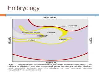
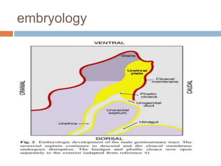
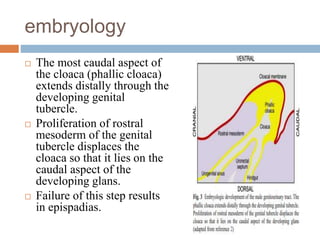
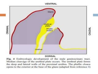
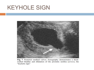
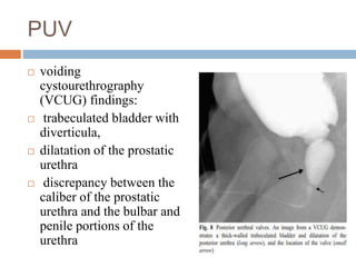
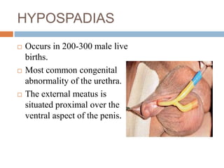
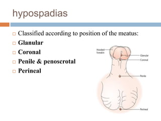
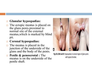
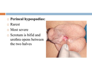
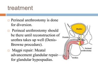
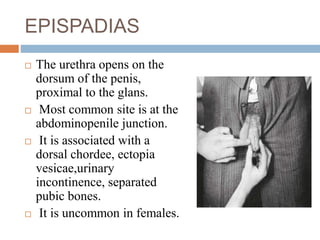
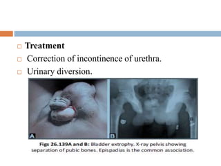
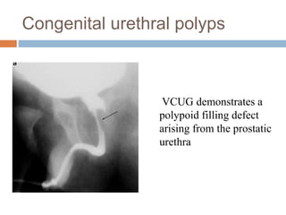
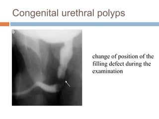
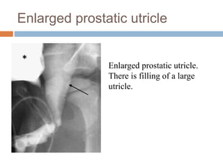
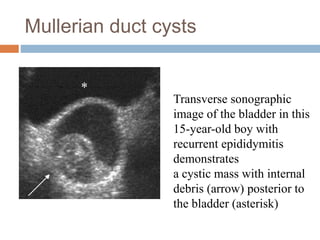
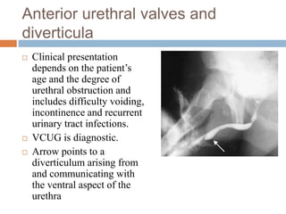
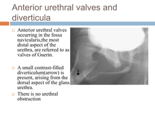
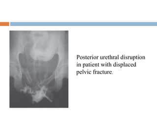
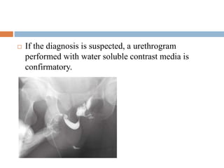
This document discusses the embryology and various congenital anomalies of the male urethra. It begins with the embryological development of the male urethra from the cloaca. It then describes several congenital anomalies in more detail, including posterior urethral valves, hypospadias, epispadias, prune belly syndrome, congenital urethral strictures, congenital urethral polyps, enlarged prostatic utricles, Mullerian duct cysts, and Cowper's syringocele. For each anomaly, it discusses features, diagnosis using imaging like VCUG, and potential treatments.
Hello Guys,
This presentation talks about diagnosis and management of Antenatally detected hydronephrosis. We have discussed evidence based fetal hydronephrosis management including - antenatal followup schedule, fetal interventions, postnatal screening and follow up proforma, MCU, Functional renal scans, prophylactic antibiotics and available surgical management options.
POSTERIOR URETHRAL VALVES- Pediatric Surgery
• Dear viewers,
• Greetings from “ Surgical Educator”
• Today I have uploaded one more video in Pediatric Surgery/Pediatric Urology- “ Posterior Urethral Valves”
• Posterior Urethral Valves is the congenital cause for Bladder Outlet Obstruction, resulting in abnormal development of the kidneys as well as the bladder.
• In this video, I talked about the learning outcomes, introduction, etiopathogenesis, clinical features, investigations, differential diagnosis, treatment, follow-up and prognosis of “ Posterior Urethral Valves”
• I hope you will enjoy the video for its educational value.
• You can watch all my teaching videos in the following links
• surgicaleducator.blogspot.com youtube.com/c/surgicaleducator
• Thank you for watching the video.
Congenital absence of anal opening can happen both in male and female babies. Since they can't pass stool, this is a neonatal emergency surgical problem. Exact type of anomaly should be ascertained and appropriate surgical correction should be done precisely.
Presentation delivered at a paediatric clinical meeting of the Federal Medical Center, Lokoja. Nigeria
This presentation doesn't serve as a substitute for texts and/or journals.
2024.06.01 Introducing a competency framework for languag learning materials ...Sandy Millin
http://sandymillin.wordpress.com/iateflwebinar2024
Published classroom materials form the basis of syllabuses, drive teacher professional development, and have a potentially huge influence on learners, teachers and education systems. All teachers also create their own materials, whether a few sentences on a blackboard, a highly-structured fully-realised online course, or anything in between. Despite this, the knowledge and skills needed to create effective language learning materials are rarely part of teacher training, and are mostly learnt by trial and error.
Knowledge and skills frameworks, generally called competency frameworks, for ELT teachers, trainers and managers have existed for a few years now. However, until I created one for my MA dissertation, there wasn’t one drawing together what we need to know and do to be able to effectively produce language learning materials.
This webinar will introduce you to my framework, highlighting the key competencies I identified from my research. It will also show how anybody involved in language teaching (any language, not just English!), teacher training, managing schools or developing language learning materials can benefit from using the framework.
Welcome to TechSoup New Member Orientation and Q&A (May 2024).pdfTechSoup
In this webinar you will learn how your organization can access TechSoup's wide variety of product discount and donation programs. From hardware to software, we'll give you a tour of the tools available to help your nonprofit with productivity, collaboration, financial management, donor tracking, security, and more.
Model Attribute Check Company Auto PropertyCeline George
In Odoo, the multi-company feature allows you to manage multiple companies within a single Odoo database instance. Each company can have its own configurations while still sharing common resources such as products, customers, and suppliers.
Palestine last event orientationfvgnh .pptxRaedMohamed3
An EFL lesson about the current events in Palestine. It is intended to be for intermediate students who wish to increase their listening skills through a short lesson in power point.
Unit 8 - Information and Communication Technology (Paper I).pdfThiyagu K
This slides describes the basic concepts of ICT, basics of Email, Emerging Technology and Digital Initiatives in Education. This presentations aligns with the UGC Paper I syllabus.
We all have good and bad thoughts from time to time and situation to situation. We are bombarded daily with spiraling thoughts(both negative and positive) creating all-consuming feel , making us difficult to manage with associated suffering. Good thoughts are like our Mob Signal (Positive thought) amidst noise(negative thought) in the atmosphere. Negative thoughts like noise outweigh positive thoughts. These thoughts often create unwanted confusion, trouble, stress and frustration in our mind as well as chaos in our physical world. Negative thoughts are also known as “distorted thinking”.
The Roman Empire A Historical Colossus.pdfkaushalkr1407
The Roman Empire, a vast and enduring power, stands as one of history's most remarkable civilizations, leaving an indelible imprint on the world. It emerged from the Roman Republic, transitioning into an imperial powerhouse under the leadership of Augustus Caesar in 27 BCE. This transformation marked the beginning of an era defined by unprecedented territorial expansion, architectural marvels, and profound cultural influence.
The empire's roots lie in the city of Rome, founded, according to legend, by Romulus in 753 BCE. Over centuries, Rome evolved from a small settlement to a formidable republic, characterized by a complex political system with elected officials and checks on power. However, internal strife, class conflicts, and military ambitions paved the way for the end of the Republic. Julius Caesar’s dictatorship and subsequent assassination in 44 BCE created a power vacuum, leading to a civil war. Octavian, later Augustus, emerged victorious, heralding the Roman Empire’s birth.
Under Augustus, the empire experienced the Pax Romana, a 200-year period of relative peace and stability. Augustus reformed the military, established efficient administrative systems, and initiated grand construction projects. The empire's borders expanded, encompassing territories from Britain to Egypt and from Spain to the Euphrates. Roman legions, renowned for their discipline and engineering prowess, secured and maintained these vast territories, building roads, fortifications, and cities that facilitated control and integration.
The Roman Empire’s society was hierarchical, with a rigid class system. At the top were the patricians, wealthy elites who held significant political power. Below them were the plebeians, free citizens with limited political influence, and the vast numbers of slaves who formed the backbone of the economy. The family unit was central, governed by the paterfamilias, the male head who held absolute authority.
Culturally, the Romans were eclectic, absorbing and adapting elements from the civilizations they encountered, particularly the Greeks. Roman art, literature, and philosophy reflected this synthesis, creating a rich cultural tapestry. Latin, the Roman language, became the lingua franca of the Western world, influencing numerous modern languages.
Roman architecture and engineering achievements were monumental. They perfected the arch, vault, and dome, constructing enduring structures like the Colosseum, Pantheon, and aqueducts. These engineering marvels not only showcased Roman ingenuity but also served practical purposes, from public entertainment to water supply.
2. EMBRYOLOGY OF MALE
URETHRA
Between four to five weeks of fetal life, the
cloaca is separated from the amniotic cavity
by the cloacal membrane.
The cloaca is divided into the urogenital sinus
and the hindgut by the urorectal septum, which
descends in a rostral-to-caudal fashion.
The urogenital sinus is the precursor of the
bladder and posterior urethra.
4. The urorectal septum is formed by proliferation,
migration and fusion of mesodermal folds that arise
from the lateral walls of the cloaca.
When the urorectal septum reaches the caudal
aspect of the cloaca, the cloacal folds, which lie on
the dorsal portion of the cloacal membrane, fuse to
form the rudimentary perineum.
The cloacal membrane subsequently undergoes
disruption such that the hindgut and phallic cloaca
open separately to the exterior, divided by the
urorectal septum
6. embryology
The most caudal aspect of
the cloaca (phallic cloaca)
extends distally through the
developing genital
tubercle.
Proliferation of rostral
mesoderm of the genital
tubercle displaces the
cloaca so that it lies on the
caudal aspect of the
developing glans.
Failure of this step results
in epispadias.
7. embryology
The endodermal lining of the phallic cloaca appose
to form the urethral plate.
Portions of the cloacal membrane also contribute
to the urethral plate.
The urethral plate undergoes median cleavage.
Following mesodermal proliferation and fold
fusion extending proximally to distally, the shaft
urethra and proximal two-thirds of the glandular
urethra are formed.
This is lined by endoderm.
8. embryology
The distal urethra at the glans penis is formed from
fusion of distal folds that arise from the apical part
of the glans distal to the cloaca and subsequent
urethral plate
It is lined by ectoderm.
Discontinuity in the urethral plate or abnormality of
fusion of the urethral folds can lead to hypospadias
9.
10. Posterior urethral valves
Posterior urethral valves (PUV), first described in
1717 by Morgagni, occur in one of every 5,000–
8,000 male infants .
In 1919, Young proposed a classification system
consisting of three types of valves.
. Type I valves (the most common) arise from the
verumontanum and extend distally to attach to the
lateral walls of the urethra as two leaflets
11. Posterior urethral valves
Type II valves extend proximally from the
verumontanum to the bladder neck.
Type III valves represent a diaphragm attached to
the circumference of the urethra with a central hole,
distal or proximal to the verumontanum.
The valves are composed of connective tissue
interspersed with smooth muscle.
12. Posterior urethral valves
The anomaly is caused by abnormal integration of
the Wolffian ducts into the urethra or due to result
of persistence of the cloacal membrane .
Many cases of PUV are detected by antenatal
sonography-bilateral hydronephrosis above a
distended bladder.
Intrauterine intervention, though improves
hydronephrosis, does not prevent the development
of renal dysplasia
13. PUV
Newborns might present with pulmonary
hypoplasia secondary to oligohydramnios, or an
abdominal mass caused by hydronephrosis or
bladder distension.
In older patients clinical signs include failure to
thrive, poor urinary stream, sepsis, poor renal
function or salt-losing nephropathy.
14. PUV
Initial treatment is catheterisation to relieve
obstruction.
Treatment is by fulguration of the valve with
continuing lifelong supportive treatment of the dilated
urinary tract, recurrent UTI & and the uraemia.
In infants vesicostomy or pyeloureterostomy might be
performed prior to valve ablation.
Posterior urethral dilatation persists after fulguration in
20% of patients. Despite therapy, 40% of patients have
poor renal growth and another 40% develop end-stage
renal disease.
15. PUV
A normal or near normal serum creatinine level or
GFR at 1 year of life, following decompression of
the urinary tract, indicates good prognosis.
And the development of proteinuria is associated
with a poor prognosis.
Ultrasonography reveals a thick bladder wall and
bilateral but asymmetric hydronephrosis.
Transperineal sonography can demonstrate
dilatation of the posterior urethra (the “keyhole”
sign)
17. PUV
voiding
cystourethrography
(VCUG) findings:
trabeculated bladder with
diverticula,
dilatation of the prostatic
urethra
discrepancy between the
caliber of the prostatic
urethra and the bulbar and
penile portions of the
urethra
18. HYPOSPADIAS
Occurs in 200-300 male live
births.
Most common congenital
abnormality of the urethra.
The external meatus is
situated proximal over the
ventral aspect of the penis.
20. Glanular hypospadias:
The ectopic meatus is placed on
the glans penis.proximal to
normal site of the external
meatus,which is marked by blind
pit.
Coronal hypospadias:
The meatus is placed in the
junction of the underside of the
glans and the body of the penis.
Penile & penoscrotal : The
meatus is on the underside of the
penile shaft.
21. Perineal hypospadias:
Rarest
Most severe
Scrotum is bifid and
urethra opens between
the two halves
22. features
Absence of urethra and corpus spongiosum distal to
abnormal urethral orifice.
Bowing or bending of penis distal to abnormal
urethral opening (chordee), with poorly developed
prepuce over inferior aspect.
Urine soakage over the scrotum with dermatitis
and infection.
Associated congenital anomalies are known to
exist.
23. Treatment
At the age of one and half years, surgical correction
of the chordee is done.
At the age of 5-7 years, reconstruction of urethra is
done using prepucial skin (ideal) or scrotal skin if
the patient has been circumcised. Best is prepucial
skin.
In hypospadias, circumcision is contraindicated as
prepucial skin is required for future urethroplasty.
24. treatment
Perineal urethrostomy is done
for diversion.
Perineal urethrostomy should
be there until reconstruction of
urethra takes up well (Denis-
Browne procedure).
Magpi repair: Meatal
advancement glandular repair—
for glandular hypospadias.
25. EPISPADIAS
The urethra opens on the
dorsum of the penis,
proximal to the glans.
Most common site is at the
abdominopenile junction.
It is associated with a
dorsal chordee, ectopia
vesicae,urinary
incontinence, separated
pubic bones.
It is uncommon in females.
27. Prune belly syndrome
Prune-belly syndrome (PBS) was first
described in 1950 by Eagle and Barret as a
triad of deficient abdominal wall muscles,
urinary tract anomalies and cryptorchidism.
Associated abnormalities include cardiac,
limb and intestinal abnormalities (30% of
patients have malrotation).
primarily seen in males, it has been described
in females having genital abnormalities
including bicornuate uterus and vaginal
atresia
28. Prune belly synrome
Urinary tract abnormalities can be evaluated by
sonography and VCUG.
Hydroureter, is caused by poor ureteral peristalsis
and poor bladder emptying .
renal dysplasia with or without cystic change might
be present.
Characteristic findings on VCUG include a large,
smooth-walled bladder without trabeculations and
marked bilateral vesicoureteral reflux into tortuous
and laterally positioned ureters.
29. Prune belly syndrome
urachal diverticulum might be
present, resulting in anterior
and midline tethering of the
bladder dome.
VCUG show the characteristic
smooth-walled bladder that is
tethered anteriorly by a urachal
remnant (long arrow).
utricle (short arrow) arising
from the dilated prostatic
urethra, which is positioned
dorsal to the wide bladder neck.
30. Prune belly syndrome
• Voiding film shows
dilatation of the
proximal urethra
consistent with
megalourethra.
Megalourethra can
coexist in
these patients.
31. Congenital urethral stricture
Most urethral strictures in males are post traumatic.
Rarely congenital urethral strictures of the bulbous
urethra in neonates and older children occur.
Might be secondary to a failure of canalization of
the cloacal membrane during fetal development.
And have also been referred to as Cobb’s collar,
Moormann’s ring and Young’s type III valve
32. Congenital urethral stricture
Patients present with urinary tract infection.
Older children might have diurnal enuresis.
Diagnosis is by VCUG or retrograde urethrography.
VCUG will demonstrate focal narrowing of the
bulbous urethra.
retrograde urethrography will confirm a normal
penile urethra.
The site of urethral narrowing in congenital
urethral stricture is distal to the external urethral
sphincter,
33. Congenital urethral stricture
Treatment is by transurethral incision. Other causes
of urethral stricture,including trauma, must be
excluded.
34. Congenital urethral polyps
Congenital urethral polyps are
benign and arise from the prostatic
urethra near the verumontanum.
They are composed of vascular
connective tissue, although
glandular and nerve tissue have
been described.
These polyps are mobile and can
move proximally into the bladder or
distally into the bulbous urethra.
35. Congenital urethral polyps
Can be a cause of urethral obstruction or bleeding.
VCUG is diagnostic and demonstrates a mobile
filling defect in the bladder neck or below the
verumontanum
Endoscopic resection is the treatment of choice.
38. Mullerian duct remnants: enlarged
prostatic utricle
and Mullerian duct cyst
The normal prostatic utricle is a minute, blind opening
located at the Verumontanum.
It is a glandular grouping lined with epithelial cells with
differentiation.
Midline cystic structures arising at the dorsal aspect of
the prostatic urethra represent two distinct categories of
Mullerian duct remnants.
They are described as enlarged prostatic utricles when
they communicate with the urethra and as Mullerian duct
cysts when they do not.
39. Enlarged prostatic utricle
An enlarged prostatic utricle is a congenital
abnormality,may be secondary to an error in the
production or sensitivity to Mullerian regression factor.
Patients present with urinary tract infection,epididymitis,
and postvoid dribbling .
Diagnosis is made by VCUG.
During voiding, filling of a structure arising from the
dorsal aspect of the prostatic urethra is seen.
Retrograde urethrography is also diagnostic.
In some cases, urethroscopy demonstrates absence of the
verumontanum .
41. Enlarged prostatic utricle
Enlarged prostatic utricles are classified according to
a grading system.
In grade 0, the opening is located in the prostatic
urethra but the utricle does not extend over the
verumontanum.
In grade I the utricle is larger but does not extend to
the bladder neck.
In grade II, it extends over the bladder neck.
In grade III, the opening of the utricle opens into the
bulbous urethra rather than the prostatic Urethra.
42. Enlarged prostatic utricle
Enlarged prostatic utricles are often seen in patients
with intersex, hypospadias (11–14%), and
cryptorchidism.
Mullerian duct cysts:
Present later in life & are not associated with
abnormal genitalia and do not communicate with the
urethra.
Occurs secondary to narrowing or obstruction of the
communication between the normal utricle and the
urethra.
43. Mullerian duct cysts
Patients present with an incidental rectal mass,
constipation, urinary retention, hematuria or
obstructive azospermia .
On US, CT or MRI a cystic mass is identified dorsal
to the prostatic urethra.
This may contain debris.
VCUG is not helpful, since these cysts do not
communicate with the urethra.
Surgical excision is curative.
44. Mullerian duct cysts
Transverse sonographic
image of the bladder in this
15-year-old boy with
recurrent epididymitis
demonstrates
a cystic mass with internal
debris (arrow) posterior to
the bladder (asterisk)
45. Cowper’s syringocele
Cowper’s glands are small paired glands located
dorsal to and on either side of the membranous
urethra.
They secrete a mucous substance during ejaculation
that acts as a lubricant.
The main duct draining Cowper’s glands drain below
the urogenital diaphragm into the ventral aspect of
the bulbous urethra
47. Cowper’s syringocele
Cowper’s syringocele, a rare anomaly, occurs when
there is dilatation of the main draining duct.
four types of Cowper’s syringocele:
Simple syringocele- in which there is reflux into a
minimally dilated duct.
Imperforate syringocele,-in which the orifice
draining the dilated duct is closed and there is cystic
dilatation of the distal duct at the level of the
bulbourethra;
48. Cowper’s syringocele
Perforate syringocele, -in which the orifice draining
the duct is patulous and there is free reflux into the
duct resembling a diverticulum.
Ruptured syringocele,- in which the distal portion of
the duct is dilated but is not in communication with
the more proximal portion of the main duct .
Cowper’s syringocele can also be classified as open
or closed, open if it communicates with the urethra
and closed if it does not.
49. Cowper’s syringocele
Clinically, patients present with frequency,
urgency,dysuria, post-void incontinence, hematuria
or urinary tract infection.
Diagnosis is made by VCUG, retrograde
urethrographyor urethrocystoscopy.
Treatment requires marsupialization of the
syringocele.
50. Anterior urethral valves and
diverticula
Anterior urethral valves and diverticula are rare.
Most anterior valves (40%) are located in the bulbous
urethra, but they can also be located at the penoscrotal
junction (30%)and penile urethra (30%) .
They are composed of folds located on the ventral aspect
of the urethra that rise during voiding, resulting in
urethral obstruction .
Anterior urethral diverticula communicate with the
urethra and are found on the ventral aspect of the urethra
between the bulbous and mid-penile urethra .
51. Anterior urethral valves and
diverticula
valves cause proximal urethral dilatation with the
formation of a saccular diverticulum.
progressive enlargement of a diverticulum can result
in a distal valve-like flap.
Embryologic theories of anterior urethral diverticula
formation include a developmental defect in the
corpus spongiosum leading to formation of a
diverticulum, cystic dilatation of urethral glands, and
sequestration of an epithelial rest.
52. Anterior urethral valves and
diverticula
Clinical presentation
depends on the patient’s
age and the degree of
urethral obstruction and
includes difficulty voiding,
incontinence and recurrent
urinary tract infections.
VCUG is diagnostic.
Arrow points to a
diverticulum arising from
and communicating with
the ventral aspect of the
urethra
53. Anterior urethral valves and
diverticula
Sonography has also been used for diagnosis .
Stone formation within a diverticulum has been
reported.
Treatment consists of transurethral valve ablation
or,in the case of diverticula, transurethral or open
diverticulectomy and urethroplasty.
54. Anterior urethral valves and
diverticula
Anterior urethral valves
occurring in the fossa
navicularis,the most
distal aspect of the
urethra, are referred to as
valves of Guerin.
A small contrast-filled
diverticulum(arrow) is
present, arising from the
dorsal aspect of the glans
urethra.
There is no urethral
obstruction
55. Anterior urethral valves and
diverticula
Valves of Guerin might have a different embryologic
basis.
failure of alignment between the glans urethra and
penile urethra.
Valves of Guerin have been associated with urethral
bleeding.
VCUG is diagnostic and will demonstrate a small
collection of contrast material at the dorsal aspect of
the distal urethra.
Marsupialization of the valve into the urethral lumen
is the treatment of choice.
56. Megalourethra
Megalourethra is caused by defective formation of
the corpus spongiosum and corpora cavernosa
secondary to a mesodermal defect.
There are two types of megalourethra:
(1) scaphoid- ventral urethral dilatation hypoplasia
of the corpus spongiosum
(2) fusiform,in which there is circumferential
urethral dilatation and hypoplasia of the corpus
spongiosa and corpora cavernosa.
57. Megalourethra
often associated with other congenital abnormalities
including cryptorchidism, renal agenesis
hypospadias, primary megaureter &PBS.
Diagnosis is by VCUG .
Reconstructive surgery is required.
58. Megalourethra
VCUG in a boy with
partial sacral agenesis
demonstrates focal
dilatation of the urethra
(arrow). Note the wide
bladder neck
59. Urethral duplication
seen in association with other anomalies including
hypospadias,epispadias, cleft lip and palate,
congenital heart disease,tracheoesophageal fistula,
imperforate anus and musculoskeletal anomalies.
Duplication commonly occurs along the sagittal
plane.
Urethral duplication can be classified into three types
using Effmann’s classification
60. Urethral duplication
Type I -partial duplication of the urethra.
Type II- there is complete duplication of the urethra.
Type IIA1- if both urethras arise from separate
bladder necks.
Type IIA2 -if one channel arises from the other
IIA2 Y-duplication occurs when one urethra arising
from the bladder neck or posterior urethra opens to the
perineum.
type IIB- if there is duplication with one meatus.
61. Urethral duplication
Type III urethral duplication consists of complete
duplication of the urethra and bladder.
urethral duplication can be caused by abnormal Mullerian
duct termination and growth arrest of the urogenital sinus
.
misalignment of the termination of the cloacal membrane
with the genital tubercle.
Symptoms include urinary tract infection,epididymitis,
and incontinence .
Diagnosis can be made by VCUG or retrograde
urethrography
63. Urethral duplication
There is partial duplication
of the urethra in this patient
with a history of
hypospadias repair.
The two channels join to a
single channel,
64. Urethral Injuries
The urethra is not a common source of urologic
trauma.
Broadly, the urethra is divided into the anterior and
posterior segments.
Each segment has a different cause for injury and
different management options based on the
mechanism of injury, the involvement of surrounding
structures, and the medical condition of the patient.
65. Urethral Injuries
The anterior segment of the urethra most commonly
injured is the bulbar urethra,which accounts for 85%
of urethral injuries.
6% of posterior urethral injuries are associated with
pelvic fracture, the so-called pelvic fracture urethral
injury.
The classic triad of physical examination findings for
urethral injury is blood at the urethral meatus,
inability to void, and a palpably distended bladder
66. Blood at the urethral meatus occurs in 37% to 93%
of patients with urethral injury.
In urethral trauma perineal or “butterfly” hematoma
occurs due to rupture of Buck fascia.
This can spread into the scrotum or up the
abdomen along the layers of dartos and Scarpa
fascia.
68. Classification
I. Depending on site of rupture:
1. Rupture of the membranous urethra.
2. Rupture of the bulbous urethra
II. Depending on circumference of the urethral wall
involved:
1. Complete.
2. Incomplete.
III. Depending on the thickness of the urethra
involved:
1. Total.
2. Partial.
69. Rupture of the bulbar urethra
Usually, due to a fall astride a projecting object, like
in sailing ships, cycling, over loose manhole cover,
gymnasium
Extravasation of urine is common and the
extravasated urine is confined in front of the
midperineal point.
Extravasated urine collects in the scrotum and penis
and beneath the deep layer of superficial fascia in the
abdominal wall.
70. Rupture of the bulbar urethra
The signs of a ruptured bulbar urethra are perineal
bruising and haematoma, typically with a butterfly
distribution.
Bleeding from the urethral meatus and retention of
urine is also typically present.
retrograde urethrography should be performed before
attempting the insertion of a Foley catheter.
Proper performance of retrograde urethrography
involves adequate filling of the entire urethra with
passage of contrast material into the bladder.
71. Rupture of the bulbar urethra
Extravasation of contrast material occurs when
continuity of the urethra has been lost because of the
injury.
The patient should be treated with appropriate
analgesia and antibiotics should be administered.
Patient should be discouraged from passing urine.
A full bladder should be drained with a catheter
placed by percutaneous suprapubic puncture.
This reduces urinary extravasation and allows
investigations to establish the extent of the urethral
injury.
72. Rupture of the bulbar urethra
Percutaneous puncture of bladder with passage of a guidewire into the
bladder followed by dilatation of track over the guidewire (b), thereby
allowing placement of catheter into the bladder
73. treatment
If the urethral tear is complete, the suprapubic
catheter should remain in situ while the bruising and
extravasation settle down.
A stricture will typically develop at the site of the
injury.
The optimal treatment is delayed anastomotic
urethroplasty after the swelling and bruising have
settled down (typically 8–12 weeks later), with
excision of the traumatised section and spatulated
end-to-end reanastomosis of the urethra
74. Rupture of the membranous
urethra
occurs in association with a fractured pelvis & may
be associated with an extraperitoneal rupture of the
bladder.
When the pelvis fractures, the membranous urethra is
ruptured as it passes through the bony ring of the
pelvis.
The urethra is elastic at this point, and if the fracture
results in only a minor displacement, then the tear
may only be partial, but more typically the rupture is
complete.
75. Rupture of the membranous
urethra
Two ends are completely displaced, with the
development of a significant interposing haematoma.
The most common causes of pelvic fracture are road
traffic accidents, severe crush injuries and falls.
Injury can also occur during instrumentation,
Calculus passage and catheterisation
In prolonged labour, due to long-standing pressure
on the urethra by foetal head
76. Prostate is attached to pubis by puboprostatic
ligament and disruption of puboprostatic ligament
with complete rupture of urethra can lead to floating
prostate—Vermooten’s sign.
Injury can lead to incomplete rupture of urethra or
may be associated with extraperitoneal rupture of
bladder.
77. Based on ascending urethrogram, posterior urethral
injury is classified as (McCallum-Colapinto
classification).
Type I: Elongation of posterior urethra, but intact
Type II: Prostate “plucked off’’ membranous urethra
with extravasation of urine above sphincter only—
Floating prostate—Vermooten’s sign
Type III: Total disruption of urethra with
extravasation of urineboth above and below the
sphincter
78. Blood in external meatus. Failure or difficulty in
passing urine.
Extravasation of urine to scrotum, perineum and
abdominal wall.
Shock with pallor, tachycardia, hypotension
On P/R examination, prostate may be felt high or
may not be palpable at all.
Signifies floating prostate.
80. If the diagnosis is suspected, a urethrogram
performed with water soluble contrast media is
confirmatory.
81. A suprapubic catheter should be inserted as soon as
practicable.
The distended bladder may be palpable making
suprapubic catheterisation straightforward, but
often the bruising and swelling associated with the
fracture makes this difficult and ultrasound
guidance is required.
82. In the presence of a coexistent extraperitoneal
bladder injury, no bladder will be apparenton
ultrasound examination, and surgical exploration,
bladder repair, suprapubic catheter placement and
drainage of the retropubic space are needed.
84. references
Bailey and love’s short practice of surgery,27th
edition.
Sabiston textbook of surgery ,20th edition.
Le Mcgregor’s synopsis of surgical anatomy.
Congenital anomalies of the male urethra byTerry
L. Levin & Bokyung Han & Brent P. LittlePediatr
Radiol (2007) 37:851–862 DOI 10.1007/s00247-
007-0495-0.
Perineal urethrostomy ,from journal of british
association of urological surgeons.
GHAI essential pediatrics,9th edition.
Campbell wash urology.11th edition