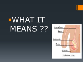
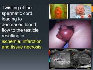
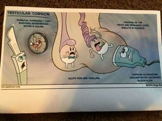
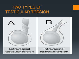
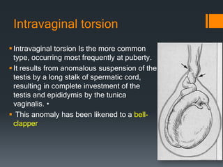
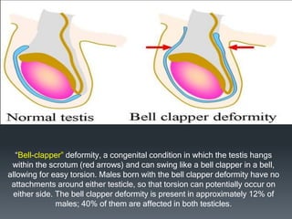
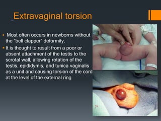
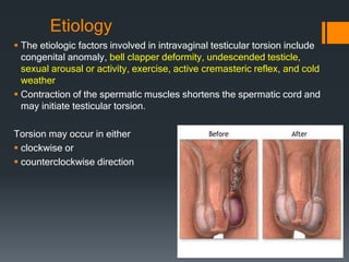
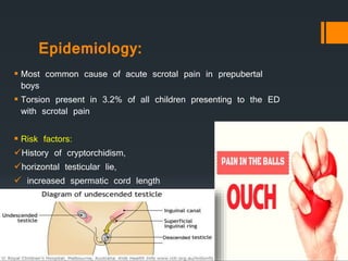
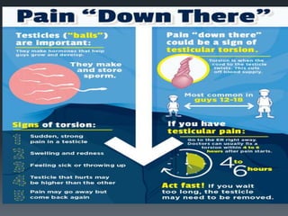
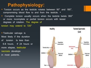
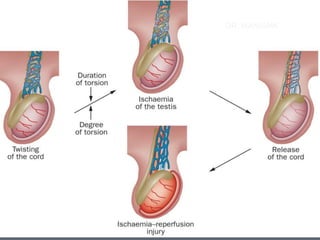
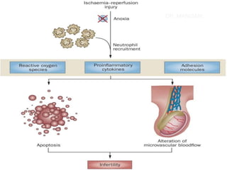
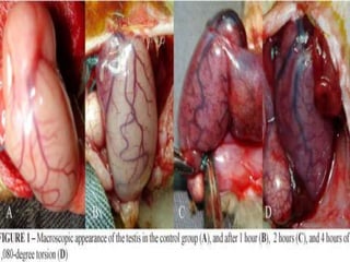
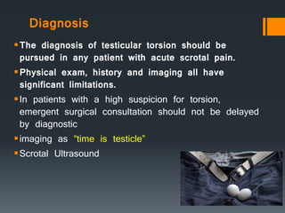
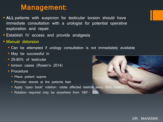
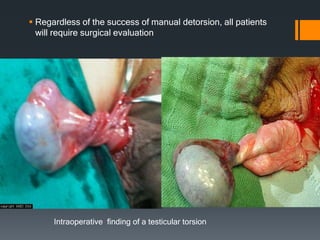
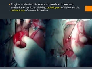
Testicular Torsion
DR. MANISMK
WHAT IT
MEANS ??
Twisting of the
spermatic cord
leading to
decreased blood
flow to the testicle
resulting in
ischemia, infarction
and tissue necrosis.
NO FLOW
TWO TYPES OF
TESTICULAR TORSION
Intravaginal torsion
Intravaginal torsion Is the more common
type, occurring most frequently at puberty.
It results from anomalous suspension of the
testis by a long stalk of spermatic cord,
resulting in complete investment of the
testis and epididymis by the tunica
vaginalis. •
 This anomaly has been likened to a bell-
clapper
“Bell-clapper” deformity, a congenital condition in which the testis hangs
within the scrotum (red arrows) and can swing like a bell clapper in a bell,
allowing for easy torsion. Males born with the bell clapper deformity have no
attachments around either testicle, so that torsion can potentially occur on
either side. The bell clapper deformity is present in approximately 12% of
males; 40% of them are affected in both testicles.
Extravaginal torsion
 Most often occurs in newborns without
the “bell clapper” deformity.
 It is thought to result from a poor or
absent attachment of the testis to the
scrotal wall, allowing rotation of the
testis, epididymis, and tunica vaginalis
as a unit and causing torsion of the cord
at the level of the external ring
Etiology
 The etiologic factors involved in intravaginal testicular torsion include
congenital anomaly, bell clapper deformity, undescended testicle,
sexual arousal or activity, exercise, active cremasteric reflex, and cold
weather
 Contraction of the spermatic muscles shortens the spermatic cord and
may initiate testicular torsion.
Torsion may occur in either
 clockwise or
 counterclockwise direction
Epidemiology:
 Most common cause of acute scrotal pain in prepubertal
boys
 Torsion present in 3.2% of all children presenting to the ED
with scrotal pain
 Risk factors:
History of cryptorchidism,
horizontal testicular lie,
 increased spermatic cord length
Pathophysiology:
 Torsion occurs as the testicle rotates between 90° and 180°,
compromising blood flow to and from the testicle. •
 Complete torsion usually occurs when the testicle twists 360°
or more; incomplete or partial torsion occurs with lesser
degrees of rotation. The degree of
torsion may extend to 720°
 Testicular salvage is
Most likely if the duration
of torsion Is less than
6-8 hours. If 24 hours or
more elapse, testicular
necrosis develops
in most patients.
DR. MANISMK
DR. MANISMK
Differential Diagnosis
 Hydrocele
 Epididymitis
 Trauma
 Inguinal hernia
 Testicular tumor
Hydrocele
Epididymitis
HERNIA
Scrotal abscess
Pay attention to history, clinical
picture and pain expression of
patient
History
 Sudden onset of scrotal pain (less
frequently, abdominal or inguinal pain)
 Nausea and vomiting
 History of blunt trauma (~ 10% of
patients)
 History of similar pain in the past
 Duration of symptoms should NOT guide
management
 Historically, believed that symptoms > 24
hours inconsistent with salvageable tissue
 However, testicle may torse + detorse
making it difficult to know how long
ischemia present
Physical Examination
Unilateral tender, firm testicle
Scrotal erythema, edema and swelling
Affected testicle typically higher than the unaffected one.
- Loss of cremasteric reflex
30% of males with normal testicles will have an absent
cremasteric reflex
 stimulation of the skin on the front and inner thigh (over Scarpa's triangle)
retracts the testis on the same side. Stimulus usually causes cremasteric
muscle contraction.
 Normal: Cremasteric reflex present (testicle rises). Seen in Epididymitis;
 Abnormal: Cremasteric reflex absent (no testicle rise). Suggests Testicular
Torsion
Diagnosis
The diagnosis of testicular torsion should be
pursued in any patient with acute scrotal pain.
Physical exam, history and imaging all have
significant limitations.
In patients with a high suspicion for torsion,
emergent surgical consultation should not be delayed
by diagnostic
imaging as “time is testicle”
Scrotal Ultrasound
Right Testicle with Decreased Flow on Color Doppler -
Left Testicle Hypoechoic and Swollen -
Management:
 ALL patients with suspicion for testicular torsion should have
immediate consultation with a urologist for potential operative
exploration and repair.
 Establish IV access and provide analgesia
 Manual detorsion
 Can be attempted if urology consultation is not immediately available
 May be successful in
 25-80% of testicular
 torsion cases (Rosen’s 2014)
 Procedure
 Place patient supine
 Provider stands at the patients feet
 Apply “open book” rotation: rotate affected testicle away from midline
 Rotation required may be anywhere from 180o – 720o
DR. MANISMK
 Regardless of the success of manual detorsion, all patients
will require surgical evaluation
Intraoperative finding of a testicular torsion
 Surgical exploration via scrotal approach with detorsion,
evaluation of testicular viability, orchidopexy of viable testicle,
orchiectomy of nonviable testicle
THANK YOU !!!

TESTICLULAR TORSION

  • 1.
  • 2.
  • 3.
    Twisting of the spermaticcord leading to decreased blood flow to the testicle resulting in ischemia, infarction and tissue necrosis. NO FLOW
  • 5.
  • 6.
    Intravaginal torsion Intravaginal torsionIs the more common type, occurring most frequently at puberty. It results from anomalous suspension of the testis by a long stalk of spermatic cord, resulting in complete investment of the testis and epididymis by the tunica vaginalis. •  This anomaly has been likened to a bell- clapper
  • 7.
    “Bell-clapper” deformity, acongenital condition in which the testis hangs within the scrotum (red arrows) and can swing like a bell clapper in a bell, allowing for easy torsion. Males born with the bell clapper deformity have no attachments around either testicle, so that torsion can potentially occur on either side. The bell clapper deformity is present in approximately 12% of males; 40% of them are affected in both testicles.
  • 8.
    Extravaginal torsion  Mostoften occurs in newborns without the “bell clapper” deformity.  It is thought to result from a poor or absent attachment of the testis to the scrotal wall, allowing rotation of the testis, epididymis, and tunica vaginalis as a unit and causing torsion of the cord at the level of the external ring
  • 9.
    Etiology  The etiologicfactors involved in intravaginal testicular torsion include congenital anomaly, bell clapper deformity, undescended testicle, sexual arousal or activity, exercise, active cremasteric reflex, and cold weather  Contraction of the spermatic muscles shortens the spermatic cord and may initiate testicular torsion. Torsion may occur in either  clockwise or  counterclockwise direction
  • 10.
    Epidemiology:  Most commoncause of acute scrotal pain in prepubertal boys  Torsion present in 3.2% of all children presenting to the ED with scrotal pain  Risk factors: History of cryptorchidism, horizontal testicular lie,  increased spermatic cord length
  • 12.
    Pathophysiology:  Torsion occursas the testicle rotates between 90° and 180°, compromising blood flow to and from the testicle. •  Complete torsion usually occurs when the testicle twists 360° or more; incomplete or partial torsion occurs with lesser degrees of rotation. The degree of torsion may extend to 720°  Testicular salvage is Most likely if the duration of torsion Is less than 6-8 hours. If 24 hours or more elapse, testicular necrosis develops in most patients.
  • 13.
  • 14.
  • 16.
    Differential Diagnosis  Hydrocele Epididymitis  Trauma  Inguinal hernia  Testicular tumor Hydrocele Epididymitis HERNIA Scrotal abscess
  • 17.
    Pay attention tohistory, clinical picture and pain expression of patient
  • 18.
    History  Sudden onsetof scrotal pain (less frequently, abdominal or inguinal pain)  Nausea and vomiting  History of blunt trauma (~ 10% of patients)  History of similar pain in the past  Duration of symptoms should NOT guide management  Historically, believed that symptoms > 24 hours inconsistent with salvageable tissue  However, testicle may torse + detorse making it difficult to know how long ischemia present
  • 19.
    Physical Examination Unilateral tender,firm testicle Scrotal erythema, edema and swelling Affected testicle typically higher than the unaffected one. - Loss of cremasteric reflex 30% of males with normal testicles will have an absent cremasteric reflex
  • 20.
     stimulation ofthe skin on the front and inner thigh (over Scarpa's triangle) retracts the testis on the same side. Stimulus usually causes cremasteric muscle contraction.  Normal: Cremasteric reflex present (testicle rises). Seen in Epididymitis;  Abnormal: Cremasteric reflex absent (no testicle rise). Suggests Testicular Torsion
  • 21.
    Diagnosis The diagnosis oftesticular torsion should be pursued in any patient with acute scrotal pain. Physical exam, history and imaging all have significant limitations. In patients with a high suspicion for torsion, emergent surgical consultation should not be delayed by diagnostic imaging as “time is testicle” Scrotal Ultrasound
  • 22.
    Right Testicle withDecreased Flow on Color Doppler -
  • 23.
  • 24.
    Management:  ALL patientswith suspicion for testicular torsion should have immediate consultation with a urologist for potential operative exploration and repair.  Establish IV access and provide analgesia  Manual detorsion  Can be attempted if urology consultation is not immediately available  May be successful in  25-80% of testicular  torsion cases (Rosen’s 2014)  Procedure  Place patient supine  Provider stands at the patients feet  Apply “open book” rotation: rotate affected testicle away from midline  Rotation required may be anywhere from 180o – 720o DR. MANISMK
  • 25.
     Regardless ofthe success of manual detorsion, all patients will require surgical evaluation Intraoperative finding of a testicular torsion
  • 26.
     Surgical explorationvia scrotal approach with detorsion, evaluation of testicular viability, orchidopexy of viable testicle, orchiectomy of nonviable testicle
  • 27.