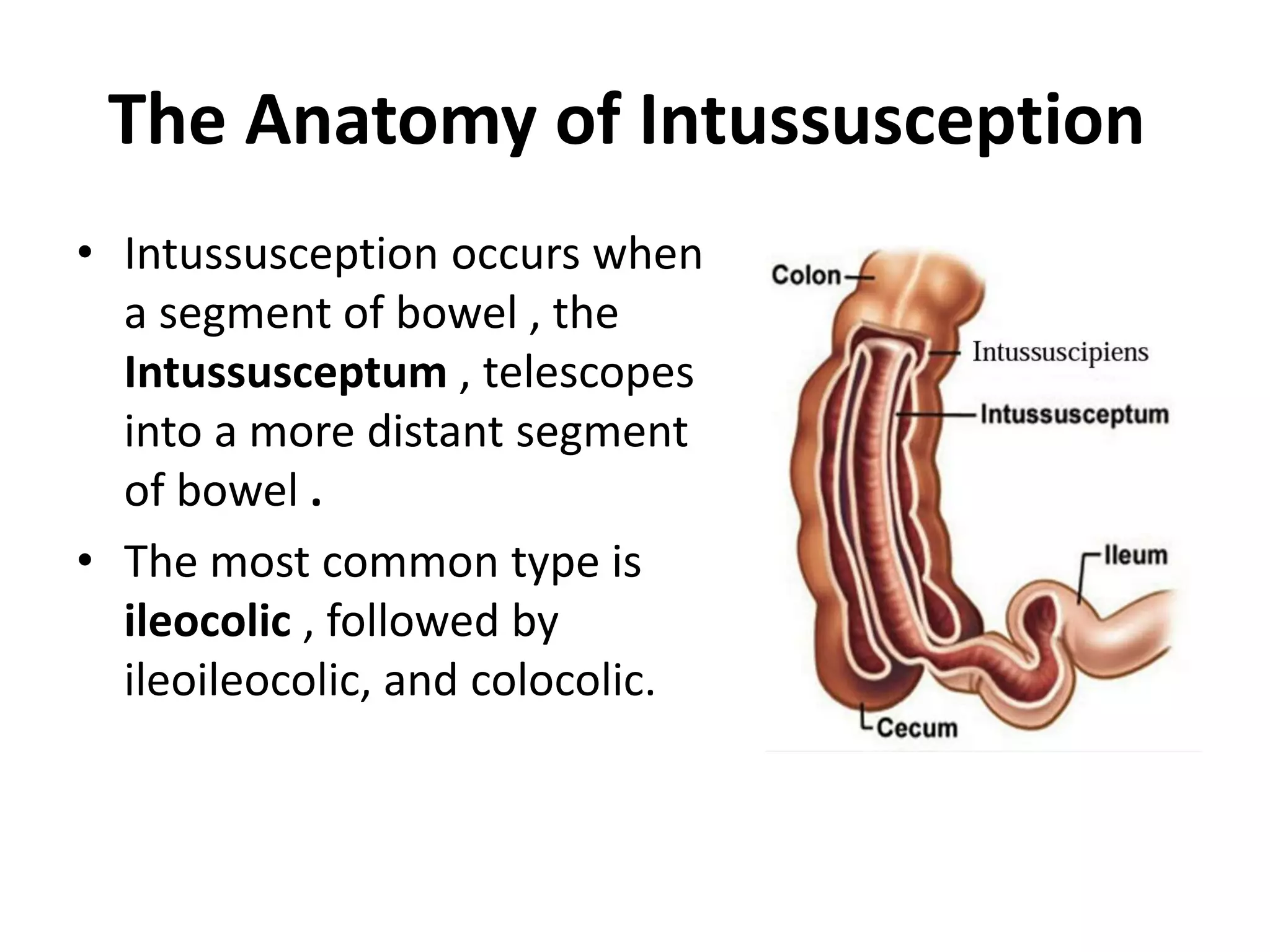
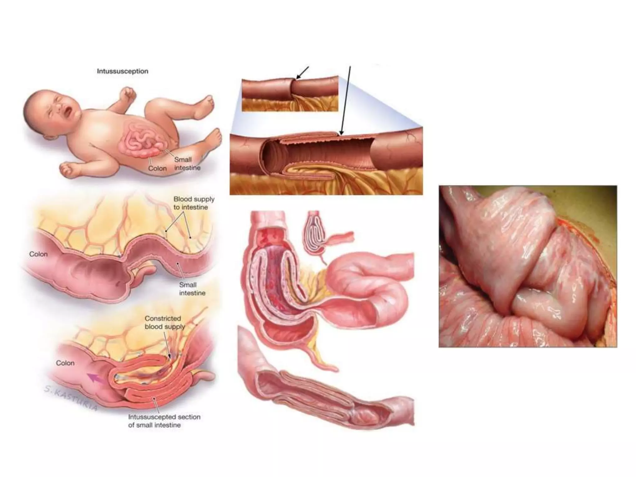
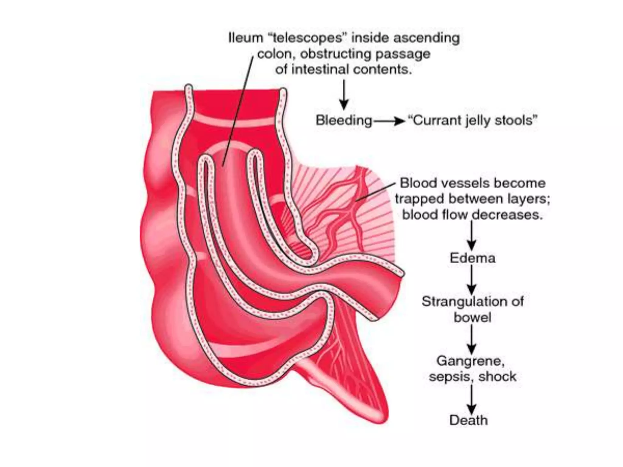
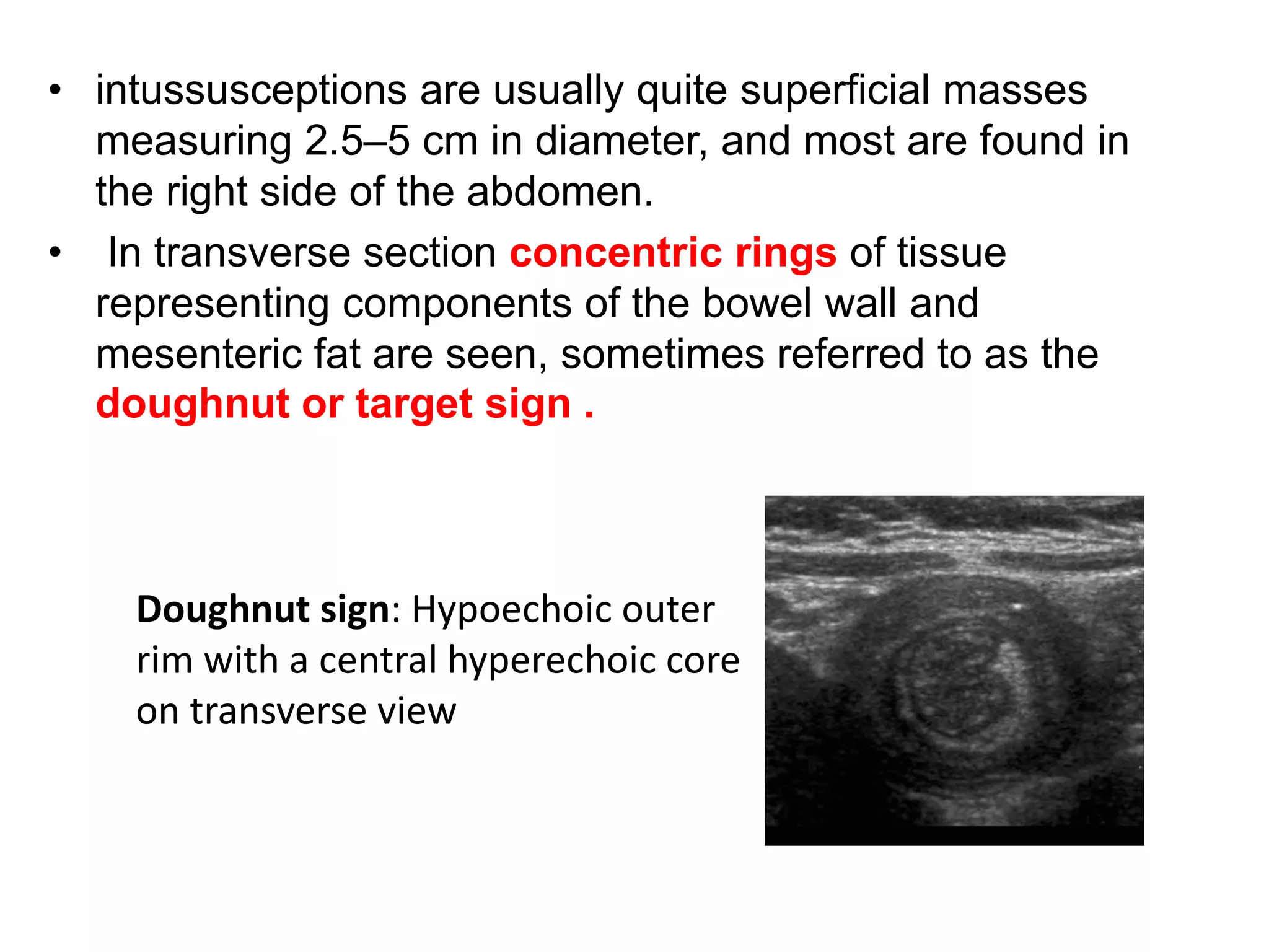
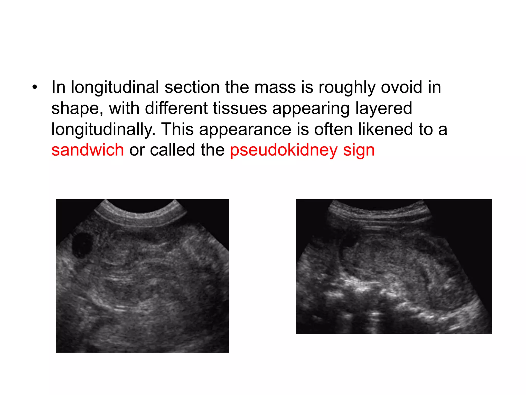
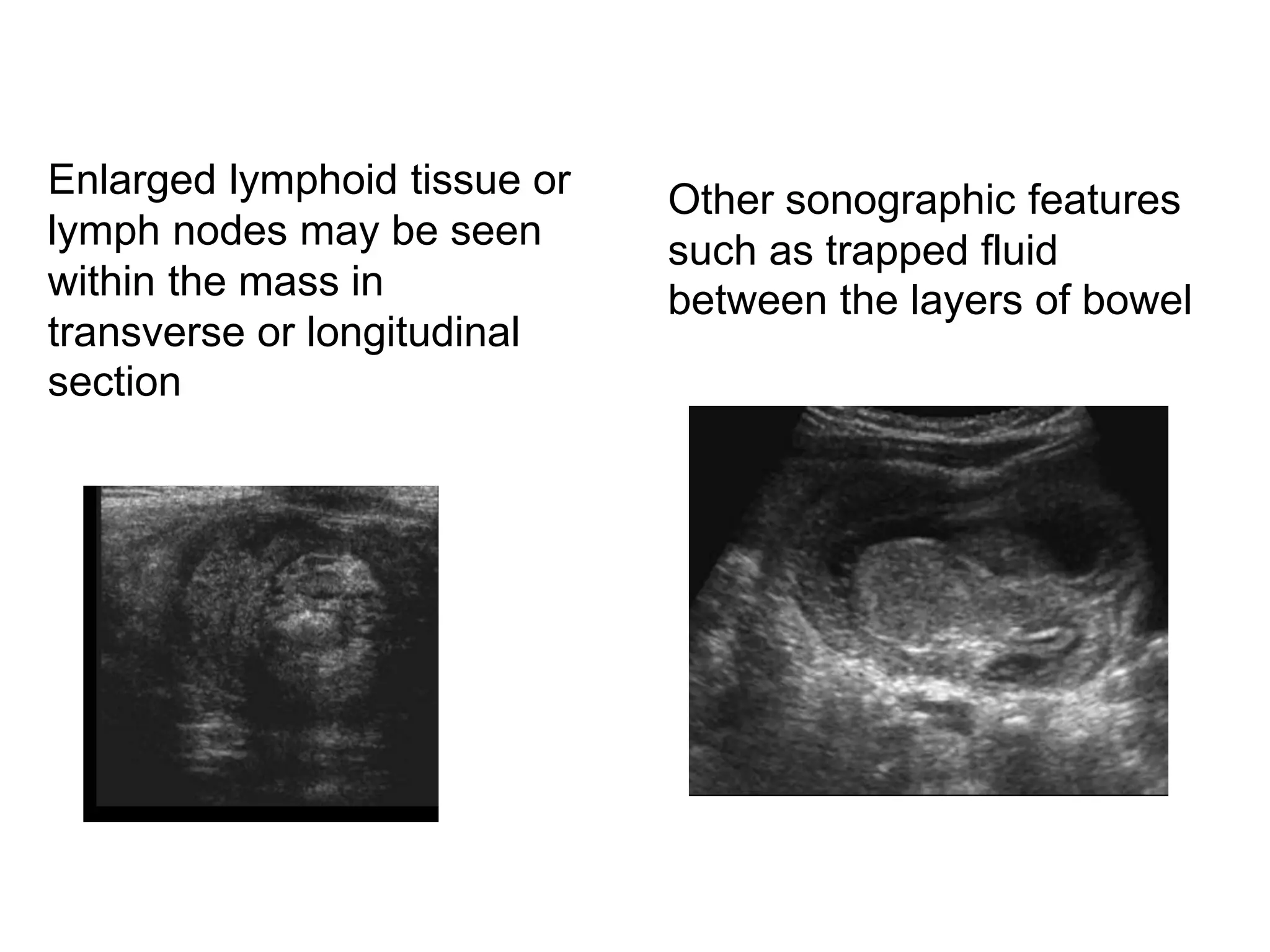
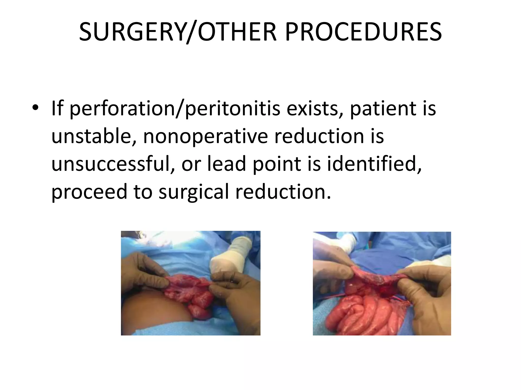
Intussusception
Dr. Leen Doya
Department of pediatric
Tishreen university
The Anatomy of Intussusception
• Intussusception occurs when
a segment of bowel , the
Intussusceptum , telescopes
into a more distant segment
of bowel .
• The most common type is
ileocolic , followed by
ileoileocolic, and colocolic.
Demographics
• Most common acute abdominal disorder of early
childhood (56 children from 100000 year in US).
• Boys 4× ‘s more frequently than girls.
• Majority of patients between 3 mon and 3 year.
- Pick incidence between 5 and 9 months.
- 75% under 2 years.
• Seasonal peaks in spring and autumn.
• 95%no pathologic lead point.
• 5-10% recognizable lead point.
Etiologies of Intussusception
• Idiopathic (<3): no defined lead point.
- Association with viral illness( adenovirus).
- Hypertrophy of lymphoid tissue.
• Recognizable cause for lead point:( > 3)
- Meckel ‘s diverticulum(the most common)..
- Intestinal polype.
- Enteric duplication.
- Lymphoma.
- Intramural hematoma.
- Henoch-Shonlein purpura.
Clinical Presentation : Variable
• Intermittent , colicky cramping , pain .
• Later development of lethargy and
somnolence.
• Vomiting (may be bile-stained).
• Current jelly stool (blood and mucus).
• Sausage shaped mass.
• Distention and tenderness.
• Additional signs : irritability, nausea ,fever ,
and anorexia.
• Classic traid : abdominal pain , currant jelly
stool, vomiting
( 50%)
PHYSICAL EXAM
• Lethargic with colicky pattern of
abdominal pain
• Mass in the RUQ may be palpated (“RUQ
sausage”)
• Absence of bowel contents in right
lower quadrant (Dance sign)
• Abdominal distention
• Rectal exam: Blood-tinged mucous or
currant jelly stool; occasionally the
intussusception can be felt
• Peritoneal signs if intestinal perforation
has occurred.
Dance sign
DIFFERENTIAL DIAGNOSIS
• Infection: Gastroenteritis, enterocolitis.....
• Immunologic: Henoch-Scho nlein purpura
• Miscellaneous:
- Appendicitis
- Meckel diverticulum: May act as a lead point in
• the absence of bleeding
– Incarcerated hernia
– Crohn disease
– Celiac disease
– Henoch Scho¨ nlein purpura
• Obstruction: Adhesions, hernia, volvulus,
stricture, bezoar, foreign body, polyp, tumor
• Other causes of lethargy:
– Hypoglycemia
– Meningitis
– Encephalitis
– Sepsis
– Seizure
– Infantile botulism
– Toxic ingestion
– Metabolic disorder
DIAGNOSTIC TESTS & INTERPRETATION
• Lab:
 No lab testing is routinely necessary.
 Consider serum electrolytes, glucose, and CBC
with appropriate symptoms
 Consider routine preoperative lab assays as
per institutional protocol.
Imaging: Abdominal x-ray
• Not sensitive or specific.
• Normal in early stages 25%.
• later can have absence of gas in right lower
quadrant (RLQ) and RUQ, as well as RUQ soft
tissue mass; with obstruction, will have air-
fluid levels, paucity of distal gas.
2. The meniscus sign is a
crescent of gas within the
colonic lumen outlining the
apex of the intussusception
1. The target sign is a rounded
soft tissue mass representing
the intussusception, with
concentric lucencies due to the
presence of mesenteric fat
within the mass
4. Nonspecific signs of
intussusception on AXR
that may suggest or
support the diagnosis
include soft tissue density
and absence of gas in the
right lower quadrant
5. and signs of small bowel
obstruction
Abdominal ultrasound
Abdominal US has high sensitivity (98–100%) and
Specificity (88–100%) for intussusception .
• intussusceptions are usually quite superficial masses
measuring 2.5–5 cm in diameter, and most are found in
the right side of the abdomen.
• In transverse section concentric rings of tissue
representing components of the bowel wall and
mesenteric fat are seen, sometimes referred to as the
doughnut or target sign .
Doughnut sign: Hypoechoic outer
rim with a central hyperechoic core
on transverse view
• In longitudinal section the mass is roughly ovoid in
shape, with different tissues appearing layered
longitudinally. This appearance is often likened to a
sandwich or called the pseudokidney sign
Enlarged lymphoid tissue or
lymph nodes may be seen
within the mass in
transverse or longitudinal
section
Other sonographic features
such as trapped fluid
between the layers of bowel
CT
• most useful in evaluating for a pathologic
leadpoint.
Barium enema
• was the gold standard for diagnosis of
intussusception until the mid-1980s.
Diagnostic and therapeutic with reduction often
achieved; air enema preferred because less
perforation risk than barium; can miss a lead point
the coiled spring sign which is
produced when small amounts
of contrast material accumulate
between the intussusceptum
and intussuscipiens.
The classic signs of
intussusception on contrast
enema are the meniscus
sign where the apex of the
intussusception projects
into the contrast material
intussusception
Air contrast or barium enema reduction is the
standard nonoperative treatment for
intussusception:
– 70–85% success with barium enema
– Up to 90% success with air enema Barium or air
contrast enema exam is useful for both
diagnosis and therapy.
Complication
• Typically do not occur within the first 24 hrs….
• Bowel obstruction.
• Intestinal ischemia.
• GI bleeding
• Perforation.
• Shock.
• Sepsis.
• dehydration.
Thus we have a window of opportunity in which to
treat and avoid surgery.
• Successful management of intussusception
depends on early recognition and diagnosis,
adequate fluid resuscitation and prompt
reduction.
• the longer the duration of symptoms (particularly
if .24 h) the lower the likelihood of successful
nonoperative reduction.
• Decreased reduction rates are also reported
when the intussusception is situated in the
rectum, in children with small bowel obstruction
and those under 3 months of age.
Treatment of Intussusception
• Conservative management:
NG drainage , resuscitation with IV fluid ,antibiotics
• Non operative management:
Air or barium enema performed if there are no
signs of peritonitis perforation.
• Operative management:
Reducible intussusception.
Irreducible intussusception.
Resection with primary anastomosis.
• Non-surgical reduction of intussusception has
a long history, with enema treatment for ileus
described for centuries
• Non-operative reduction techniques using
enemas may be hydrostatic (using barium,
water soluble contrast, saline or Hartmann’s
solution) or pneumatic (using either air, or
medical gases )
SURGERY/OTHER PROCEDURES
• If perforation/peritonitis exists, patient is
unstable, nonoperative reduction is
unsuccessful, or lead point is identified,
proceed to surgical reduction.
Thank you

Intussusception

  • 1.
    Intussusception Dr. Leen Doya Departmentof pediatric Tishreen university
  • 2.
    The Anatomy ofIntussusception • Intussusception occurs when a segment of bowel , the Intussusceptum , telescopes into a more distant segment of bowel . • The most common type is ileocolic , followed by ileoileocolic, and colocolic.
  • 4.
    Demographics • Most commonacute abdominal disorder of early childhood (56 children from 100000 year in US). • Boys 4× ‘s more frequently than girls. • Majority of patients between 3 mon and 3 year. - Pick incidence between 5 and 9 months. - 75% under 2 years. • Seasonal peaks in spring and autumn. • 95%no pathologic lead point. • 5-10% recognizable lead point.
  • 5.
    Etiologies of Intussusception •Idiopathic (<3): no defined lead point. - Association with viral illness( adenovirus). - Hypertrophy of lymphoid tissue. • Recognizable cause for lead point:( > 3) - Meckel ‘s diverticulum(the most common).. - Intestinal polype. - Enteric duplication. - Lymphoma. - Intramural hematoma. - Henoch-Shonlein purpura.
  • 6.
    Clinical Presentation :Variable • Intermittent , colicky cramping , pain . • Later development of lethargy and somnolence. • Vomiting (may be bile-stained). • Current jelly stool (blood and mucus). • Sausage shaped mass. • Distention and tenderness. • Additional signs : irritability, nausea ,fever , and anorexia. • Classic traid : abdominal pain , currant jelly stool, vomiting ( 50%)
  • 7.
    PHYSICAL EXAM • Lethargicwith colicky pattern of abdominal pain • Mass in the RUQ may be palpated (“RUQ sausage”) • Absence of bowel contents in right lower quadrant (Dance sign) • Abdominal distention • Rectal exam: Blood-tinged mucous or currant jelly stool; occasionally the intussusception can be felt • Peritoneal signs if intestinal perforation has occurred.
  • 8.
  • 10.
    DIFFERENTIAL DIAGNOSIS • Infection:Gastroenteritis, enterocolitis..... • Immunologic: Henoch-Scho nlein purpura • Miscellaneous: - Appendicitis - Meckel diverticulum: May act as a lead point in • the absence of bleeding – Incarcerated hernia – Crohn disease – Celiac disease – Henoch Scho¨ nlein purpura
  • 11.
    • Obstruction: Adhesions,hernia, volvulus, stricture, bezoar, foreign body, polyp, tumor • Other causes of lethargy: – Hypoglycemia – Meningitis – Encephalitis – Sepsis – Seizure – Infantile botulism – Toxic ingestion – Metabolic disorder
  • 12.
    DIAGNOSTIC TESTS &INTERPRETATION • Lab:  No lab testing is routinely necessary.  Consider serum electrolytes, glucose, and CBC with appropriate symptoms  Consider routine preoperative lab assays as per institutional protocol.
  • 13.
    Imaging: Abdominal x-ray •Not sensitive or specific. • Normal in early stages 25%. • later can have absence of gas in right lower quadrant (RLQ) and RUQ, as well as RUQ soft tissue mass; with obstruction, will have air- fluid levels, paucity of distal gas.
  • 14.
    2. The meniscussign is a crescent of gas within the colonic lumen outlining the apex of the intussusception 1. The target sign is a rounded soft tissue mass representing the intussusception, with concentric lucencies due to the presence of mesenteric fat within the mass
  • 15.
    4. Nonspecific signsof intussusception on AXR that may suggest or support the diagnosis include soft tissue density and absence of gas in the right lower quadrant 5. and signs of small bowel obstruction
  • 16.
    Abdominal ultrasound Abdominal UShas high sensitivity (98–100%) and Specificity (88–100%) for intussusception .
  • 17.
    • intussusceptions areusually quite superficial masses measuring 2.5–5 cm in diameter, and most are found in the right side of the abdomen. • In transverse section concentric rings of tissue representing components of the bowel wall and mesenteric fat are seen, sometimes referred to as the doughnut or target sign . Doughnut sign: Hypoechoic outer rim with a central hyperechoic core on transverse view
  • 18.
    • In longitudinalsection the mass is roughly ovoid in shape, with different tissues appearing layered longitudinally. This appearance is often likened to a sandwich or called the pseudokidney sign
  • 19.
    Enlarged lymphoid tissueor lymph nodes may be seen within the mass in transverse or longitudinal section Other sonographic features such as trapped fluid between the layers of bowel
  • 20.
    CT • most usefulin evaluating for a pathologic leadpoint.
  • 21.
    Barium enema • wasthe gold standard for diagnosis of intussusception until the mid-1980s. Diagnostic and therapeutic with reduction often achieved; air enema preferred because less perforation risk than barium; can miss a lead point
  • 22.
    the coiled springsign which is produced when small amounts of contrast material accumulate between the intussusceptum and intussuscipiens. The classic signs of intussusception on contrast enema are the meniscus sign where the apex of the intussusception projects into the contrast material intussusception
  • 23.
    Air contrast orbarium enema reduction is the standard nonoperative treatment for intussusception: – 70–85% success with barium enema – Up to 90% success with air enema Barium or air contrast enema exam is useful for both diagnosis and therapy.
  • 24.
    Complication • Typically donot occur within the first 24 hrs…. • Bowel obstruction. • Intestinal ischemia. • GI bleeding • Perforation. • Shock. • Sepsis. • dehydration. Thus we have a window of opportunity in which to treat and avoid surgery.
  • 26.
    • Successful managementof intussusception depends on early recognition and diagnosis, adequate fluid resuscitation and prompt reduction. • the longer the duration of symptoms (particularly if .24 h) the lower the likelihood of successful nonoperative reduction. • Decreased reduction rates are also reported when the intussusception is situated in the rectum, in children with small bowel obstruction and those under 3 months of age.
  • 27.
    Treatment of Intussusception •Conservative management: NG drainage , resuscitation with IV fluid ,antibiotics • Non operative management: Air or barium enema performed if there are no signs of peritonitis perforation. • Operative management: Reducible intussusception. Irreducible intussusception. Resection with primary anastomosis.
  • 28.
    • Non-surgical reductionof intussusception has a long history, with enema treatment for ileus described for centuries • Non-operative reduction techniques using enemas may be hydrostatic (using barium, water soluble contrast, saline or Hartmann’s solution) or pneumatic (using either air, or medical gases )
  • 29.
    SURGERY/OTHER PROCEDURES • Ifperforation/peritonitis exists, patient is unstable, nonoperative reduction is unsuccessful, or lead point is identified, proceed to surgical reduction.
  • 31.