Pelvic organ prolapse
Etiology of pelvic organ prolapse
Vaginal vault prolapse
Etiological factors of vault prolapse
Signs and symptoms of vaginal vault prolapse
Diagnosis of vaginal vault prolapse
Treatment measures
Bladder exstrophy is a congenital (present at birth) abnormality of the bladder. It happens when the skin over the lower abdominal wall (bottom part of the tummy) does not form properly, so the bladder is open and exposed on the outside of the abdomen. In epispadias, the urethra does not form properly.
The prostate is an exocrine gland of the male mammalian reproductive system
It is a walnut-sized gland that forms part of the male reproductive system and is located in front of the rectum and just below the urinary bladder
Function is to store and secrete a clear, slightly alkaline fluid that constitutes 10-30% of the volume of the seminal fluid that along with the spermatozoa, constitutes semen
A healthy human prostate measures (4cm-vertical, by 3cm-horizontal, 2cm ant-post ).
It surrounds the urethra just below the urinary bladder. It has anterior, median, posterior and two lateral lobes
It’s work is regulated by androgens which are responsible for male sex characteristics
Generalised disease of the prostate due to hormonal derangement which leads to non malignant enlargement of the gland (increase in the number of epithelial cells and stromal tissue)to cause compression of the urethra leading to symptoms (LUTS
New Directions in Targeted Therapeutic Approaches for Older Adults With Mantl...i3 Health
i3 Health is pleased to make the speaker slides from this activity available for use as a non-accredited self-study or teaching resource.
This slide deck presented by Dr. Kami Maddocks, Professor-Clinical in the Division of Hematology and
Associate Division Director for Ambulatory Operations
The Ohio State University Comprehensive Cancer Center, will provide insight into new directions in targeted therapeutic approaches for older adults with mantle cell lymphoma.
STATEMENT OF NEED
Mantle cell lymphoma (MCL) is a rare, aggressive B-cell non-Hodgkin lymphoma (NHL) accounting for 5% to 7% of all lymphomas. Its prognosis ranges from indolent disease that does not require treatment for years to very aggressive disease, which is associated with poor survival (Silkenstedt et al, 2021). Typically, MCL is diagnosed at advanced stage and in older patients who cannot tolerate intensive therapy (NCCN, 2022). Although recent advances have slightly increased remission rates, recurrence and relapse remain very common, leading to a median overall survival between 3 and 6 years (LLS, 2021). Though there are several effective options, progress is still needed towards establishing an accepted frontline approach for MCL (Castellino et al, 2022). Treatment selection and management of MCL are complicated by the heterogeneity of prognosis, advanced age and comorbidities of patients, and lack of an established standard approach for treatment, making it vital that clinicians be familiar with the latest research and advances in this area. In this activity chaired by Michael Wang, MD, Professor in the Department of Lymphoma & Myeloma at MD Anderson Cancer Center, expert faculty will discuss prognostic factors informing treatment, the promising results of recent trials in new therapeutic approaches, and the implications of treatment resistance in therapeutic selection for MCL.
Target Audience
Hematology/oncology fellows, attending faculty, and other health care professionals involved in the treatment of patients with mantle cell lymphoma (MCL).
Learning Objectives
1.) Identify clinical and biological prognostic factors that can guide treatment decision making for older adults with MCL
2.) Evaluate emerging data on targeted therapeutic approaches for treatment-naive and relapsed/refractory MCL and their applicability to older adults
3.) Assess mechanisms of resistance to targeted therapies for MCL and their implications for treatment selection
Title: Sense of Taste
Presenter: Dr. Faiza, Assistant Professor of Physiology
Qualifications:
MBBS (Best Graduate, AIMC Lahore)
FCPS Physiology
ICMT, CHPE, DHPE (STMU)
MPH (GC University, Faisalabad)
MBA (Virtual University of Pakistan)
Learning Objectives:
Describe the structure and function of taste buds.
Describe the relationship between the taste threshold and taste index of common substances.
Explain the chemical basis and signal transduction of taste perception for each type of primary taste sensation.
Recognize different abnormalities of taste perception and their causes.
Key Topics:
Significance of Taste Sensation:
Differentiation between pleasant and harmful food
Influence on behavior
Selection of food based on metabolic needs
Receptors of Taste:
Taste buds on the tongue
Influence of sense of smell, texture of food, and pain stimulation (e.g., by pepper)
Primary and Secondary Taste Sensations:
Primary taste sensations: Sweet, Sour, Salty, Bitter, Umami
Chemical basis and signal transduction mechanisms for each taste
Taste Threshold and Index:
Taste threshold values for Sweet (sucrose), Salty (NaCl), Sour (HCl), and Bitter (Quinine)
Taste index relationship: Inversely proportional to taste threshold
Taste Blindness:
Inability to taste certain substances, particularly thiourea compounds
Example: Phenylthiocarbamide
Structure and Function of Taste Buds:
Composition: Epithelial cells, Sustentacular/Supporting cells, Taste cells, Basal cells
Features: Taste pores, Taste hairs/microvilli, and Taste nerve fibers
Location of Taste Buds:
Found in papillae of the tongue (Fungiform, Circumvallate, Foliate)
Also present on the palate, tonsillar pillars, epiglottis, and proximal esophagus
Mechanism of Taste Stimulation:
Interaction of taste substances with receptors on microvilli
Signal transduction pathways for Umami, Sweet, Bitter, Sour, and Salty tastes
Taste Sensitivity and Adaptation:
Decrease in sensitivity with age
Rapid adaptation of taste sensation
Role of Saliva in Taste:
Dissolution of tastants to reach receptors
Washing away the stimulus
Taste Preferences and Aversions:
Mechanisms behind taste preference and aversion
Influence of receptors and neural pathways
Impact of Sensory Nerve Damage:
Degeneration of taste buds if the sensory nerve fiber is cut
Abnormalities of Taste Detection:
Conditions: Ageusia, Hypogeusia, Dysgeusia (parageusia)
Causes: Nerve damage, neurological disorders, infections, poor oral hygiene, adverse drug effects, deficiencies, aging, tobacco use, altered neurotransmitter levels
Neurotransmitters and Taste Threshold:
Effects of serotonin (5-HT) and norepinephrine (NE) on taste sensitivity
Supertasters:
25% of the population with heightened sensitivity to taste, especially bitterness
Increased number of fungiform papillae
Tom Selleck Health: A Comprehensive Look at the Iconic Actor’s Wellness Journeygreendigital
Tom Selleck, an enduring figure in Hollywood. has captivated audiences for decades with his rugged charm, iconic moustache. and memorable roles in television and film. From his breakout role as Thomas Magnum in Magnum P.I. to his current portrayal of Frank Reagan in Blue Bloods. Selleck's career has spanned over 50 years. But beyond his professional achievements. fans have often been curious about Tom Selleck Health. especially as he has aged in the public eye.
Follow us on: Pinterest
Introduction
Many have been interested in Tom Selleck health. not only because of his enduring presence on screen but also because of the challenges. and lifestyle choices he has faced and made over the years. This article delves into the various aspects of Tom Selleck health. exploring his fitness regimen, diet, mental health. and the challenges he has encountered as he ages. We'll look at how he maintains his well-being. the health issues he has faced, and his approach to ageing .
Early Life and Career
Childhood and Athletic Beginnings
Tom Selleck was born on January 29, 1945, in Detroit, Michigan, and grew up in Sherman Oaks, California. From an early age, he was involved in sports, particularly basketball. which played a significant role in his physical development. His athletic pursuits continued into college. where he attended the University of Southern California (USC) on a basketball scholarship. This early involvement in sports laid a strong foundation for his physical health and disciplined lifestyle.
Transition to Acting
Selleck's transition from an athlete to an actor came with its physical demands. His first significant role in "Magnum P.I." required him to perform various stunts and maintain a fit appearance. This role, which he played from 1980 to 1988. necessitated a rigorous fitness routine to meet the show's demands. setting the stage for his long-term commitment to health and wellness.
Fitness Regimen
Workout Routine
Tom Selleck health and fitness regimen has evolved. adapting to his changing roles and age. During his "Magnum, P.I." days. Selleck's workouts were intense and focused on building and maintaining muscle mass. His routine included weightlifting, cardiovascular exercises. and specific training for the stunts he performed on the show.
Selleck adjusted his fitness routine as he aged to suit his body's needs. Today, his workouts focus on maintaining flexibility, strength, and cardiovascular health. He incorporates low-impact exercises such as swimming, walking, and light weightlifting. This balanced approach helps him stay fit without putting undue strain on his joints and muscles.
Importance of Flexibility and Mobility
In recent years, Selleck has emphasized the importance of flexibility and mobility in his fitness regimen. Understanding the natural decline in muscle mass and joint flexibility with age. he includes stretching and yoga in his routine. These practices help prevent injuries, improve posture, and maintain mobilit
Factory Supply Best Quality Pmk Oil CAS 28578–16–7 PMK Powder in Stockrebeccabio
Factory Supply Best Quality Pmk Oil CAS 28578–16–7 PMK Powder in Stock
Telegram: bmksupplier
signal: +85264872720
threema: TUD4A6YC
You can contact me on Telegram or Threema
Communicate promptly and reply
Free of customs clearance, Double Clearance 100% pass delivery to USA, Canada, Spain, Germany, Netherland, Poland, Italy, Sweden, UK, Czech Republic, Australia, Mexico, Russia, Ukraine, Kazakhstan.Door to door service
Hot Selling Organic intermediates
MANAGEMENT OF ATRIOVENTRICULAR CONDUCTION BLOCK.pdfJim Jacob Roy
Cardiac conduction defects can occur due to various causes.
Atrioventricular conduction blocks ( AV blocks ) are classified into 3 types.
This document describes the acute management of AV block.
Report Back from SGO 2024: What’s the Latest in Cervical Cancer?bkling
Are you curious about what’s new in cervical cancer research or unsure what the findings mean? Join Dr. Emily Ko, a gynecologic oncologist at Penn Medicine, to learn about the latest updates from the Society of Gynecologic Oncology (SGO) 2024 Annual Meeting on Women’s Cancer. Dr. Ko will discuss what the research presented at the conference means for you and answer your questions about the new developments.
These lecture slides, by Dr Sidra Arshad, offer a quick overview of physiological basis of a normal electrocardiogram.
Learning objectives:
1. Define an electrocardiogram (ECG) and electrocardiography
2. Describe how dipoles generated by the heart produce the waveforms of the ECG
3. Describe the components of a normal electrocardiogram of a typical bipolar leads (limb II)
4. Differentiate between intervals and segments
5. Enlist some common indications for obtaining an ECG
Study Resources:
1. Chapter 11, Guyton and Hall Textbook of Medical Physiology, 14th edition
2. Chapter 9, Human Physiology - From Cells to Systems, Lauralee Sherwood, 9th edition
3. Chapter 29, Ganong’s Review of Medical Physiology, 26th edition
4. Electrocardiogram, StatPearls - https://www.ncbi.nlm.nih.gov/books/NBK549803/
5. ECG in Medical Practice by ABM Abdullah, 4th edition
6. ECG Basics, http://www.nataliescasebook.com/tag/e-c-g-basics
micro teaching on communication m.sc nursing.pdfAnurag Sharma
Microteaching is a unique model of practice teaching. It is a viable instrument for the. desired change in the teaching behavior or the behavior potential which, in specified types of real. classroom situations, tends to facilitate the achievement of specified types of objectives.
Prix Galien International 2024 Forum ProgramLevi Shapiro
June 20, 2024, Prix Galien International and Jerusalem Ethics Forum in ROME. Detailed agenda including panels:
- ADVANCES IN CARDIOLOGY: A NEW PARADIGM IS COMING
- WOMEN’S HEALTH: FERTILITY PRESERVATION
- WHAT’S NEW IN THE TREATMENT OF INFECTIOUS,
ONCOLOGICAL AND INFLAMMATORY SKIN DISEASES?
- ARTIFICIAL INTELLIGENCE AND ETHICS
- GENE THERAPY
- BEYOND BORDERS: GLOBAL INITIATIVES FOR DEMOCRATIZING LIFE SCIENCE TECHNOLOGIES AND PROMOTING ACCESS TO HEALTHCARE
- ETHICAL CHALLENGES IN LIFE SCIENCES
- Prix Galien International Awards Ceremony
Title: Sense of Smell
Presenter: Dr. Faiza, Assistant Professor of Physiology
Qualifications:
MBBS (Best Graduate, AIMC Lahore)
FCPS Physiology
ICMT, CHPE, DHPE (STMU)
MPH (GC University, Faisalabad)
MBA (Virtual University of Pakistan)
Learning Objectives:
Describe the primary categories of smells and the concept of odor blindness.
Explain the structure and location of the olfactory membrane and mucosa, including the types and roles of cells involved in olfaction.
Describe the pathway and mechanisms of olfactory signal transmission from the olfactory receptors to the brain.
Illustrate the biochemical cascade triggered by odorant binding to olfactory receptors, including the role of G-proteins and second messengers in generating an action potential.
Identify different types of olfactory disorders such as anosmia, hyposmia, hyperosmia, and dysosmia, including their potential causes.
Key Topics:
Olfactory Genes:
3% of the human genome accounts for olfactory genes.
400 genes for odorant receptors.
Olfactory Membrane:
Located in the superior part of the nasal cavity.
Medially: Folds downward along the superior septum.
Laterally: Folds over the superior turbinate and upper surface of the middle turbinate.
Total surface area: 5-10 square centimeters.
Olfactory Mucosa:
Olfactory Cells: Bipolar nerve cells derived from the CNS (100 million), with 4-25 olfactory cilia per cell.
Sustentacular Cells: Produce mucus and maintain ionic and molecular environment.
Basal Cells: Replace worn-out olfactory cells with an average lifespan of 1-2 months.
Bowman’s Gland: Secretes mucus.
Stimulation of Olfactory Cells:
Odorant dissolves in mucus and attaches to receptors on olfactory cilia.
Involves a cascade effect through G-proteins and second messengers, leading to depolarization and action potential generation in the olfactory nerve.
Quality of a Good Odorant:
Small (3-20 Carbon atoms), volatile, water-soluble, and lipid-soluble.
Facilitated by odorant-binding proteins in mucus.
Membrane Potential and Action Potential:
Resting membrane potential: -55mV.
Action potential frequency in the olfactory nerve increases with odorant strength.
Adaptation Towards the Sense of Smell:
Rapid adaptation within the first second, with further slow adaptation.
Psychological adaptation greater than receptor adaptation, involving feedback inhibition from the central nervous system.
Primary Sensations of Smell:
Camphoraceous, Musky, Floral, Pepperminty, Ethereal, Pungent, Putrid.
Odor Detection Threshold:
Examples: Hydrogen sulfide (0.0005 ppm), Methyl-mercaptan (0.002 ppm).
Some toxic substances are odorless at lethal concentrations.
Characteristics of Smell:
Odor blindness for single substances due to lack of appropriate receptor protein.
Behavioral and emotional influences of smell.
Transmission of Olfactory Signals:
From olfactory cells to glomeruli in the olfactory bulb, involving lateral inhibition.
Primitive, less old, and new olfactory systems with different path
- Video recording of this lecture in English language: https://youtu.be/lK81BzxMqdo
- Video recording of this lecture in Arabic language: https://youtu.be/Ve4P0COk9OI
- Link to download the book free: https://nephrotube.blogspot.com/p/nephrotube-nephrology-books.html
- Link to NephroTube website: www.NephroTube.com
- Link to NephroTube social media accounts: https://nephrotube.blogspot.com/p/join-nephrotube-on-social-media.html
Hemodialysis: Chapter 3, Dialysis Water Unit - Dr.Gawad
Anorectal malformations.pdf
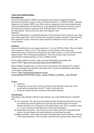
1. Anorectal malformation
Introduction
Anorectal malformations (ARMs) are among the more frequent congenital anomalies
encountered in pediatric surgery, with an estimated incidence 1 in 5000 live births. Antenatal
diagnosis of an isolated ARM is rare. Most cases are diagnosed in the early neonatal period.
Anorectal malformations are birth defects or developmental deformities of the lower end of
the alimentary tract that is anorectal canal. With this defect, the anus and rectum don’t
develop properly. They are the lower part of the digestive tract.
Definition
Anorectal malformation is congenital anomalies of the anorectal canal or anal area may range
from simple imperforate anal to include other associated complex anomalies of genitourinary
(GU) and pelvic organs, which may require extensive treatment for faecal, urinary, and
sexual function.
Incidence
Anorectal malformations occur approximately in 1 in every 5000 live births. They are slightly
more common in males (1.2 to 1). The majority of male patients with an anorectal
malformation have some form of connection to the urinary system, or a recto-urethral fistula
(approximately 70% of this patient population). The most common type of anorectal
malformation in female patients is a recto-vestibular fistula.
Source author smith ac,avansino topic anorectal malformation last update date
august12,2020 https://www.ncbi.nlm.nih.gov/books/NBK542275/
Kanti Children's hospitals have served a total of 103 patients in the Surgical ICU of Kanti
Children’s Hospital, from October to December 2018. Anorectal malformation 9(October),
7 (November), 6(December)
Source:- https://realmedicinefoundation.org/wp-
content/uploads/2019/09/RMF_Nepal___Kanti_Children_s_Hospital___Q4_2018.pdf
Causes
a. Unknown
b. Congenital:-it occurs due to arrest in embryonic development of the anus, lower
rectum and uro-genital tract at the 8th
week of embryonic life.
c. It may be related with down syndrome and familial incidence
Classification
Depending on the relations with the levator muscles, ano-rectal malformation is classified
into:-
(i) Low anomalies: The rectum has decended normally through the puborectalis muscle,
the internal and external sphincters are present and well developed with normal
function and there is no connection to the genitourinary tract. In low anomalies, there
is 1.5 cm or less between the blind end of colon and anal dimple.
(ii) Intermediate anomalies:- The rectum is at the or below the level of the puborectalis
muscle; the anal dimple and external sphincter are positioned normally.
(iii)High anomalies:- The rectum ends above the puborectalis muscle and there is absence
of the internal sphincter. This is usually associated with a genitourinary fistula (recto
2. urethral in male or rectovaginal in fistula). In high anal agenesis, there is more than
1.5cm between the blind end of colon and anal dimple.
Pathophysiology
The ano- rectal and genitourinary tract originates from an embryologic structure called
cloaca. By 6 weeks of gestation, cloaca is divided into the anterior urogenital sinus and
posterior ano-rectal canal by a septum. The rectum and urinary tract separate completely by
the 7th
week of gestation. further development of the ano-rectal and uro-genital organs occur
up to 12 weeks of gestation.
Developmental failure during those period results several form of malformation with or
without an obvious anal opening. Anomalies reflect the stage of the development of these
processes. Failure to develop the urorectal septum results in a fistula between the bowel and
urinary tract(in boys) or the vagina (in girls). Many have a fistula from the distal rectum to
the perineum or genitourinary system.
Clinical presentation
a) Absence of anal opening or abnormally formed anal opening at birth (during perineal
examination)
b) Absence of meconium
c) Meconium discharge from other site than anus
d) Progressive distension of abdomen that may associate with vomiting.
e) Fistula formation and passage of stool through the fistula (on examination)
f) Ribbon like stool
g) Male:- fistula formed between rectum and vagina or perineum.
h) Female:- fistula formed between rectum and vagina or perineum.
Feature according to type of defects.
a. Imperforated anus:- Infants fails to pass meconium, and greenish bulging membrane
is seen on examination.
b. Anal stenosis :- Infants passes ribbon like stools with difficulty
c. Anal agenesis: failure to develop anus so presence of only anal dimple. Intestinal
obstruction develops, if there is absence of any fistula. Usually fistulas are found to
the perineum or urethra in male and perineum or vulva in female.
d. Rectal agenesis: it presents with fistula. In male baby, fistula may communicate with
posterior urethra and in female with upper vagina.
e. Rectoperineal fistula: it is manifested as small orifice in the perineum. In male baby it
is found close to the scrotum and in female at the vulva.
f. Rectovaginal fistula: it is the communication between rectum and vagina and stool
passes through the vagina.
Diagnosis Procedure
a. History and clinical presentations that is absence of anal opening at birth
b. Neonatal examination: assess patency of anus and rectum and perineal area of the
newborn for passage of stool at abnormal site.
c. Observation of the newborn regarding passage of meconium.
d. Abdominal USG: evaluate anatomic malformation
e. Urine examination for presence of meconium and epithelial debris in urine
f. An intravenous pyelogram (IVP) and cystourethrography are done for the infant with
suspected anomalies of the urinary tract.
g. Evaluation of the child for presence of other anomalies in the body
3. h. Electrocardiogram to rule out cardiac problem
Management
The reconstructive operation is needed to correct or repair the malformations. It depends
upon the sex of the baby and type of anomalies presence. General measures include:
a. Admit the child in tertiary level hospital.
b. With hold oral feeding.
c. Start intravenous fluid therapy.
d. Reassure the parents and explain the nature of anomalies as well as treatment process.
e. Administer vitamin 'k' injection and antibiotics as per prescription.
f. Maintain warmth to prevent baby from hypothermia.
g. Nutrition- feedings is started soon after surgical repairment and breast feeding is
encouraged to minimize the risk of constipation.
h. Monitoring vitals and record accurately.
i. Maintain strict intake output record.
Surgery
Surgery is indicated as a definitive treatment: Surgery for low ano-rectal malformations
includes rectal cutback anoplasty or y- v plasty for male and dilation of fistula with definitive
repairment and perineal anoplasty for female child. Dilation initiated when the child returns
for the second week's follow up visit.
In case of high ano-rectal malformations: Initial colostomy is done during neonatal period
and definitive reconstructive surgery as posterior sagittal ano-rectoplasty at the age of 10-
12months with infant is having weight7-9 kg. Thereafter colostomy closure is done 10-
12weeks of successful definitive surgery.
Note: anoplasty - moving the fistula opening to the center of the sphincter and enlarging the
rectal opening.
Type specific management
Anal stenosis: Manual dilation.
Imperforated anal membrane: Excision followed by daily dilatation.
Prophylactic antibiotics therapy.
Nursing Management
Assessment: Nursing assessment should focus on pass of meconium within 48 hours of birth,
and stool come from any abnormal opening.
Nursing Diagnoses
Fluid volume deficit related to excessive loss through vomiting.
Impaired skin integrity related to the colostomy.
Risk for infection related to surgical procedures
Nursing Interventions
Avoid infection.
Teach the caregivers to keep the area around the colostomy clean with soap and
water
To diaper the baby in the usual way;
Monitor white blood cell (WBC) count;
4. wash hands and teach patient and
Wash hands before contact with patients and between procedures with the patient.
Protect skin integrity.
A protective ointment is useful to protect the skin around the colostomy;
Monitor site of impaired tissue integrity at least once daily for colour changes,
redness, swelling, warmth, pain, or other signs of infection; and
Keep a sterile dressing technique during wound care.
Restore balanced fluid volume.
Administer parenteral fluids as prescribed;
Immediate administered infusion of fluids for patients with abnormal vital signs
Monitor both intake and output.
Teach family members how to monitor output in the home;
Pre operative care
a. Inform parents about baby's condition
b. Maintenance of warmth.
c. Fluid and electrolytes therapy and NG decompression.
d. Written Consent.
e. Parental measurement instruction on surgical procedure,
f. Measurement of abdominal surgical girth.
g. Vitals monitoring and proper recording.
h. Physical preparation, pre-medications, etc.
i. Diagnostic care and collection of report.
j. Keep baby nil per orally
Post operative care
a. Maintain airway status.
b. Pain management
c. Care of soakage from suture site and other immediate post surgery care.
d. Measure to prevent from infection
e. Positioning of baby on side lying position with the hip elevated or a supine position
with the legs suspended at 90º to the trunk to prevent pressure over suture area.
f. Postoperative nasogastric decompression and drainage care.
g. Colostomy care and check for return of peristalsis.
h. Provide post-operative medication
i. Emotional support for family
j. Parents teaching on colostomy care at home, breastfeeding and other feeding, skin
care, bowel and bladder care, prevention of fecal impaction, regular bowel habit
training and follow up visit and keep perineal area dry and clean.
Complication
Urinary infection
Intestinal obstruction
Faecal impaction
Colostomy infection
Fistula and anal stenosis
REFERENCE OF ANORECTAL MALFORMATION
Uprety K, Child Health Nursing, fourth Edition (2071 Bhadra), Tara Books and
Stationery, Chhetrapati, Kathmandu, pg 138- 140
5. Shrestha T. Essential Child Health Nursing. first Edition 2015,August. Medhavi
Publication; Jamal, Kathmandu Page no.286-290
WilsonD, Rodgers CC, Hockenberry M, Wongs Essential of pediatric Nursing 10 th
edition, ELSEVIER, page no 1422-1425
Adhikari T, Essential of Pediatric Nursing, first 2014 edition, vidyarthi pustak
bhadar, bhotahity, Kathmandu, page no177-182
https://www.urmc.rochester.edu/encyclopedia/content.aspx?contenttypeid=90&conte
ntid=P01980#:~:text=Anorectal%20malformations%20are%20birth%20defects,end%
20of%20the%20large%20intestine
https://realmedicinefoundation.org/wp-
content/uploads/2019/09/RMF_Nepal___Kanti_Children_s_Hospital___Q4_2018.pdf
https://www.ncbi.nlm.nih.gov/books/NBK542275/