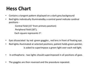
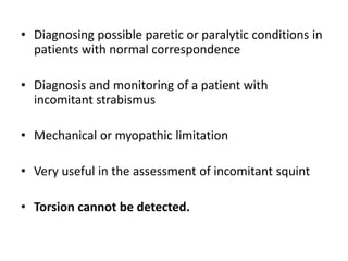
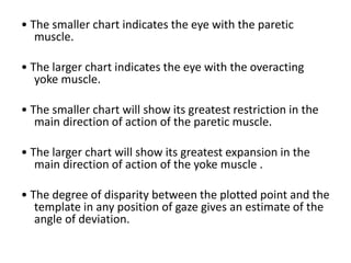
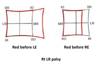
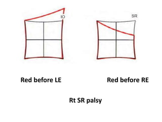
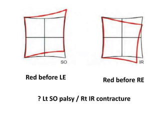
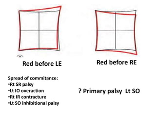
Hess Chart
- Dr. Swathi.N
Diplopia
Acquired misalignment of the visual axes - image
falls simultaneously on the fovea of 1 eye and
on a non-foveal point in the other eye.
The object that falls on these non-corresponding
points must be outside Panum's area to
appear double.
Pannum’s area
The region in front and back of the horopter in which single vision is
present - Panum’s fusional area.
In Panum’s area visual objects are seen stereoscopically.
Horopter - the locus of all object points that are imaged
on corresponding retinal elements at a given fixation
distance.
All object points lying on the horopter curve
stimulate corresponding retinal elements.
By definition, all points on the horopter curve
are seen singly.
Also by definition, all points not lying on the
horopter curve are imaged disparately and,
with certain qualifications, are seen double.
The diplopia elicited by object points off the
horopter is called physiologic diplopia.
The same object is seen as having 2 locations in
subjective space, and the foveal image is
always clearer than the nonfoveal image of
the nonfixating eye.
The perception of diplopia depends on
• the age at onset,
• its duration,
• the patient's subjective awareness of it.
The younger the child, the greater the ability to
suppress, or inhibit, the nonfoveal image.
Diplopia test
Determines the subjective
localization of
a single object point
imaged on the fovea of
the fixating eye
and an extrafoveal retinal
area in the other eye
No horizontal phoria
Esophoria
Exophoria
RE LE
The two visual fields must be differentiated
Haploscopic Tests
• Two test objects rather than one are
presented to the patient, who is required to
place them in such a fashion that they appear
superimposed.
• Again assuming that correspondence is
normal, the two objects are placed to
stimulate the foveae of the two eyes.
• Each eye may be presented with a different target,
• complementary colors may be placed in the visual
field of the patient,
• each eye may be provided with a corresponding
colored filter.
Hess Chart
• Contains a tangent pattern displayed on a dark grey background
• Red lights individually illuminatedby a control panel indicate cardinal
positions -
Central field (15° from primary position)
Peripheral field (30°);
Each square represents 5°.
• Eyes dissociated by red -green goggles , red lens in front of fixating eye.
• Red lights illuminated at selected positions; patient holds green pointer,
is asked to superimpose a green light over each red light.
• In orthophoria - two lights should superimposed in all positions of gaze.
• The goggles are then reversed and the procedure repeated.
• Diagnosing possible paretic or paralytic conditions in
patients with normal correspondence
• Diagnosis and monitoring of a patient with
incomitant strabismus
• Mechanical or myopathic limitation
• Very useful in the assessment of incomitant squint
• Torsion cannot be detected.
• The smaller chart indicates the eye with the paretic
muscle.
• The larger chart indicates the eye with the overacting
yoke muscle.
• The smaller chart will show its greatest restriction in the
main direction of action of the paretic muscle.
• The larger chart will show its greatest expansion in the
main direction of action of the yoke muscle .
• The degree of disparity between the plotted point and the
template in any position of gaze gives an estimate of the
angle of deviation.
Red before RERed before LE
Rt LR palsy
Red before LE Red before RE
Rt SR palsy
Red before LE Red before RE
? Lt SO palsy / Rt IR contracture
Red before LE Red before RE
Spread of commitance:
•Rt SR palsy
•Lt IO overaction
•Rt IR contracture
•Lt SO inhibitional palsy
? Primary palsy Lt SO
Thank You

Diplopia

  • 1.
  • 2.
    Diplopia Acquired misalignment ofthe visual axes - image falls simultaneously on the fovea of 1 eye and on a non-foveal point in the other eye.
  • 3.
    The object thatfalls on these non-corresponding points must be outside Panum's area to appear double.
  • 4.
    Pannum’s area The regionin front and back of the horopter in which single vision is present - Panum’s fusional area. In Panum’s area visual objects are seen stereoscopically.
  • 5.
    Horopter - thelocus of all object points that are imaged on corresponding retinal elements at a given fixation distance.
  • 6.
    All object pointslying on the horopter curve stimulate corresponding retinal elements. By definition, all points on the horopter curve are seen singly. Also by definition, all points not lying on the horopter curve are imaged disparately and, with certain qualifications, are seen double. The diplopia elicited by object points off the horopter is called physiologic diplopia.
  • 7.
    The same objectis seen as having 2 locations in subjective space, and the foveal image is always clearer than the nonfoveal image of the nonfixating eye.
  • 8.
    The perception ofdiplopia depends on • the age at onset, • its duration, • the patient's subjective awareness of it. The younger the child, the greater the ability to suppress, or inhibit, the nonfoveal image.
  • 9.
    Diplopia test Determines thesubjective localization of a single object point imaged on the fovea of the fixating eye and an extrafoveal retinal area in the other eye No horizontal phoria Esophoria Exophoria RE LE
  • 10.
    The two visualfields must be differentiated
  • 11.
    Haploscopic Tests • Twotest objects rather than one are presented to the patient, who is required to place them in such a fashion that they appear superimposed. • Again assuming that correspondence is normal, the two objects are placed to stimulate the foveae of the two eyes.
  • 12.
    • Each eyemay be presented with a different target, • complementary colors may be placed in the visual field of the patient, • each eye may be provided with a corresponding colored filter.
  • 13.
    Hess Chart • Containsa tangent pattern displayed on a dark grey background • Red lights individually illuminatedby a control panel indicate cardinal positions - Central field (15° from primary position) Peripheral field (30°); Each square represents 5°. • Eyes dissociated by red -green goggles , red lens in front of fixating eye. • Red lights illuminated at selected positions; patient holds green pointer, is asked to superimpose a green light over each red light. • In orthophoria - two lights should superimposed in all positions of gaze. • The goggles are then reversed and the procedure repeated.
  • 14.
    • Diagnosing possibleparetic or paralytic conditions in patients with normal correspondence • Diagnosis and monitoring of a patient with incomitant strabismus • Mechanical or myopathic limitation • Very useful in the assessment of incomitant squint • Torsion cannot be detected.
  • 15.
    • The smallerchart indicates the eye with the paretic muscle. • The larger chart indicates the eye with the overacting yoke muscle. • The smaller chart will show its greatest restriction in the main direction of action of the paretic muscle. • The larger chart will show its greatest expansion in the main direction of action of the yoke muscle . • The degree of disparity between the plotted point and the template in any position of gaze gives an estimate of the angle of deviation.
  • 16.
    Red before RERedbefore LE Rt LR palsy
  • 17.
    Red before LERed before RE Rt SR palsy
  • 18.
    Red before LERed before RE ? Lt SO palsy / Rt IR contracture
  • 19.
    Red before LERed before RE Spread of commitance: •Rt SR palsy •Lt IO overaction •Rt IR contracture •Lt SO inhibitional palsy ? Primary palsy Lt SO
  • 20.