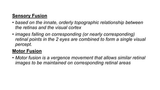
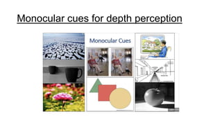
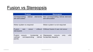
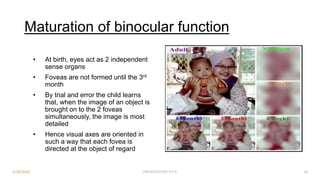
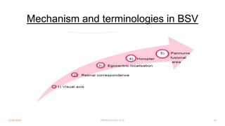
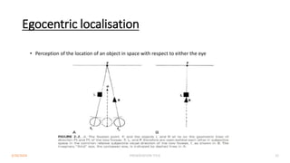
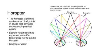
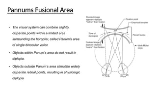
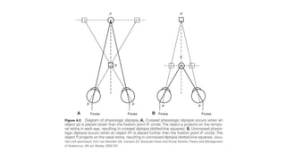
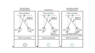
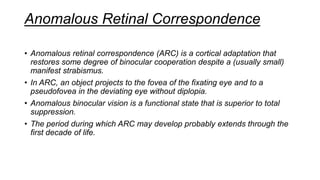
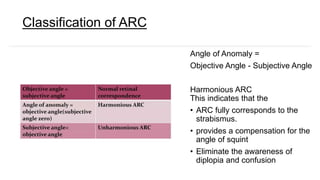
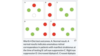
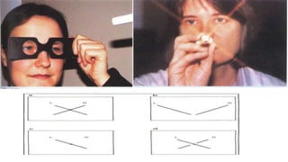
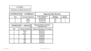
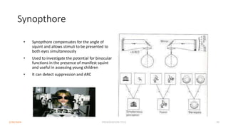
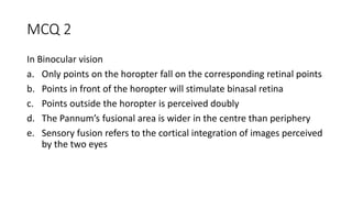
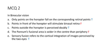
Binocular Single Vision
Presenter : Dr Vishnu Prasad Rao
Supervisor : Prof Dr. Shatriah Ismail
01 Definition
02 Grading
03 Fusion vs Stereopsis
04 Development of BSV
05 Retinal Correspondence
06 Horopter/Pannums Fusional Area
07 Anomalies of BSV
08 Investigations
09 MCQ
Table Of
Contents
Definition
State of simultaneous vision which is achieved by coordinated
use of both eyes so that separate and slight dissimilar images
arising in each eye appreciated as single image by process of
vision
Grading
1. SIMULTANEOUS PERCEPTION
2. FUSION
3. STEREOPSIS
Simultaneous Perception
• Simultaneous perception exists
when signals transmitted from
the two eyes to the visual
cortex are perceived at the
same time.
• It consists of the ability to see
two dissimilar objects
simultaneously.
Fusion
• Fusion is the cortical unification of 2 images of an object, 1 from
each eye, into a single percept.
• For retinal images to be fused, they must be similar in size and
shape.
• For fusion of macular images (central fusion) to occur, there can
be very little dissimilarity between the images in each eye
• More image dissimilarity is tolerated in the periphery (peripheral
fusion), where the receptive fields are larger.
Sensory Fusion
• based on the innate, orderly topographic relationship between
the retinas and the visual cortex
• images falling on corresponding (or nearly corresponding)
retinal points in the 2 eyes are combined to form a single visual
percept.
Motor Fusion
• Motor fusion is a vergence movement that allows similar retinal
images to be maintained on corresponding retinal areas
Stereopsis
• Stereopsis is a binocular sensation of relative depth caused by horizontal
disparity of retinal images.
• It is the highest form of binocular cooperation.
• The ability to fuse images that stimulates horizontally disparate retinal
elements within Panum’s fusional area resulting in binocular appreciation
of visual object in depth i.e. in the third dimension
• Able to perceive width, depth, and height ,when look at an object.
How it occurs?
3/30/2024 PRESENTATION TITLE 10
 Retinal disparity (fixation disparity) is the
basis of 3D perception
 Stereopsis occurs when retinal disparity
is too great to permit simple
superimposition of the two retinal
images, but not great enough to elicit
diplopia
Stereopsis and depth perception
• Stereopsis and depth perception are not synonymous.
• Monocular cues— which include object overlap, relative object size,
highlights and shadows, motion parallax, and perspective also contribute
to depth perception.
• Monocular patients can have excellent depth perception using these cues.
Monocular cues for depth perception
Fusion vs Stereopsis
3/30/2024 PRESENTATION TITLE 14
Advantanges of BSV
● Results in stereopsis
● Optical defects in one eye is compensated by the
normal eye
● Enhanced visual acuity, contrast sensitivity
● Defective vision in any part of one visual field is
masked, e.g. blind spot
● Wider visual field than monocular
• Normal monocular visual field
• 60̊ superior & nasal
• 75̊ inferiorly
• 95 temporally
• With BSV : 160-200̊
3/30/2024 PRESENTATION TITLE 15
Development of BSV
3/30/2024 PRESENTATION TITLE 16
• Basic visual functions are innate and therefore present at birth
• Their coordination, maturation, & refinement take place during
early postnatal period
BSV milestones
AGE2 MILESTONES
Birth no bifoveal fixation. Monocular fixation is present at birth, but
poor
2-3 weeks infant begins to make movements, turning his eyes to fixate an
object
4-5 weeks can sustain monocular fixation of large near objects
6 weeks fixation alternates rapidly between two eyes & child begins to
fixate binocularly with conjugate pursuit movements which
are saccadic initially but become smooth and gliding by 3-
5mts of age.
3-6 months conjugate movements and disconjugate vergence movements
1 year fusional movements are firmly established.
2-3 years adult level of visual acuity is reached.
3/30/2024 PRESENTATION TITLE 17
Maturation of binocular function
• At birth, eyes act as 2 independent
sense organs
• Foveas are not formed until the 3rd
month
• By trial and error the child learns
that, when the image of an object is
brought on to the 2 foveas
simultaneously, the image is most
detailed
• Hence visual axes are oriented in
such a way that each fovea is
directed at the object of regard
3/30/2024 PRESENTATION TITLE 18
Mechanism and terminologies in BSV
3/30/2024 PRESENTATION TITLE 19
Visual Axis
• A visual axis is defined as a line that
connects an object point with its
image on the retina
• Types;
a) Principal
• - Fovea – area of highest VA, carries
principal visual axis
a) Secondary
• - Other retinal elements, secondary
visual axis
3/30/2024 PRESENTATION TITLE 20
Retinal Correspondence
• Retinal correspondence is the term
used when a viewed target
stimulates paired retinal areas in an
individual’s 2 eyes. These retinal
locations are said to be
corresponding.
• When the image of an object in
space falls on corresponding points,
it is perceived as a single object
located in the same subjective or
egocentric direction.
Egocentric localisation
• Perception of the location of an object in space with respect to either the eye
3/30/2024 PRESENTATION TITLE 22
Horopter
• The horopter is defined
as the locus of all points
in space that stimulate
corresponding retinal
points
• Double vision would be
expected when the
target does not lie on the
horopter
• Horizon of vision
Pannums Fusional Area
• The visual system can combine slightly
disparate points within a limited area
surrounding the horopter, called Panum’s area
of single binocular vision
• Objects within Panum’s area do not result in
diplopia.
• Objects outside Panum’s area stimulate widely
disparate retinal points, resulting in physiologic
diplopia
Anomalies of BSV
• When a manifest deviation of the eyes occurs, the corresponding retinal
elements of the eyes are no longer directed at the same object.
• This places the patient at risk for 2 distinct visual phenomena: visual
confusion and diplopia.
Sensory Adaptations in Strabismus
• To avoid visual confusion and diplopia, the visual system uses the
mechanisms of suppression and anomalous retinal correspondence.
• Pathologic suppression and anomalous retinal correspondence develop
only in the immature visual system under binocular conditions.
Suppression
• Suppression is the alteration of visual sensation that occurs when an eye’s
retinal image is prevented from reaching consciousness during binocular
visual activity
• Can be classified as such
- Central vs peripheral
- Non alternating vs alternating
- Facultative vs constant
Anomalous Retinal Correspondence
• Anomalous retinal correspondence (ARC) is a cortical adaptation that
restores some degree of binocular cooperation despite a (usually small)
manifest strabismus.
• In ARC, an object projects to the fovea of the fixating eye and to a
pseudofovea in the deviating eye without diplopia.
• Anomalous binocular vision is a functional state that is superior to total
suppression.
• The period during which ARC may develop probably extends through the
first decade of life.
Classification of ARC
Angle of Anomaly =
Objective Angle - Subjective Angle
Harmonious ARC
This indicates that the
• ARC fully corresponds to the
strabismus.
• provides a compensation for the
angle of squint
• Eliminate the awareness of
diplopia and confusion
Unharmonious ARC
- angle of anomaly < objective angle of deviation
- subjective angle # 0
Paradoxical
- when the angle of anomaly > objective angle of deviation.
- It occurs mainly after overcorrection of the squint surgery
Investigations
Test for fusion
- Bagollini striated glass test
- Worth 4 dots test
Test for stereopsis
- TNO test
- Frisby test
- Lang’s stereo test
- Synaptophore
WORTH FOUR-DOT TEST
This is a dissociation test which can be used with both distance and near
fixation, and differentiates between BSV, or ARC and suppression .
Procedure:
- The patient wears a green lens in front of the left eye
- And a red lens in front of the right eye
- The patient then views a box with four lights ; one red , two green and one
white
 If BSV is present all four lights are seen.
 If all four lights are seen in the presence of a manifest deviation, harmonious
ARC is present.
 If two red lights are seen , left suppression is present .
 If three green lights are seen , right suppression is present.
 If two red and three green lights are seen , diplopia is present.
 If the green and red lights alternate , alternating suppression is present.
Results
This is a test for detecting BSV , ARC or suppression . Each lens have fine
striations which convert a point source of light into a line, as with the
Maddox rod .
Procedure:
- The two lenses are placed at 45°and 135° in front of each eye and the
patient fixates a small light source .
- Each eye perceives an oblique line of light , perpendicular to that
perceived by the fellow eye.
- Dissimilar images are thus presented to each eye under binocular
viewing conditions .
BAGOLINI STRIATED GLASSES
 If the two streaks intersect at their centers in the form of an oblique cross ( an
X ), the patient has BSV if the eyes are straight , or harmonious ARC in the
presence of manifest strabismus .
 If the two lines are seen but they do not form a cross , diplopia is present .
 If only one streak is seen , there is no simultaneous perception and
suppression is present .
 If a small gap is seen in one of the streaks , a central suppression
scotoma ( as found in microtropia ) is present .
Results:
TNO test
● A booklet consisting of 7 plates
● Viewed with red- green spectacles
● Each plates contain various shape (squares, dots, crosses)
created by computer generated random dot analgyphs
● Analgyphs is a stereogram in which two disparate views are
printed in red and green on white background
● The eye looking through the red filter sees only green picture
as black
● The eye looking through the green filter sees the red picture
as black
● Two views fuse to give a stereoscopic effect
● 480- 15 sec of arc
● No monocular clues
3/30/2024 PRESENTATION TITLE 41
3/30/2024 PRESENTATION TITLE 42
Frisby test
● Assess fine stereopsis
● Degree of disparity ranges from 600 to 15 seconds of
• arc
● Stereogram consists of 3 plates each containing 4 squares of small
random shape
● The three plates are of differing thickness, 6mm, 3mm and
• 1.5mm (55, 170, 340sec of arc , 40cm viewing distance)
● By adjusting the viewing distance, the test can give a disparity
of 600 to 15 sec of arc
● One of the squares contain one hidden circle which is seen disparately.
● Doesn’t require use of polarized glasses
3/30/2024 PRESENTATION TITLE 43
Titmus test
1) WIRT FLYTEST
• Assess gross stereopsis
• Utilizes the principle of vectograph
• Useful in young children
• Made up by 2 plates in the form of booklet
• The plates are reviewed with polaroid glasses
• The right side of booklet – large housefly with threshold 3000 sec of arc
• Stereoscopic ,will reach above the plate (child is encouraged to pick up the
tip of the wings between finger and thumb).
• Performed at a distance of 16 inches (40cm)
3/30/2024 PRESENTATION TITLE 44
2) Animal test
● Assess fine stereopsis
● 3 rows of 5 animals each
● 1 animal from each row is imaged disparately (appear
forward).
● In each row, one of the animals imaged in two eyes is
printed heavily (misleading clue)
3/30/2024 PRESENTATION TITLE 45
3) Circles test
● Consist of 9 squares
● Each containing 4 circles arranged in the form of a lozenge
● Only 1 of the circle in each square is
• disparately imaged at random
3/30/2024 PRESENTATION TITLE 46
3/30/2024 PRESENTATION TITLE 47
Lang stereotest
• Targets are made up of fine vertical lines which are seen
alternatively by each eye when focused through built in
cylindrical lens elements
• Lang I: Disparity = Car 550", Star 600", Cat 1200"
• Lang II: Disparity = Moon 200", Car 400", Elephant 600", Star
monocular visible
• Unique feature – No polaroid glasses need to be worn for
viewing the test plate
3/30/2024 PRESENTATION TITLE 48
Synopthore
• Synopthore compensates for the angle of
squint and allows stimuli to be presented to
both eyes simultaneously
• Used to investigate the potential for binocular
functions in the presence of manifest squint
and useful in assessing young children
• It can detect suppression and ARC
3/30/2024 PRESENTATION TITLE 49
Two pencil test
• Gross stereopsis
• Examiner holds the pencil vertically in front of patients eye
• Patient touch the upper tip of pencil
• Having stereopsis – Pass test with BE open
• One eye closed – patient fails the test
• Disparity range : 3000-5000 secs of arc
3/30/2024 PRESENTATION TITLE 50
Conclusion
• BSV is not inborn,but develops in the first decade of life
• Abnormal visual experience results in poor or no BSV
• Reversible only if intervened in the plastic period of
development
• A good understanding of mechanism of BSV is
fundamental in successive treatment of its anamolies
References
Reference
• Adler’s physiology of eye: clinical application, 10th Ed. St
Louis: Mosby, 2003.
• Khurana A. K.: Theory and Practise of Squint and Orthoptics;
first ed
• AAO Pediatric Ophthalmology and strabismus 2022- 2023
MCQ 1
1. Requirements for Binocular Single Vision include
a. Normal retinal correspondence
b. Good vision in one eye
c. Precise coordination of the eyes at all times
d. Ability of fusional areas of brain to fuse slightly dissimilar images
e. Normal macular function
MCQ 1
1. Requirements for Binocular Single Vision include
a. Normal retinal correspondence T
b. Good vision in one eye F
c. Precise coordination of the eyes at all times T
d. Ability of fusional areas of brain to fuse slightly dissimilar images T
e. Normal macular function T
MCQ 2
In Binocular vision
a. Only points on the horopter fall on the corresponding retinal points
b. Points in front of the horopter will stimulate binasal retina
c. Points outside the horopter is perceived doubly
d. The Pannum’s fusional area is wider in the centre than periphery
e. Sensory fusion refers to the cortical integration of images perceived
by the two eyes
MCQ 2
In Binocular vision
a. Only points on the horopter fall on the corresponding retinal points T
b. Points in front of the horopter will stimulate binasal retina F
c. Points outside the horopter is perceived doubly T
d. The Pannum’s fusional area is wider in the centre than periphery F
e. Sensory fusion refers to the cortical integration of images perceived by
the two eyes T
MCQ 3
Advantages of binocular single vision include
a. A larger field of vision
b. Elimination of blind spot
c. Better binocular visual acuity
d. Accurate depth perception
e. Multiple mental impression
MCQ 3
Advantages of binocular single vision include
a. A larger field of vision T
b. Elimination of blind spot T
c. Better binocular visual acuity T
d. Accurate depth perception T
e. Multiple mental impression F
THANK YOU

Binocular Single Vision Ophthalmology .pptx

  • 1.
    Binocular Single Vision Presenter: Dr Vishnu Prasad Rao Supervisor : Prof Dr. Shatriah Ismail
  • 2.
    01 Definition 02 Grading 03Fusion vs Stereopsis 04 Development of BSV 05 Retinal Correspondence 06 Horopter/Pannums Fusional Area 07 Anomalies of BSV 08 Investigations 09 MCQ Table Of Contents
  • 3.
    Definition State of simultaneousvision which is achieved by coordinated use of both eyes so that separate and slight dissimilar images arising in each eye appreciated as single image by process of vision
  • 4.
  • 5.
    Simultaneous Perception • Simultaneousperception exists when signals transmitted from the two eyes to the visual cortex are perceived at the same time. • It consists of the ability to see two dissimilar objects simultaneously.
  • 6.
    Fusion • Fusion isthe cortical unification of 2 images of an object, 1 from each eye, into a single percept. • For retinal images to be fused, they must be similar in size and shape. • For fusion of macular images (central fusion) to occur, there can be very little dissimilarity between the images in each eye • More image dissimilarity is tolerated in the periphery (peripheral fusion), where the receptive fields are larger.
  • 7.
    Sensory Fusion • basedon the innate, orderly topographic relationship between the retinas and the visual cortex • images falling on corresponding (or nearly corresponding) retinal points in the 2 eyes are combined to form a single visual percept. Motor Fusion • Motor fusion is a vergence movement that allows similar retinal images to be maintained on corresponding retinal areas
  • 9.
    Stereopsis • Stereopsis isa binocular sensation of relative depth caused by horizontal disparity of retinal images. • It is the highest form of binocular cooperation. • The ability to fuse images that stimulates horizontally disparate retinal elements within Panum’s fusional area resulting in binocular appreciation of visual object in depth i.e. in the third dimension • Able to perceive width, depth, and height ,when look at an object.
  • 10.
    How it occurs? 3/30/2024PRESENTATION TITLE 10  Retinal disparity (fixation disparity) is the basis of 3D perception  Stereopsis occurs when retinal disparity is too great to permit simple superimposition of the two retinal images, but not great enough to elicit diplopia
  • 11.
    Stereopsis and depthperception • Stereopsis and depth perception are not synonymous. • Monocular cues— which include object overlap, relative object size, highlights and shadows, motion parallax, and perspective also contribute to depth perception. • Monocular patients can have excellent depth perception using these cues.
  • 12.
    Monocular cues fordepth perception
  • 14.
    Fusion vs Stereopsis 3/30/2024PRESENTATION TITLE 14
  • 15.
    Advantanges of BSV ●Results in stereopsis ● Optical defects in one eye is compensated by the normal eye ● Enhanced visual acuity, contrast sensitivity ● Defective vision in any part of one visual field is masked, e.g. blind spot ● Wider visual field than monocular • Normal monocular visual field • 60̊ superior & nasal • 75̊ inferiorly • 95 temporally • With BSV : 160-200̊ 3/30/2024 PRESENTATION TITLE 15
  • 16.
    Development of BSV 3/30/2024PRESENTATION TITLE 16 • Basic visual functions are innate and therefore present at birth • Their coordination, maturation, & refinement take place during early postnatal period
  • 17.
    BSV milestones AGE2 MILESTONES Birthno bifoveal fixation. Monocular fixation is present at birth, but poor 2-3 weeks infant begins to make movements, turning his eyes to fixate an object 4-5 weeks can sustain monocular fixation of large near objects 6 weeks fixation alternates rapidly between two eyes & child begins to fixate binocularly with conjugate pursuit movements which are saccadic initially but become smooth and gliding by 3- 5mts of age. 3-6 months conjugate movements and disconjugate vergence movements 1 year fusional movements are firmly established. 2-3 years adult level of visual acuity is reached. 3/30/2024 PRESENTATION TITLE 17
  • 18.
    Maturation of binocularfunction • At birth, eyes act as 2 independent sense organs • Foveas are not formed until the 3rd month • By trial and error the child learns that, when the image of an object is brought on to the 2 foveas simultaneously, the image is most detailed • Hence visual axes are oriented in such a way that each fovea is directed at the object of regard 3/30/2024 PRESENTATION TITLE 18
  • 19.
    Mechanism and terminologiesin BSV 3/30/2024 PRESENTATION TITLE 19
  • 20.
    Visual Axis • Avisual axis is defined as a line that connects an object point with its image on the retina • Types; a) Principal • - Fovea – area of highest VA, carries principal visual axis a) Secondary • - Other retinal elements, secondary visual axis 3/30/2024 PRESENTATION TITLE 20
  • 21.
    Retinal Correspondence • Retinalcorrespondence is the term used when a viewed target stimulates paired retinal areas in an individual’s 2 eyes. These retinal locations are said to be corresponding. • When the image of an object in space falls on corresponding points, it is perceived as a single object located in the same subjective or egocentric direction.
  • 22.
    Egocentric localisation • Perceptionof the location of an object in space with respect to either the eye 3/30/2024 PRESENTATION TITLE 22
  • 23.
    Horopter • The horopteris defined as the locus of all points in space that stimulate corresponding retinal points • Double vision would be expected when the target does not lie on the horopter • Horizon of vision
  • 24.
    Pannums Fusional Area •The visual system can combine slightly disparate points within a limited area surrounding the horopter, called Panum’s area of single binocular vision • Objects within Panum’s area do not result in diplopia. • Objects outside Panum’s area stimulate widely disparate retinal points, resulting in physiologic diplopia
  • 26.
    Anomalies of BSV •When a manifest deviation of the eyes occurs, the corresponding retinal elements of the eyes are no longer directed at the same object. • This places the patient at risk for 2 distinct visual phenomena: visual confusion and diplopia.
  • 27.
    Sensory Adaptations inStrabismus • To avoid visual confusion and diplopia, the visual system uses the mechanisms of suppression and anomalous retinal correspondence. • Pathologic suppression and anomalous retinal correspondence develop only in the immature visual system under binocular conditions.
  • 29.
    Suppression • Suppression isthe alteration of visual sensation that occurs when an eye’s retinal image is prevented from reaching consciousness during binocular visual activity • Can be classified as such - Central vs peripheral - Non alternating vs alternating - Facultative vs constant
  • 30.
    Anomalous Retinal Correspondence •Anomalous retinal correspondence (ARC) is a cortical adaptation that restores some degree of binocular cooperation despite a (usually small) manifest strabismus. • In ARC, an object projects to the fovea of the fixating eye and to a pseudofovea in the deviating eye without diplopia. • Anomalous binocular vision is a functional state that is superior to total suppression. • The period during which ARC may develop probably extends through the first decade of life.
  • 31.
    Classification of ARC Angleof Anomaly = Objective Angle - Subjective Angle Harmonious ARC This indicates that the • ARC fully corresponds to the strabismus. • provides a compensation for the angle of squint • Eliminate the awareness of diplopia and confusion
  • 32.
    Unharmonious ARC - angleof anomaly < objective angle of deviation - subjective angle # 0 Paradoxical - when the angle of anomaly > objective angle of deviation. - It occurs mainly after overcorrection of the squint surgery
  • 33.
    Investigations Test for fusion -Bagollini striated glass test - Worth 4 dots test Test for stereopsis - TNO test - Frisby test - Lang’s stereo test - Synaptophore
  • 34.
    WORTH FOUR-DOT TEST Thisis a dissociation test which can be used with both distance and near fixation, and differentiates between BSV, or ARC and suppression . Procedure: - The patient wears a green lens in front of the left eye - And a red lens in front of the right eye - The patient then views a box with four lights ; one red , two green and one white
  • 35.
     If BSVis present all four lights are seen.  If all four lights are seen in the presence of a manifest deviation, harmonious ARC is present.  If two red lights are seen , left suppression is present .  If three green lights are seen , right suppression is present.  If two red and three green lights are seen , diplopia is present.  If the green and red lights alternate , alternating suppression is present. Results
  • 37.
    This is atest for detecting BSV , ARC or suppression . Each lens have fine striations which convert a point source of light into a line, as with the Maddox rod . Procedure: - The two lenses are placed at 45°and 135° in front of each eye and the patient fixates a small light source . - Each eye perceives an oblique line of light , perpendicular to that perceived by the fellow eye. - Dissimilar images are thus presented to each eye under binocular viewing conditions . BAGOLINI STRIATED GLASSES
  • 39.
     If thetwo streaks intersect at their centers in the form of an oblique cross ( an X ), the patient has BSV if the eyes are straight , or harmonious ARC in the presence of manifest strabismus .  If the two lines are seen but they do not form a cross , diplopia is present .  If only one streak is seen , there is no simultaneous perception and suppression is present .  If a small gap is seen in one of the streaks , a central suppression scotoma ( as found in microtropia ) is present . Results:
  • 41.
    TNO test ● Abooklet consisting of 7 plates ● Viewed with red- green spectacles ● Each plates contain various shape (squares, dots, crosses) created by computer generated random dot analgyphs ● Analgyphs is a stereogram in which two disparate views are printed in red and green on white background ● The eye looking through the red filter sees only green picture as black ● The eye looking through the green filter sees the red picture as black ● Two views fuse to give a stereoscopic effect ● 480- 15 sec of arc ● No monocular clues 3/30/2024 PRESENTATION TITLE 41
  • 42.
  • 43.
    Frisby test ● Assessfine stereopsis ● Degree of disparity ranges from 600 to 15 seconds of • arc ● Stereogram consists of 3 plates each containing 4 squares of small random shape ● The three plates are of differing thickness, 6mm, 3mm and • 1.5mm (55, 170, 340sec of arc , 40cm viewing distance) ● By adjusting the viewing distance, the test can give a disparity of 600 to 15 sec of arc ● One of the squares contain one hidden circle which is seen disparately. ● Doesn’t require use of polarized glasses 3/30/2024 PRESENTATION TITLE 43
  • 44.
    Titmus test 1) WIRTFLYTEST • Assess gross stereopsis • Utilizes the principle of vectograph • Useful in young children • Made up by 2 plates in the form of booklet • The plates are reviewed with polaroid glasses • The right side of booklet – large housefly with threshold 3000 sec of arc • Stereoscopic ,will reach above the plate (child is encouraged to pick up the tip of the wings between finger and thumb). • Performed at a distance of 16 inches (40cm) 3/30/2024 PRESENTATION TITLE 44
  • 45.
    2) Animal test ●Assess fine stereopsis ● 3 rows of 5 animals each ● 1 animal from each row is imaged disparately (appear forward). ● In each row, one of the animals imaged in two eyes is printed heavily (misleading clue) 3/30/2024 PRESENTATION TITLE 45
  • 46.
    3) Circles test ●Consist of 9 squares ● Each containing 4 circles arranged in the form of a lozenge ● Only 1 of the circle in each square is • disparately imaged at random 3/30/2024 PRESENTATION TITLE 46
  • 47.
  • 48.
    Lang stereotest • Targetsare made up of fine vertical lines which are seen alternatively by each eye when focused through built in cylindrical lens elements • Lang I: Disparity = Car 550", Star 600", Cat 1200" • Lang II: Disparity = Moon 200", Car 400", Elephant 600", Star monocular visible • Unique feature – No polaroid glasses need to be worn for viewing the test plate 3/30/2024 PRESENTATION TITLE 48
  • 49.
    Synopthore • Synopthore compensatesfor the angle of squint and allows stimuli to be presented to both eyes simultaneously • Used to investigate the potential for binocular functions in the presence of manifest squint and useful in assessing young children • It can detect suppression and ARC 3/30/2024 PRESENTATION TITLE 49
  • 50.
    Two pencil test •Gross stereopsis • Examiner holds the pencil vertically in front of patients eye • Patient touch the upper tip of pencil • Having stereopsis – Pass test with BE open • One eye closed – patient fails the test • Disparity range : 3000-5000 secs of arc 3/30/2024 PRESENTATION TITLE 50
  • 51.
    Conclusion • BSV isnot inborn,but develops in the first decade of life • Abnormal visual experience results in poor or no BSV • Reversible only if intervened in the plastic period of development • A good understanding of mechanism of BSV is fundamental in successive treatment of its anamolies
  • 52.
    References Reference • Adler’s physiologyof eye: clinical application, 10th Ed. St Louis: Mosby, 2003. • Khurana A. K.: Theory and Practise of Squint and Orthoptics; first ed • AAO Pediatric Ophthalmology and strabismus 2022- 2023
  • 53.
    MCQ 1 1. Requirementsfor Binocular Single Vision include a. Normal retinal correspondence b. Good vision in one eye c. Precise coordination of the eyes at all times d. Ability of fusional areas of brain to fuse slightly dissimilar images e. Normal macular function
  • 54.
    MCQ 1 1. Requirementsfor Binocular Single Vision include a. Normal retinal correspondence T b. Good vision in one eye F c. Precise coordination of the eyes at all times T d. Ability of fusional areas of brain to fuse slightly dissimilar images T e. Normal macular function T
  • 55.
    MCQ 2 In Binocularvision a. Only points on the horopter fall on the corresponding retinal points b. Points in front of the horopter will stimulate binasal retina c. Points outside the horopter is perceived doubly d. The Pannum’s fusional area is wider in the centre than periphery e. Sensory fusion refers to the cortical integration of images perceived by the two eyes
  • 56.
    MCQ 2 In Binocularvision a. Only points on the horopter fall on the corresponding retinal points T b. Points in front of the horopter will stimulate binasal retina F c. Points outside the horopter is perceived doubly T d. The Pannum’s fusional area is wider in the centre than periphery F e. Sensory fusion refers to the cortical integration of images perceived by the two eyes T
  • 57.
    MCQ 3 Advantages ofbinocular single vision include a. A larger field of vision b. Elimination of blind spot c. Better binocular visual acuity d. Accurate depth perception e. Multiple mental impression
  • 58.
    MCQ 3 Advantages ofbinocular single vision include a. A larger field of vision T b. Elimination of blind spot T c. Better binocular visual acuity T d. Accurate depth perception T e. Multiple mental impression F
  • 59.

Editor's Notes

  • #12 Perspective : linear and aerial
  • #13 Texture gradient, relative size, motion parallax, linear and aerial perspective, shading and interposition, blurring
  • #21 Optical axis: limbus centre Pupillary axis: centre of pupil Visual axis: centre of purkinje image coaxial illumination ( point of fixation to the fovea passing through the nodal point ) Line of sight: point of fixation to centre of pupil Angle Alpha: between optical axis and visual axis ( OVA ) – used in IOL study and placement in cataract Angle Kappa: between pupillary axis and visual axis – used in corneal refractive procedure - Normal is +5 degrees Angle Lambda: between line of sight and pupillary angle // similar to angle kappa ( used interchangeably ) Positive angle kappa because the fovea lies slightly temporal to the pupillary axis
  • #23 Egocentric localization is defined as localization of objects with respect to one's body
  • #25 This theoretical horopter is known as the Vieth-Müller circle. When the horopter is determined experimentally, the locus of points that are seen singly falls not on a circle but on a curve called the empirical horopter
  • #27 Visual confusion occurs when 2 objects separated in visual space both project images to corresponding retinal areas. Clinically significant visual confusion is rare. Confusion may be a phenomenon of extrafoveal retinal areas only because the 2 foveal areas are physiologically incapable of simultaneous perception of dissimilar objects. The closest foveal equivalent to confusion is retinal rivalry, wherein there is rapid alternation of the 2 perceived images Double vision, or diplopia, usually results from an acquired misalignment of the visual axes that causes an image to fall simultaneously on the fovea of 1 eye and on a nonfoveal point in the other eye. As stated earlier, the object that falls on these disparate retinal points must be outside Panum’s area to appear double. The same object is perceived as having 2 locations in subjective space, and the foveal image is always clearer than the nonfoveal image of the nonfixating eye.
  • #30 suppression can be regarded as an active binocular adaptation within the immature visual system to avoid diplopia. If a patient with strabismus and normal retinal correspondence (NRC) does not have diplopia, suppression is present, provided the sensory pathways are intact
  • #31 Pre requisite for ARC -Early onset squint -Constant angle of deviation -Small angle squint -Unilateral squint
  • #32 synoptophore can be used to assess the subjective and objective angle of deviation The interpretation of this test is as follows- Angle of Anomaly = Objective Angle - Subjective Angle If Subjective Angle = Objective Angle → NRC If Subjective Angle < Objective Angle → ARC If Angle of Anomaly = Objective Angle Harmonious ARC (full sensory adaptation) If Angle of Anomaly < Objective Angle Unharmonious ARC
  • #33 The interpretation of this test is as follows- Angle of Anomaly = Objective Angle - Subjective Angle If Subjective Angle = Objective Angle → NRC If Subjective Angle < Objective Angle → ARC If Angle of Anomaly = Objective Angle Harmonious ARC (full sensory adaptation) If Angle of Anomaly < Objective Angle Unharmonious ARC Usually the angle of anomaly is equal or lesser than than the objective angle
  • #54 BSV is established at 6 months, continues to mature next few years of life
  • #55 BSV is established at 6 months, continues to mature next few years of life