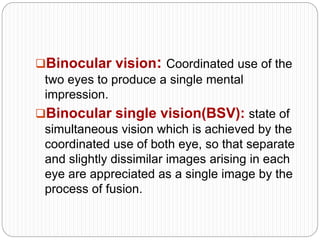
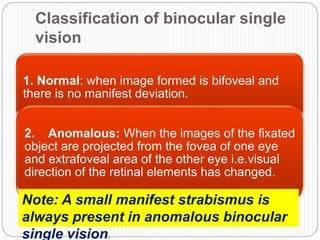
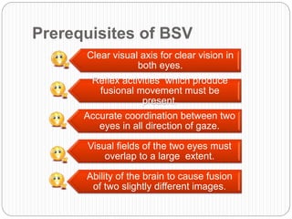
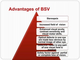
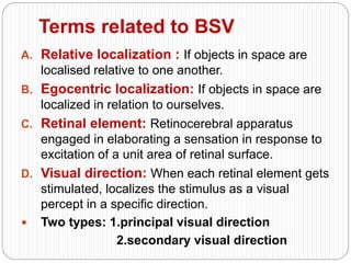
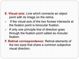
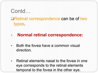
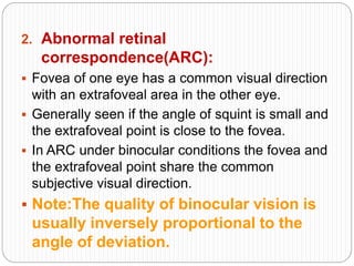
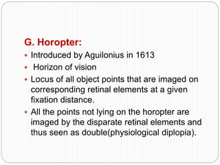
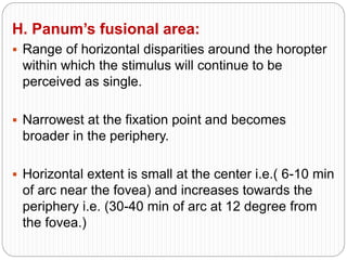
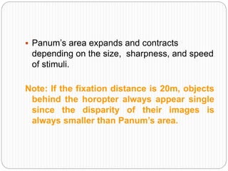
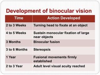
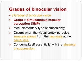
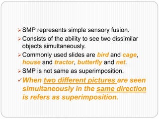
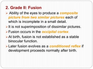
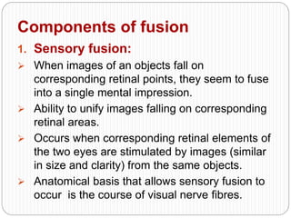
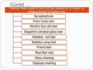
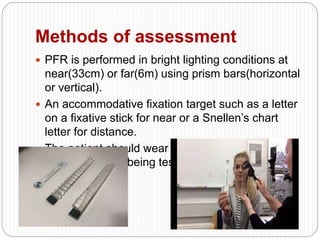
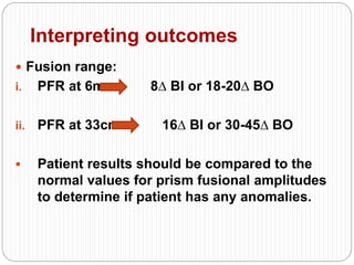
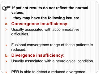
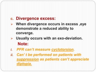
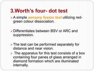
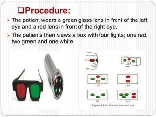
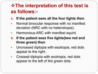
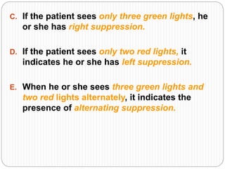
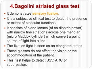
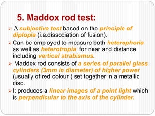
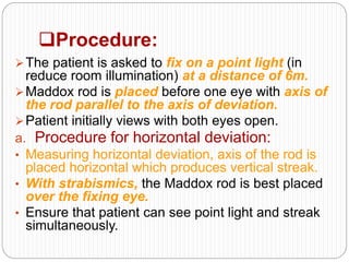
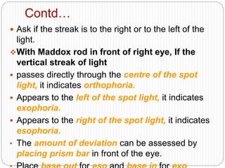
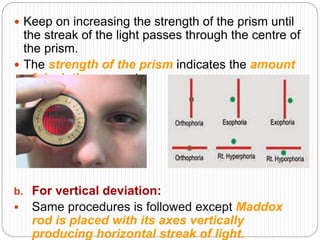
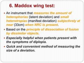
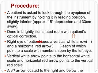
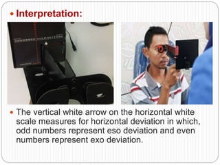
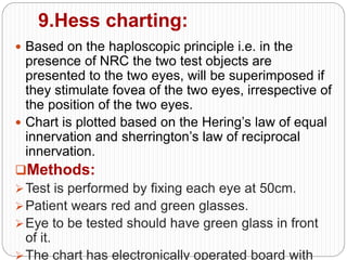
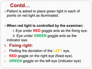
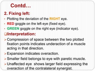
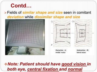
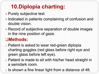
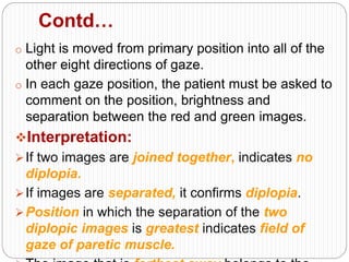
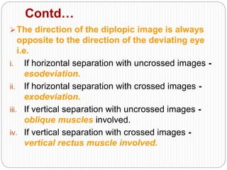
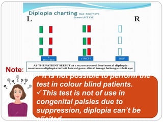
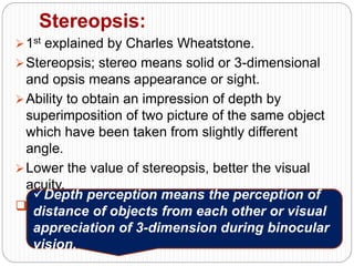
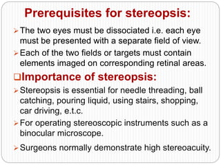
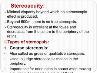
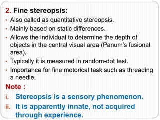
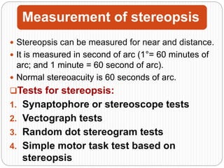
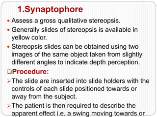
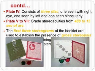
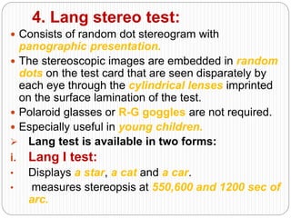
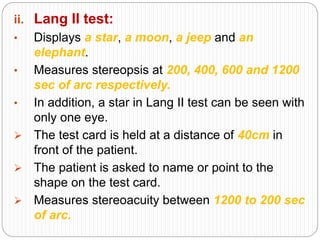
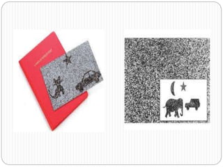
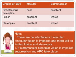
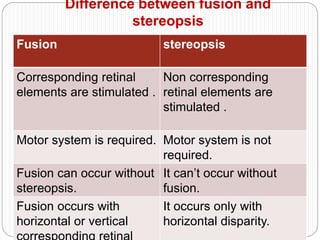
Measurement of fusion
and stereopsis
Binocular vision: Coordinated use of the
two eyes to produce a single mental
impression.
Binocular single vision(BSV): state of
simultaneous vision which is achieved by the
coordinated use of both eye, so that separate
and slightly dissimilar images arising in each
eye are appreciated as a single image by the
process of fusion.
Classification of binocular single
vision
1. Normal: when image formed is bifoveal and
there is no manifest deviation.
2. Anomalous: When the images of the fixated
object are projected from the fovea of one eye
and extrafoveal area of the other eye i.e.visual
direction of the retinal elements has changed.
Note: A small manifest strabismus is
always present in anomalous binocular
single vision.
Prerequisites of BSV
Clear visual axis for clear vision in
both eyes.
Reflex activities which produce
fusional movement must be
present.
Accurate coordination between two
eyes in all direction of gaze.
Visual fields of the two eyes must
overlap to a large extent.
Ability of the brain to cause fusion
of two slightly different images.
Advantages of BSV
Stereopsis
Increased field of vision
Enhanced visual acuity,
contrast sensitivity and
visual motor skills.
Optical defects in one eye
are made less obvious by
normal image of other eye.
Defective vision in any part
of one visual field is
masked.
Safety factor against partial
or complete loss of vision.
Terms related to BSV
A. Relative localization : If objects in space are
localised relative to one another.
B. Egocentric localization: If objects in space are
localized in relation to ourselves.
C. Retinal element: Retinocerebral apparatus
engaged in elaborating a sensation in response to
excitation of a unit area of retinal surface.
D. Visual direction: When each retinal element gets
stimulated, localizes the stimulus as a visual
percept in a specific direction.
 Two types: 1.principal visual direction
2.secondary visual direction
E. Visual axis: Line which connects an object
point with its image on the retina.
 If the visual axis of the two foveas intersects at
the fixation point is binocular fixation.
 If only one principle line of direction goes
through the fixation point called as mocular
fixation.
F. Retinal correspondence: Retinal elements of
the two eyes that share a common subjective
visual direction.
Contd…
Retinal correspondence can be of two
types.
1. Normal retinal correspondence:
 Both the fovea have a common visual
direction.
 Retinal elements nasal to the fovea in one
eye corresponds to the retinal elements
temporal to the fovea in the other eye.
2. Abnormal retinal
correspondence(ARC):
 Fovea of one eye has a common visual direction
with an extrafoveal area in the other eye.
 Generally seen if the angle of squint is small and
the extrafoveal point is close to the fovea.
 In ARC under binocular conditions the fovea and
the extrafoveal point share the common
subjective visual direction.
 Note:The quality of binocular vision is
usually inversely proportional to the
angle of deviation.
G. Horopter:
 Introduced by Aguilonius in 1613
 Horizon of vision
 Locus of all object points that are imaged on
corresponding retinal elements at a given
fixation distance.
 All the points not lying on the horopter are
imaged by the disparate retinal elements and
thus seen as double(physiological diplopia).
H. Panum’s fusional area:
 Range of horizontal disparities around the horopter
within which the stimulus will continue to be
perceived as single.
 Narrowest at the fixation point and becomes
broader in the periphery.
 Horizontal extent is small at the center i.e.( 6-10 min
of arc near the fovea) and increases towards the
periphery i.e. (30-40 min of arc at 12 degree from
the fovea.)
 Panum’s area expands and contracts
depending on the size, sharpness, and speed
of stimuli.
Note: If the fixation distance is 20m, objects
behind the horopter always appear single
since the disparity of their images is
always smaller than Panum’s area.
Development of binocular vision
Time Action Developed
2 to 3 Weeks Turning head to fixate at an object
4 to 5 Weeks Sustain monocular fixation of large
near objects
3 Months Binocular fusion
3 to 6 Months Stereopsis
1 Year Fusional movements firmly
established
2 to 3 Year Adult level visual acuity reached
Grades of binocular vision
 3 Grades of binocular vision
1. Grade I: Simultaneous macular
perception (SMP)
 Most elementary type of binocularity.
 Occurs when the visual cortex perceive
separate stimuli from the two eyes at the
same time.
 Concerns itself essentially with the absence
of suppression.
SMP represents simple sensory fusion.
Consists of the ability to see two dissimilar
objects simultaneously.
Commonly used slides are bird and cage,
house and tractor, butterfly and net.
SMP is not same as superimposition.
When two different pictures are seen
simultaneously in the same direction
is refers as superimposition.
2. Grade II: Fusion
 Ability of the eyes to produce a composite
picture from two similar pictures each of
which is incomplete in a small detail.
It is not superimposition of dissimilar pictures.
Fusion occurs in the occipital cortex.
At birth, fusion is not established as a stable
binocular function.
Later fusion evolves as a conditioned reflex if
development proceeds normally after birth.
Components of fusion
1. Sensory fusion:
 When images of an objects fall on
corresponding retinal points, they seem to fuse
into a single mental impression.
 Ability to unify images falling on corresponding
retinal areas.
 Occurs when corresponding retinal elements of
the two eyes are stimulated by images (similar
in size and clarity) from the same objects.
 Anatomical basis that allows sensory fusion to
occur is the course of visual nerve fibres.
2.Motor fusion:
 Vergence movement that causes similar retinal
images to fall and be maintained on corresponding
retinal areas.
Ability to align the eyes in such a manner that
sensory fusion can be maintained.
Stimulus for these fusional eye movements is
retinal disparity.
It is the exclusive function of the extra foveal retinal
periphery.
Contd…
Diplopia preventing mechanism.
Note:
I. The normal fusional range is 35-40 PD base
out and 16 PD base in on near reading.
II. 16 PD base out and 8 PD base in on distance
testing.
III. Fusion (whether sensory or motor) is always
a central process i.e. it takes place in the
visual cortex.
Measurement of fusion
 It is essential to assess the following
components
before any test is undertaken.
 Visual acuity
 Fixation in the squinting eye
 direction and size of deviation
Fusion assessment is essential both for the
prognosis and management of strabismus.
Fusion is essential for the restoration of BSV.
Contd….
Various tests used to find out the presence of fusion or
measurement of fusion are:
Synaptophore
Prism fusion test
Worth’s four dot test
Bagolini’s striated glass test
Maddox rod test
Maddox wing test
Friend test
Red filter test
Hess charting
Diplopia charting
1.Synaptophore
Also known as major amblyoscope.
In Britain known as synaptophore, but in
USA as troposkop.
 based on haploscopic principle (i.e. division
of physical space into two separate area of
visual space each of which is visible to one
eye only.)
Measures the angles of deviation.
It treats binocular vision anomalies by
conventional orthoptic method.
Test of fusion with
synaptophore
 Fusion can be tested using two similar
pictures each of which incomplete in one
small detail. E.g.
Two rabbits each lacking either a tail or a
bunch of flowers. If fusion is present, one
rabbit complete with tail and holding a bunch
of flower will be seen.
Ways to measure components of
fusion using synaptophore
1. Sensory fusion:
 One tube of synaptophore is locked
 patient is asked to create a composite image
 finally the position of sensory fusion is
achieved
2. Motor fusion:
 Lock the column at their real position
angle
 Then adduction knob is adjusted to
measure positive fusion
Contd…
And abduction knob is adjusted to measure
negative fusion
Then finally note the value when the image split
into two
Fusional exercise:
 Fusional exercises is given on synaptophore
with the fusion slide.
 Fusion range can be increased by gradually
converging both the tubes of synaptophore till
the fusion breaks.
 Exercises is given daily or on alternate days for
2.Prism fusion test
 known as prism fusion range or fusional
vergence amplitude test (objective test).
Assess motor fusion.
Specially measures the extent to which a
patient can maintain BSV in the presence of
increasing vergence demands.
Involves placing a prism bar in front of an eye.
A natural shift of the eye occurs in a patient
with BSV.
Contd…
When measuring horizontal fusion ranges,
base in prisms assess fusional divergence
while base out prisms assess fusional
covergence.
Vertical fusional vergence amplitude can also
be measured with base up and base down
prisms.
While performing the test, prism strength is
increased placing greater demand on the
vergence system eventually resulting in a
break point accompanying diplopia.
Break point, recovery and blur are key
aspects of this assessment.
Indications of use
This test is designed to assess the following:
 The limits of a patient’s BSV.
 Fusional convergence with a base out prism.
 Fusional divergence with a base in prism.
 Vertical amplitude with base up and base
down prisms.
 Positive and negative relative fusional
convergence by asking patient to report when
blur is appreciated.
 Progress of a patient undergoing
management for ocular misalignments.
Methods of assessment
 PFR is performed in bright lighting conditions at
near(33cm) or far(6m) using prism bars(horizontal
or vertical).
 An accommodative fixation target such as a letter
on a fixative stick for near or a Snellen’s chart
letter for distance.
 The patient should wear their refractive correction
for the distance being tested.
Interpreting outcomes
 Fusion range:
i. PFR at 6m 8∆ BI or 18-20∆ BO
ii. PFR at 33cm 16∆ BI or 30-45∆ BO
 Patient results should be compared to the
normal values for prism fusional amplitudes
to determine if patient has any anomalies.
 If patient results do not reflect the normal
values,
 they may have the following issues:
a. Convergence insufficiency:
 Usually associated with accommodative
difficulties.
 Fusional convergence range of these patients is
reduced.
b. Divergence insufficiency:
 Usually associated with a neurological condition.
 PFR is able to detect a reduced divergence
c. Divergence excess:
 When divergence occurs in excess ,eye
demonstrate a reduced ability to
converge.
 Usually occurs with an exo-deviation.
Note:
i. PFR can’t measure cyclotorsion.
ii. Can’ t be performed on patients with
suppression as patients can’t appreciate
diplopia.
3.Worth’s four- dot test
A simple sensory fusion test utilizing red-
green colour dissociation.
Differentiates between BSV or ARC and
suppression.
The test can be performed separately for
distance and near vision.
 The apparatus for this test consists of a box
containing four panes of glass arranged in
diamond formation which are illuminated
internally.
Procedure:
 The patient wears a green glass lens in front of the left
eye and a red lens in front of the right eye.
 The patients then views a box with four lights; one red,
two green and one white
The interpretation of this test is
as follows:-
A. If the patient sees all the four lights then
 Normal binocular response with no manifest
deviation (NRC with no heterotropia).
 Harmonious ARC with manifest squint.
B. If the patient sees five lights(two red and
three green) then
 Uncrossed diplopia with esotropia, red dots
appear to the right .
 Crossed diplopia with exotropia, red dots
appear to the left of the green dots.
C. If the patient sees only three green lights, he
or she has right suppression.
D. If the patient sees only two red lights, it
indicates he or she has left suppression.
E. When he or she sees three green lights and
two red lights alternately, it indicates the
presence of alternating suppression.
4.Bagolini striated glass test
 It demonstrates sensory fusion.
 It is a subjective clinical test to detect the presence
or extent of binocular functions.
 It consists of plano lenses (of no dioptric power)
with narrow fine striations across one meridian
(micro Maddox cylinder) which convert a point
source of light into a line.
 The fixation light is seen as an elongated streak.
 These glasses do not affect the vision or the
accommodation of the patient.
 This test helps to detect BSV, ARC or
suppression.
Procedure:
The two lenses are usually placed at 45°(OS) and
135°(OD) in front of each eye (over refractive
correction if any) and the patient fixates a small
light source.
Each eye perceives an oblique line of light,
perpendicular to that perceived by the fellow eye.
The interpretation of this test is
as follows:
I. If the two streaks intersect at their
centers in the form of an oblique cross
(X) then
 The patients has BSV if the eyes are
straight
 Harmonious ARC in the presence of
manifest strabismus.
II. If the two lines are seen but they form a
cross, then diplopia is present.
III. If only one streak is seen, then there is
no simultaneous perception but
suppression is present.
IV. If a small gap is seen in one of the streaks,
then a central suppression scotoma (as found
in microtropia) is present.
Apart from testing binocular
functions, Bagolini striated
glasses can measure
cyclotropia.
5. Maddox rod test:
 A subjective test based on the principle of
diplopia (i.e.dissociation of fusion).
Can be employed to measure both heterophoria
as well as heterotropia for near and distance
including vertical strabismus.
 Maddox rod consists of a series of parallel glass
cylinders (3mm in diameter) of higher power
(usually of red colour ) set together in a metallic
disc.
It produces a linear images of a point light which
is perpendicular to the axis of the cylinder.
Procedure:
The patient is asked to fix on a point light (in
reduce room illumination) at a distance of 6m.
Maddox rod is placed before one eye with axis of
the rod parallel to the axis of deviation.
Patient initially views with both eyes open.
a. Procedure for horizontal deviation:
• Measuring horizontal deviation, axis of the rod is
placed horizontal which produces vertical streak.
• With strabismics, the Maddox rod is best placed
over the fixing eye.
• Ensure that patient can see point light and streak
simultaneously.
Contd…
 Ask if the streak is to the right or to the left of the
light.
With Maddox rod in front of right eye, If the
vertical streak of light
 passes directly through the centre of the spot
light, it indicates orthophoria.
 Appears to the left of the spot light, it indicates
exophoria.
 Appears to the right of the spot light, it indicates
esophoria.
• The amount of deviation can be assessed by
placing prism bar in front of the eye.
 Keep on increasing the strength of the prism until
the streak of the light passes through the centre of
the prism.
 The strength of the prism indicates the amount
of deviation present.
b. For vertical deviation:
 Same procedures is followed except Maddox
rod is placed with its axes vertically
producing horizontal streak of light.
Contd…
If the horizontal streak of light is
 Above the spot light, it indicates the presence of
hypo deviation.
 Below the spot light, it indicates the presence of
hyper deviation.
Do you know:
1.Maddox rod test can’t be
performed,
if there is suppression under
testing
condition.
2.It is useful only to measure small
deviation.
6. Maddox wing test:
 An instrument that measures the amount of
heterophorias (latent deviation) and small
heterotropias (manifest deviation) subjectively at
near (33cm) when NRC is present.
 Based on the principle of dissociation of fusion
by dissimilar objects.
 Especially helpful when patients present with
the symptoms of diplopia.
 Quick and convenient method of measuring the
size of a deviation.
Procedure:
 A patient is asked to look through the eyepiece of
the instrument by holding it in reading position,
slightly inferior (approx. 15° depression and 33cm
away).
 Done in brightly illuminated room with patient’s
optical correction.
 Right eye of patient sees a vertical white arrow( )
and a horizontal red arrow( ),each of which
point to a scale with numbers seen by the left eye.
 Vertical white arrow points to the horizontal white
scale and horizontal red arrow points to the vertical
red scale.
 A 3rd arrow located to the right and below the
 Interpretation:
 The vertical white arrow on the horizontal white
scale measures for horizontal deviation in which,
odd numbers represent eso deviation and even
numbers represent exo deviation.
Contd…
 The horizontal red arrow on the vertical red scale
measures vertical deviation, in which odd numbers
represent right hyper deviations and even numbers
represent left hyper deviations.
 In the absence of a deviated eye, both red and
white arrows point to zero.
Note:
 Cyclophoria is measured by asking the patient to
align the horizontal red arrow with the horizontal
white scale.
i. If arrow record above zero then the reading of
scale indicates the amount of incyclophoria.
ii. If arrow record below zero then the reading of
7. FRIEND test:
 In this test, the letters F, I, N are written in green
and R, E, D in red.
 It is incorporated in the Snellen’s vision box.
 Demonstrates the sensory fusion test.
Procedure:
The patient is made to sit at a distance of 6m
after wearing diplopia goggles (red glass
before right eye and green glass before left
eye).
Patient is asked to read these letter on vision
box.
Interpretation:
If the patient read FRIEND at once, it indicates the
presence of binocular single vision.
If the patient read FIN only, he or she
has right suppression.
If the patient read RED only, he or
she has left suppression.
If the patient read FIN at one time and
RED at other time, it indicates the presence
of alternating suppression.
8.Red filter test:
Contains a series of red filters of increasing density.
Usually consists of gelatin fibers, beginning
with one layer and increasing to six or eight
layers.
The more the layers, darker the filter.
Used to detect the presence and type
of diplopia (crossed or uncrossed,
detect the depth of suppression and
determine retinal correspondence.
Procedure:
o A light is projected to the eyes of the patient.
Contd…
o The red filter is placed in front of patient’s one eye.
o He/she is asked to see at the light source.
 Interpretation:
 If patient report seeing one red circle, it indicates
the presence of suppression.
 If the patient see the red and white light
alternatively, he/she has alternative suppression.
 If the patient sees one red circle and one white
circle simultaneously, they are diplopic.
Contd…
The location of red circle in relation to the
white circle will tell the examiner, the type of
strabismus or diplopia.
i. In exotropia, when red filter is placed on right eye
of patient and he/she reports the red circle on the
left, this denotes crossed diplopia.
ii. In esotropia, the patient will report the red circle
to the right which is uncrossed diplopia.
iii. If the patient reports the red circle above white
circle, it denotes a hypotropia and if the red circle
is below the white circle, it is hypertropia.
9.Hess charting:
 Based on the haploscopic principle i.e. in the
presence of NRC the two test objects are
presented to the two eyes, will be superimposed if
they stimulate fovea of the two eyes, irrespective of
the position of the two eyes.
 Chart is plotted based on the Hering’s law of equal
innervation and sherrington’s law of reciprocal
innervation.
Methods:
Test is performed by fixing each eye at 50cm.
Patient wears red and green glasses.
Eye to be tested should have green glass in front
of it.
The chart has electronically operated board with
Contd…
Patient is asked to place green light in each of
points on red light as illuminated.
When red light is controlled by the examiner;
i. Eye under RED goggle acts as the fixing eye.
ii. Eye under GREEN goggle acts as the
indicator eye.
1. Fixing right:
o Plotting the deviation of the LEFT eye.
o RED goggle on the right eye (fixed eye).
o GREEN goggle on the left eye (indicator eye)
Contd…
2. Fixing left:
• Plotting the deviation of the RIGHT eye.
• RED goggle on the left eye (fixed eye).
• GREEN goggle on the right eye (indicator eye).
Interpretation:
Compression of space between the two plotted
fixation points indicates underaction of a muscle
acting in that direction.
Expansion indicates overaction.
Smaller field belongs to eye with paretic muscle.
Unaffected eye shows larger field expressing the
overaction of the contralateral synergist.
Contd…
Fields of similar shape and size seen in comitant
deviation while dissimilar shape and size
indicate incomitance.
Note: Patient should have good vision in
both eye, central fixation and normal
10.Diplopia charting:
Purely subjective test
Indicated in patients complaining of confusion and
double vision.
Record of subjective separation of double images
in the nine position of gaze.
Methods:
o Patient is asked to wear red-green diplopia
charting goggles (red glass before right eye and
green glass before left eye).
o Patient is made to sit with his/her head straight in
a semidark room.
o Is shown a fine linear light from a distance of 4ft.
Contd…
o Light is moved from primary position into all of the
other eight directions of gaze.
o In each gaze position, the patient must be asked to
comment on the position, brightness and
separation between the red and green images.
Interpretation:
If two images are joined together, indicates no
diplopia.
If images are separated, it confirms diplopia.
Position in which the separation of the two
diplopic images is greatest indicates field of
gaze of paretic muscle.
Contd…
The direction of the diplopic image is always
opposite to the direction of the deviating eye
i.e.
i. If horizontal separation with uncrossed images -
esodeviation.
ii. If horizontal separation with crossed images -
exodeviation.
iii. If vertical separation with uncrossed images -
oblique muscles involved.
iv. If vertical separation with crossed images -
vertical rectus muscle involved.
Note:
It is not possible to perform the
test in colour blind patients.
This test is not of use in
congenital palsies due to
suppression, diplopia can’t be
elicited.
Stereopsis:
1st explained by Charles Wheatstone.
Stereopsis; stereo means solid or 3-dimensional
and opsis means appearance or sight.
Ability to obtain an impression of depth by
superimposition of two picture of the same object
which have been taken from slightly different
angle.
Lower the value of stereopsis, better the visual
acuity.
Note:
Depth perception means the perception of
distance of objects from each other or visual
appreciation of 3-dimension during binocular
vision.
Prerequisites for stereopsis:
The two eyes must be dissociated i.e. each eye
must be presented with a separate field of view.
Each of the two fields or targets must contain
elements imaged on corresponding retinal areas.
Importance of stereopsis:
Stereopsis is essential for needle threading, ball
catching, pouring liquid, using stairs, shopping,
car driving, e.t.c.
For operating stereoscopic instruments such as a
binocular microscope.
Surgeons normally demonstrate high stereoacuity.
Stereoacuity:
Minimal disparity beyond which no stereoscopic
effect is produced.
Beyond 600m, there is no true stereopsis.
Stereoacuity is excellent at the fovea and
decreases from the centre to the periphery of the
retina.
Types of stereopsis:
1. Coarse stereopsis:
• Also called as gross or qualitative stereopsis.
• Used to judge stereoscopic motion in the
periphery.
• Importance for orientation in space while moving
2. Fine stereopsis:
• Also called as quantitative stereopsis.
• Mainly based on static differences.
• Allows the individual to determine the depth of
objects in the central visual area (Panum’s fusional
area).
• Typically it is measured in random-dot test.
• Importance for fine motorical task such as threading
a needle.
Note :
i. Stereopsis is a sensory phenomenon.
ii. It is apparently innate, not acquired
through experience.
Measurement of stereopsis
 Stereopsis can be measured for near and distance.
 It is measured in second of arc (1°= 60 minutes of
arc; and 1 minute = 60 second of arc).
 Normal stereoacuity is 60 seconds of arc.
Tests for stereopsis:
1. Synaptophore or stereoscope tests
2. Vectograph tests
3. Random dot stereogram tests
4. Simple motor task test based on
stereopsis
1.Synaptophore
 Assess a gross qualitative stereopsis.
 Generally slides of stereopsis is available in
yellow color.
 Stereopsis slides can be obtained using two
images of the same object taken from slightly
different angles to indicate depth perception.
Procedure:
The slide are inserted into slide holders with the
controls of each slide positioned towards or
away from the subject.
The patient is then required to describe the
apparent effect i.e. a swing moving towards or
contd...
 The test should be repeated with slide changing
position to ensure the stereoscopic effect.
2.Vectograph test (contour-stereo
test)
 Consists of polaroid material on which the two
targets are imprinted so that each target is
polarized at 90° with respect to the other.
 Vectograph dissociates the eyes optically.
 Each target is seen separately with the two eyes
by the use of properly oriented polaroid
spectacles.
A. Titmus stereo test:
 Utilizes the principle of vectograph (contour
test).
 most widely used clinical tests of stereopsis.
 Basically made up of two plates in the form of a
booklet.
The Titmus stereo test consists of three parts.
a. The fly test
b. The animal test
c. The circle test
a. The fly test:
 The right side of the test booklet contains a large
housefly to test gross stereopsis (threshold
3000 sec of arc).
 Especially useful in young children.
 The patient is asked to pick up one of the wings
of the fly.
 If the patient sees stereoscopically, he /she will
reach above the plate.
 In the absence of gross stereopsis, the fly will
appear as an ordinary flat photograph.
b. The animal test:
 It is also performed to check the gross
stereopsis.
 Consists of 3 rows of five animals each.
 One animal from each row is imaged disparately
(threshold 100, 200 and 400 sec of arc
respectively).
 In each row , one of the animals correspondingly
imaged in the two eyes is printed heavily black
(serves as misleading clue).
 The patient is asked which one of the animals
 A patient without stereopsis will name the animal
printed heavily (misleading clues).
 While in the presence of stereopsis he/she will
name the disparately imaged animal.
c. The circle test:
 Consists of nine squares, each containing four
circles arranged in the form of a lozenge.
 Only one of the circles in each square is
disparately imaged at random (threshold ranging
from 800 to 40 sec of arc).
 The patient is asked to ‘push-down’ the circle
that stands out, beginning the first set.
Contd…
 When he/she makes mistake or finds no circle to
push down, the limit of his/her stereopsis is
presumably reached.
Do you know:
i.Circle no.5,equivalent to 100 sec of arc is
considered to be lowest limit of fine central
stereoacuity and is designed as the lowest limit of
good stereoacuity.
ii. Presence of good stereoacuity on Titmus stereo
test is indicative of good visual acuity.
iii.This test often is unreliable in differentiating
patients with amblyopia and heterotropia.
B. The Bernell stereo reindeer test
 It resembles Titmus stereo test.
 A polarized test providing stimuli for both gross
and fine stereopsis testing.
 The test has the advantage that the examiner can
make the reindeer’s nose wiggle by rotating the
picture slightly.
3.Random dot stereogram
tests
 These tests are devoid of monocular cues and the
patients can’t guess what the stereo figure is and
where it is located on the test plate.
 Provides truer measurement of stereopsis than
Titmus stereo test.
A. Random dot E-test:
 This test consists of three cards to be viewed with
polaroid spectacles.
 One card is a bas relief model of the stereotest
figure.
 The 2nd card contains the ‘E’ stereo figure with a
random dot background.
 The 3rd card is a stereoblank with an identical
random dot background.
Contd…
This test is performed after showing the bas relief
model and two cards are held 50cm in front of the
patient.
Then patient is asked to indicate which card
contains the letter ‘E’.
The patient gives a ‘pass’ or ‘fail’ response.
B. TNO random dot test:
This test is graded to provide retinal disparities
ranging from 480 to 15 sec of arc.
 It is based on the same principle as ‘Random dot
E-test’
Consists of a booklet containing 7 plates.
Each test plate consists of a stereogram in which
various shapes (square, dots, crosses) have been
created by random dots in complementary
colours.
The plates contains two types of figures,
i. One which can be perceived when viewed
binocularly with red green spectacles by normal
ii. The second set of figures can be seen with and
without spectacles even in the absence of
stereopsis.
Procedure:
The patient is asked to wear the red- green
goggles.
The patient is made to hold the booklet at about
40cm keeping the room light on.
 Plate I: consists of two butterflies; one is easily
seen monocularly and the other is only seen in
stereopsis.
 Plate II: consists of four discs; two are seen
without stereopsis and two are seen in stereopsis.
 Plate III: consists of hidden shapes (circle, square,
contd…
 Plate IV: Consists of three disc; one seen with right
eye, one seen by left and one seen binocularly.
 Plate V to VII: Grade stereoacuities from 480 to 15
sec of arc.
The first three stereograms of the booklet are
used to establish the presence of gross stereopsis
quickly, while the remaining four plates allow to
quantitate the level of stereopsis.
4. Lang stereo test:
 Consists of random dot stereogram with
panographic presentation.
 The stereoscopic images are embedded in random
dots on the test card that are seen disparately by
each eye through the cylindrical lenses imprinted
on the surface lamination of the test.
 Polaroid glasses or R-G goggles are not required.
 Especially useful in young children.
 Lang test is available in two forms:
i. Lang I test:
• Displays a star, a cat and a car.
• measures stereopsis at 550,600 and 1200 sec of
arc.
ii. Lang II test:
• Displays a star, a moon, a jeep and an
elephant.
• Measures stereopsis at 200, 400, 600 and 1200
sec of arc respectively.
• In addition, a star in Lang II test can be seen with
only one eye.
 The test card is held at a distance of 40cm in
front of the patient.
 The patient is asked to name or point to the
shape on the test card.
 Measures stereoacuity between 1200 to 200 sec
of arc.
4.Frisby test:
Consists of three plastic cards of varying thickness
(6mm, 3mm and 1.5mm respectively) each
containing four squares of small random shapes.
one of the squares in each plate contains a hidden
circle which is seen disparately.
The disparity is created by displacement of random
shapes (thickness of the plate).
This test doesn’t require use of glasses.
It is especially useful in young children.
Measures stereo acuity between 600 to 15 sec of
arc.
5. Stereoscopic contours induced optokinetic
nystagmus test and television random dot
stereo test have recently been suggested to test
stereopsis in infants.
IV. Simple motor task test based on
stereopsis
i. Two pencil test:
 It is simple primitive but an effective test for
detecting the presence or absence of gross
stereopsis (threshold value 3000 to 5000 sec of
arc).
 It was popularized by Lang in 1975A.D.
 Procedure:
 Examiner holds a pencil vertically in front of the
patient, who is asked to touch its upper tip of the
 Patient having stereopsis passes the test with both
eyes open.
 If stereopsis is absent, patients fail the test with one
eye closed or when both eyes are open.
Grades of BSV Macular Extramacular
Simultaneous
perception
none excellent
Fusion excellent limited
Stereopsis excellent limited
Note:
i. There are no adaptations if macular
binocular fusion is impaired and there will be
limited fusion and stereopsis.
ii. If extramacular binocular vision is impaired
suppression and ARC take place.
Difference between fusion and
stereopsis
Fusion stereopsis
Corresponding retinal
elements are stimulated .
Non corresponding
retinal elements are
stimulated .
Motor system is required. Motor system is not
required.
Fusion can occur without
stereopsis.
It can’t occur without
fusion.
Fusion occurs with
horizontal or vertical
It occurs only with
horizontal disparity.
References:
Fusion

Fusion

  • 1.
  • 2.
    Binocular vision: Coordinateduse of the two eyes to produce a single mental impression. Binocular single vision(BSV): state of simultaneous vision which is achieved by the coordinated use of both eye, so that separate and slightly dissimilar images arising in each eye are appreciated as a single image by the process of fusion.
  • 3.
    Classification of binocularsingle vision 1. Normal: when image formed is bifoveal and there is no manifest deviation. 2. Anomalous: When the images of the fixated object are projected from the fovea of one eye and extrafoveal area of the other eye i.e.visual direction of the retinal elements has changed. Note: A small manifest strabismus is always present in anomalous binocular single vision.
  • 4.
    Prerequisites of BSV Clearvisual axis for clear vision in both eyes. Reflex activities which produce fusional movement must be present. Accurate coordination between two eyes in all direction of gaze. Visual fields of the two eyes must overlap to a large extent. Ability of the brain to cause fusion of two slightly different images.
  • 5.
    Advantages of BSV Stereopsis Increasedfield of vision Enhanced visual acuity, contrast sensitivity and visual motor skills. Optical defects in one eye are made less obvious by normal image of other eye. Defective vision in any part of one visual field is masked. Safety factor against partial or complete loss of vision.
  • 6.
    Terms related toBSV A. Relative localization : If objects in space are localised relative to one another. B. Egocentric localization: If objects in space are localized in relation to ourselves. C. Retinal element: Retinocerebral apparatus engaged in elaborating a sensation in response to excitation of a unit area of retinal surface. D. Visual direction: When each retinal element gets stimulated, localizes the stimulus as a visual percept in a specific direction.  Two types: 1.principal visual direction 2.secondary visual direction
  • 7.
    E. Visual axis:Line which connects an object point with its image on the retina.  If the visual axis of the two foveas intersects at the fixation point is binocular fixation.  If only one principle line of direction goes through the fixation point called as mocular fixation. F. Retinal correspondence: Retinal elements of the two eyes that share a common subjective visual direction.
  • 8.
    Contd… Retinal correspondence canbe of two types. 1. Normal retinal correspondence:  Both the fovea have a common visual direction.  Retinal elements nasal to the fovea in one eye corresponds to the retinal elements temporal to the fovea in the other eye.
  • 9.
    2. Abnormal retinal correspondence(ARC): Fovea of one eye has a common visual direction with an extrafoveal area in the other eye.  Generally seen if the angle of squint is small and the extrafoveal point is close to the fovea.  In ARC under binocular conditions the fovea and the extrafoveal point share the common subjective visual direction.  Note:The quality of binocular vision is usually inversely proportional to the angle of deviation.
  • 10.
    G. Horopter:  Introducedby Aguilonius in 1613  Horizon of vision  Locus of all object points that are imaged on corresponding retinal elements at a given fixation distance.  All the points not lying on the horopter are imaged by the disparate retinal elements and thus seen as double(physiological diplopia).
  • 11.
    H. Panum’s fusionalarea:  Range of horizontal disparities around the horopter within which the stimulus will continue to be perceived as single.  Narrowest at the fixation point and becomes broader in the periphery.  Horizontal extent is small at the center i.e.( 6-10 min of arc near the fovea) and increases towards the periphery i.e. (30-40 min of arc at 12 degree from the fovea.)
  • 12.
     Panum’s areaexpands and contracts depending on the size, sharpness, and speed of stimuli. Note: If the fixation distance is 20m, objects behind the horopter always appear single since the disparity of their images is always smaller than Panum’s area.
  • 13.
    Development of binocularvision Time Action Developed 2 to 3 Weeks Turning head to fixate at an object 4 to 5 Weeks Sustain monocular fixation of large near objects 3 Months Binocular fusion 3 to 6 Months Stereopsis 1 Year Fusional movements firmly established 2 to 3 Year Adult level visual acuity reached
  • 14.
    Grades of binocularvision  3 Grades of binocular vision 1. Grade I: Simultaneous macular perception (SMP)  Most elementary type of binocularity.  Occurs when the visual cortex perceive separate stimuli from the two eyes at the same time.  Concerns itself essentially with the absence of suppression.
  • 15.
    SMP represents simplesensory fusion. Consists of the ability to see two dissimilar objects simultaneously. Commonly used slides are bird and cage, house and tractor, butterfly and net. SMP is not same as superimposition. When two different pictures are seen simultaneously in the same direction is refers as superimposition.
  • 16.
    2. Grade II:Fusion  Ability of the eyes to produce a composite picture from two similar pictures each of which is incomplete in a small detail. It is not superimposition of dissimilar pictures. Fusion occurs in the occipital cortex. At birth, fusion is not established as a stable binocular function. Later fusion evolves as a conditioned reflex if development proceeds normally after birth.
  • 17.
    Components of fusion 1.Sensory fusion:  When images of an objects fall on corresponding retinal points, they seem to fuse into a single mental impression.  Ability to unify images falling on corresponding retinal areas.  Occurs when corresponding retinal elements of the two eyes are stimulated by images (similar in size and clarity) from the same objects.  Anatomical basis that allows sensory fusion to occur is the course of visual nerve fibres.
  • 18.
    2.Motor fusion:  Vergencemovement that causes similar retinal images to fall and be maintained on corresponding retinal areas. Ability to align the eyes in such a manner that sensory fusion can be maintained. Stimulus for these fusional eye movements is retinal disparity. It is the exclusive function of the extra foveal retinal periphery.
  • 19.
    Contd… Diplopia preventing mechanism. Note: I.The normal fusional range is 35-40 PD base out and 16 PD base in on near reading. II. 16 PD base out and 8 PD base in on distance testing. III. Fusion (whether sensory or motor) is always a central process i.e. it takes place in the visual cortex.
  • 20.
    Measurement of fusion It is essential to assess the following components before any test is undertaken.  Visual acuity  Fixation in the squinting eye  direction and size of deviation Fusion assessment is essential both for the prognosis and management of strabismus. Fusion is essential for the restoration of BSV.
  • 21.
    Contd…. Various tests usedto find out the presence of fusion or measurement of fusion are: Synaptophore Prism fusion test Worth’s four dot test Bagolini’s striated glass test Maddox rod test Maddox wing test Friend test Red filter test Hess charting Diplopia charting
  • 22.
    1.Synaptophore Also known asmajor amblyoscope. In Britain known as synaptophore, but in USA as troposkop.  based on haploscopic principle (i.e. division of physical space into two separate area of visual space each of which is visible to one eye only.) Measures the angles of deviation. It treats binocular vision anomalies by conventional orthoptic method.
  • 23.
    Test of fusionwith synaptophore  Fusion can be tested using two similar pictures each of which incomplete in one small detail. E.g. Two rabbits each lacking either a tail or a bunch of flowers. If fusion is present, one rabbit complete with tail and holding a bunch of flower will be seen.
  • 24.
    Ways to measurecomponents of fusion using synaptophore 1. Sensory fusion:  One tube of synaptophore is locked  patient is asked to create a composite image  finally the position of sensory fusion is achieved 2. Motor fusion:  Lock the column at their real position angle  Then adduction knob is adjusted to measure positive fusion
  • 25.
    Contd… And abduction knobis adjusted to measure negative fusion Then finally note the value when the image split into two Fusional exercise:  Fusional exercises is given on synaptophore with the fusion slide.  Fusion range can be increased by gradually converging both the tubes of synaptophore till the fusion breaks.  Exercises is given daily or on alternate days for
  • 26.
    2.Prism fusion test known as prism fusion range or fusional vergence amplitude test (objective test). Assess motor fusion. Specially measures the extent to which a patient can maintain BSV in the presence of increasing vergence demands. Involves placing a prism bar in front of an eye. A natural shift of the eye occurs in a patient with BSV.
  • 27.
    Contd… When measuring horizontalfusion ranges, base in prisms assess fusional divergence while base out prisms assess fusional covergence. Vertical fusional vergence amplitude can also be measured with base up and base down prisms. While performing the test, prism strength is increased placing greater demand on the vergence system eventually resulting in a break point accompanying diplopia. Break point, recovery and blur are key aspects of this assessment.
  • 28.
    Indications of use Thistest is designed to assess the following:  The limits of a patient’s BSV.  Fusional convergence with a base out prism.  Fusional divergence with a base in prism.  Vertical amplitude with base up and base down prisms.  Positive and negative relative fusional convergence by asking patient to report when blur is appreciated.  Progress of a patient undergoing management for ocular misalignments.
  • 29.
    Methods of assessment PFR is performed in bright lighting conditions at near(33cm) or far(6m) using prism bars(horizontal or vertical).  An accommodative fixation target such as a letter on a fixative stick for near or a Snellen’s chart letter for distance.  The patient should wear their refractive correction for the distance being tested.
  • 30.
    Interpreting outcomes  Fusionrange: i. PFR at 6m 8∆ BI or 18-20∆ BO ii. PFR at 33cm 16∆ BI or 30-45∆ BO  Patient results should be compared to the normal values for prism fusional amplitudes to determine if patient has any anomalies.
  • 31.
     If patientresults do not reflect the normal values,  they may have the following issues: a. Convergence insufficiency:  Usually associated with accommodative difficulties.  Fusional convergence range of these patients is reduced. b. Divergence insufficiency:  Usually associated with a neurological condition.  PFR is able to detect a reduced divergence
  • 32.
    c. Divergence excess: When divergence occurs in excess ,eye demonstrate a reduced ability to converge.  Usually occurs with an exo-deviation. Note: i. PFR can’t measure cyclotorsion. ii. Can’ t be performed on patients with suppression as patients can’t appreciate diplopia.
  • 33.
    3.Worth’s four- dottest A simple sensory fusion test utilizing red- green colour dissociation. Differentiates between BSV or ARC and suppression. The test can be performed separately for distance and near vision.  The apparatus for this test consists of a box containing four panes of glass arranged in diamond formation which are illuminated internally.
  • 34.
    Procedure:  The patientwears a green glass lens in front of the left eye and a red lens in front of the right eye.  The patients then views a box with four lights; one red, two green and one white
  • 35.
    The interpretation ofthis test is as follows:- A. If the patient sees all the four lights then  Normal binocular response with no manifest deviation (NRC with no heterotropia).  Harmonious ARC with manifest squint. B. If the patient sees five lights(two red and three green) then  Uncrossed diplopia with esotropia, red dots appear to the right .  Crossed diplopia with exotropia, red dots appear to the left of the green dots.
  • 36.
    C. If thepatient sees only three green lights, he or she has right suppression. D. If the patient sees only two red lights, it indicates he or she has left suppression. E. When he or she sees three green lights and two red lights alternately, it indicates the presence of alternating suppression.
  • 37.
    4.Bagolini striated glasstest  It demonstrates sensory fusion.  It is a subjective clinical test to detect the presence or extent of binocular functions.  It consists of plano lenses (of no dioptric power) with narrow fine striations across one meridian (micro Maddox cylinder) which convert a point source of light into a line.  The fixation light is seen as an elongated streak.  These glasses do not affect the vision or the accommodation of the patient.  This test helps to detect BSV, ARC or suppression.
  • 38.
    Procedure: The two lensesare usually placed at 45°(OS) and 135°(OD) in front of each eye (over refractive correction if any) and the patient fixates a small light source. Each eye perceives an oblique line of light, perpendicular to that perceived by the fellow eye.
  • 39.
    The interpretation ofthis test is as follows: I. If the two streaks intersect at their centers in the form of an oblique cross (X) then  The patients has BSV if the eyes are straight  Harmonious ARC in the presence of manifest strabismus. II. If the two lines are seen but they form a cross, then diplopia is present. III. If only one streak is seen, then there is no simultaneous perception but suppression is present.
  • 40.
    IV. If asmall gap is seen in one of the streaks, then a central suppression scotoma (as found in microtropia) is present.
  • 41.
    Apart from testingbinocular functions, Bagolini striated glasses can measure cyclotropia.
  • 42.
    5. Maddox rodtest:  A subjective test based on the principle of diplopia (i.e.dissociation of fusion). Can be employed to measure both heterophoria as well as heterotropia for near and distance including vertical strabismus.  Maddox rod consists of a series of parallel glass cylinders (3mm in diameter) of higher power (usually of red colour ) set together in a metallic disc. It produces a linear images of a point light which is perpendicular to the axis of the cylinder.
  • 43.
    Procedure: The patient isasked to fix on a point light (in reduce room illumination) at a distance of 6m. Maddox rod is placed before one eye with axis of the rod parallel to the axis of deviation. Patient initially views with both eyes open. a. Procedure for horizontal deviation: • Measuring horizontal deviation, axis of the rod is placed horizontal which produces vertical streak. • With strabismics, the Maddox rod is best placed over the fixing eye. • Ensure that patient can see point light and streak simultaneously.
  • 44.
    Contd…  Ask ifthe streak is to the right or to the left of the light. With Maddox rod in front of right eye, If the vertical streak of light  passes directly through the centre of the spot light, it indicates orthophoria.  Appears to the left of the spot light, it indicates exophoria.  Appears to the right of the spot light, it indicates esophoria. • The amount of deviation can be assessed by placing prism bar in front of the eye.
  • 45.
     Keep onincreasing the strength of the prism until the streak of the light passes through the centre of the prism.  The strength of the prism indicates the amount of deviation present. b. For vertical deviation:  Same procedures is followed except Maddox rod is placed with its axes vertically producing horizontal streak of light.
  • 46.
    Contd… If the horizontalstreak of light is  Above the spot light, it indicates the presence of hypo deviation.  Below the spot light, it indicates the presence of hyper deviation. Do you know: 1.Maddox rod test can’t be performed, if there is suppression under testing condition. 2.It is useful only to measure small deviation.
  • 47.
    6. Maddox wingtest:  An instrument that measures the amount of heterophorias (latent deviation) and small heterotropias (manifest deviation) subjectively at near (33cm) when NRC is present.  Based on the principle of dissociation of fusion by dissimilar objects.  Especially helpful when patients present with the symptoms of diplopia.  Quick and convenient method of measuring the size of a deviation.
  • 48.
    Procedure:  A patientis asked to look through the eyepiece of the instrument by holding it in reading position, slightly inferior (approx. 15° depression and 33cm away).  Done in brightly illuminated room with patient’s optical correction.  Right eye of patient sees a vertical white arrow( ) and a horizontal red arrow( ),each of which point to a scale with numbers seen by the left eye.  Vertical white arrow points to the horizontal white scale and horizontal red arrow points to the vertical red scale.  A 3rd arrow located to the right and below the
  • 49.
     Interpretation:  Thevertical white arrow on the horizontal white scale measures for horizontal deviation in which, odd numbers represent eso deviation and even numbers represent exo deviation.
  • 50.
    Contd…  The horizontalred arrow on the vertical red scale measures vertical deviation, in which odd numbers represent right hyper deviations and even numbers represent left hyper deviations.  In the absence of a deviated eye, both red and white arrows point to zero. Note:  Cyclophoria is measured by asking the patient to align the horizontal red arrow with the horizontal white scale. i. If arrow record above zero then the reading of scale indicates the amount of incyclophoria. ii. If arrow record below zero then the reading of
  • 51.
    7. FRIEND test: In this test, the letters F, I, N are written in green and R, E, D in red.  It is incorporated in the Snellen’s vision box.  Demonstrates the sensory fusion test. Procedure: The patient is made to sit at a distance of 6m after wearing diplopia goggles (red glass before right eye and green glass before left eye). Patient is asked to read these letter on vision box.
  • 52.
    Interpretation: If the patientread FRIEND at once, it indicates the presence of binocular single vision. If the patient read FIN only, he or she has right suppression. If the patient read RED only, he or she has left suppression. If the patient read FIN at one time and RED at other time, it indicates the presence of alternating suppression.
  • 53.
    8.Red filter test: Containsa series of red filters of increasing density. Usually consists of gelatin fibers, beginning with one layer and increasing to six or eight layers. The more the layers, darker the filter. Used to detect the presence and type of diplopia (crossed or uncrossed, detect the depth of suppression and determine retinal correspondence. Procedure: o A light is projected to the eyes of the patient.
  • 54.
    Contd… o The redfilter is placed in front of patient’s one eye. o He/she is asked to see at the light source.  Interpretation:  If patient report seeing one red circle, it indicates the presence of suppression.  If the patient see the red and white light alternatively, he/she has alternative suppression.  If the patient sees one red circle and one white circle simultaneously, they are diplopic.
  • 55.
    Contd… The location ofred circle in relation to the white circle will tell the examiner, the type of strabismus or diplopia. i. In exotropia, when red filter is placed on right eye of patient and he/she reports the red circle on the left, this denotes crossed diplopia. ii. In esotropia, the patient will report the red circle to the right which is uncrossed diplopia. iii. If the patient reports the red circle above white circle, it denotes a hypotropia and if the red circle is below the white circle, it is hypertropia.
  • 56.
    9.Hess charting:  Basedon the haploscopic principle i.e. in the presence of NRC the two test objects are presented to the two eyes, will be superimposed if they stimulate fovea of the two eyes, irrespective of the position of the two eyes.  Chart is plotted based on the Hering’s law of equal innervation and sherrington’s law of reciprocal innervation. Methods: Test is performed by fixing each eye at 50cm. Patient wears red and green glasses. Eye to be tested should have green glass in front of it. The chart has electronically operated board with
  • 57.
    Contd… Patient is askedto place green light in each of points on red light as illuminated. When red light is controlled by the examiner; i. Eye under RED goggle acts as the fixing eye. ii. Eye under GREEN goggle acts as the indicator eye. 1. Fixing right: o Plotting the deviation of the LEFT eye. o RED goggle on the right eye (fixed eye). o GREEN goggle on the left eye (indicator eye)
  • 58.
    Contd… 2. Fixing left: •Plotting the deviation of the RIGHT eye. • RED goggle on the left eye (fixed eye). • GREEN goggle on the right eye (indicator eye). Interpretation: Compression of space between the two plotted fixation points indicates underaction of a muscle acting in that direction. Expansion indicates overaction. Smaller field belongs to eye with paretic muscle. Unaffected eye shows larger field expressing the overaction of the contralateral synergist.
  • 59.
    Contd… Fields of similarshape and size seen in comitant deviation while dissimilar shape and size indicate incomitance. Note: Patient should have good vision in both eye, central fixation and normal
  • 60.
    10.Diplopia charting: Purely subjectivetest Indicated in patients complaining of confusion and double vision. Record of subjective separation of double images in the nine position of gaze. Methods: o Patient is asked to wear red-green diplopia charting goggles (red glass before right eye and green glass before left eye). o Patient is made to sit with his/her head straight in a semidark room. o Is shown a fine linear light from a distance of 4ft.
  • 61.
    Contd… o Light ismoved from primary position into all of the other eight directions of gaze. o In each gaze position, the patient must be asked to comment on the position, brightness and separation between the red and green images. Interpretation: If two images are joined together, indicates no diplopia. If images are separated, it confirms diplopia. Position in which the separation of the two diplopic images is greatest indicates field of gaze of paretic muscle.
  • 62.
    Contd… The direction ofthe diplopic image is always opposite to the direction of the deviating eye i.e. i. If horizontal separation with uncrossed images - esodeviation. ii. If horizontal separation with crossed images - exodeviation. iii. If vertical separation with uncrossed images - oblique muscles involved. iv. If vertical separation with crossed images - vertical rectus muscle involved.
  • 63.
    Note: It is notpossible to perform the test in colour blind patients. This test is not of use in congenital palsies due to suppression, diplopia can’t be elicited.
  • 64.
    Stereopsis: 1st explained byCharles Wheatstone. Stereopsis; stereo means solid or 3-dimensional and opsis means appearance or sight. Ability to obtain an impression of depth by superimposition of two picture of the same object which have been taken from slightly different angle. Lower the value of stereopsis, better the visual acuity. Note: Depth perception means the perception of distance of objects from each other or visual appreciation of 3-dimension during binocular vision.
  • 65.
    Prerequisites for stereopsis: Thetwo eyes must be dissociated i.e. each eye must be presented with a separate field of view. Each of the two fields or targets must contain elements imaged on corresponding retinal areas. Importance of stereopsis: Stereopsis is essential for needle threading, ball catching, pouring liquid, using stairs, shopping, car driving, e.t.c. For operating stereoscopic instruments such as a binocular microscope. Surgeons normally demonstrate high stereoacuity.
  • 66.
    Stereoacuity: Minimal disparity beyondwhich no stereoscopic effect is produced. Beyond 600m, there is no true stereopsis. Stereoacuity is excellent at the fovea and decreases from the centre to the periphery of the retina. Types of stereopsis: 1. Coarse stereopsis: • Also called as gross or qualitative stereopsis. • Used to judge stereoscopic motion in the periphery. • Importance for orientation in space while moving
  • 67.
    2. Fine stereopsis: •Also called as quantitative stereopsis. • Mainly based on static differences. • Allows the individual to determine the depth of objects in the central visual area (Panum’s fusional area). • Typically it is measured in random-dot test. • Importance for fine motorical task such as threading a needle. Note : i. Stereopsis is a sensory phenomenon. ii. It is apparently innate, not acquired through experience.
  • 68.
    Measurement of stereopsis Stereopsis can be measured for near and distance.  It is measured in second of arc (1°= 60 minutes of arc; and 1 minute = 60 second of arc).  Normal stereoacuity is 60 seconds of arc. Tests for stereopsis: 1. Synaptophore or stereoscope tests 2. Vectograph tests 3. Random dot stereogram tests 4. Simple motor task test based on stereopsis
  • 69.
    1.Synaptophore  Assess agross qualitative stereopsis.  Generally slides of stereopsis is available in yellow color.  Stereopsis slides can be obtained using two images of the same object taken from slightly different angles to indicate depth perception. Procedure: The slide are inserted into slide holders with the controls of each slide positioned towards or away from the subject. The patient is then required to describe the apparent effect i.e. a swing moving towards or
  • 70.
    contd...  The testshould be repeated with slide changing position to ensure the stereoscopic effect.
  • 71.
    2.Vectograph test (contour-stereo test) Consists of polaroid material on which the two targets are imprinted so that each target is polarized at 90° with respect to the other.  Vectograph dissociates the eyes optically.  Each target is seen separately with the two eyes by the use of properly oriented polaroid spectacles. A. Titmus stereo test:  Utilizes the principle of vectograph (contour test).  most widely used clinical tests of stereopsis.  Basically made up of two plates in the form of a booklet.
  • 72.
    The Titmus stereotest consists of three parts. a. The fly test b. The animal test c. The circle test a. The fly test:  The right side of the test booklet contains a large housefly to test gross stereopsis (threshold 3000 sec of arc).  Especially useful in young children.  The patient is asked to pick up one of the wings of the fly.  If the patient sees stereoscopically, he /she will reach above the plate.  In the absence of gross stereopsis, the fly will appear as an ordinary flat photograph.
  • 74.
    b. The animaltest:  It is also performed to check the gross stereopsis.  Consists of 3 rows of five animals each.  One animal from each row is imaged disparately (threshold 100, 200 and 400 sec of arc respectively).  In each row , one of the animals correspondingly imaged in the two eyes is printed heavily black (serves as misleading clue).  The patient is asked which one of the animals
  • 75.
     A patientwithout stereopsis will name the animal printed heavily (misleading clues).  While in the presence of stereopsis he/she will name the disparately imaged animal. c. The circle test:  Consists of nine squares, each containing four circles arranged in the form of a lozenge.  Only one of the circles in each square is disparately imaged at random (threshold ranging from 800 to 40 sec of arc).  The patient is asked to ‘push-down’ the circle that stands out, beginning the first set.
  • 76.
    Contd…  When he/shemakes mistake or finds no circle to push down, the limit of his/her stereopsis is presumably reached. Do you know: i.Circle no.5,equivalent to 100 sec of arc is considered to be lowest limit of fine central stereoacuity and is designed as the lowest limit of good stereoacuity. ii. Presence of good stereoacuity on Titmus stereo test is indicative of good visual acuity. iii.This test often is unreliable in differentiating patients with amblyopia and heterotropia.
  • 77.
    B. The Bernellstereo reindeer test  It resembles Titmus stereo test.  A polarized test providing stimuli for both gross and fine stereopsis testing.  The test has the advantage that the examiner can make the reindeer’s nose wiggle by rotating the picture slightly.
  • 78.
    3.Random dot stereogram tests These tests are devoid of monocular cues and the patients can’t guess what the stereo figure is and where it is located on the test plate.  Provides truer measurement of stereopsis than Titmus stereo test. A. Random dot E-test:  This test consists of three cards to be viewed with polaroid spectacles.  One card is a bas relief model of the stereotest figure.  The 2nd card contains the ‘E’ stereo figure with a random dot background.  The 3rd card is a stereoblank with an identical random dot background.
  • 79.
    Contd… This test isperformed after showing the bas relief model and two cards are held 50cm in front of the patient. Then patient is asked to indicate which card contains the letter ‘E’. The patient gives a ‘pass’ or ‘fail’ response.
  • 80.
    B. TNO randomdot test: This test is graded to provide retinal disparities ranging from 480 to 15 sec of arc.  It is based on the same principle as ‘Random dot E-test’ Consists of a booklet containing 7 plates. Each test plate consists of a stereogram in which various shapes (square, dots, crosses) have been created by random dots in complementary colours. The plates contains two types of figures, i. One which can be perceived when viewed binocularly with red green spectacles by normal
  • 81.
    ii. The secondset of figures can be seen with and without spectacles even in the absence of stereopsis. Procedure: The patient is asked to wear the red- green goggles. The patient is made to hold the booklet at about 40cm keeping the room light on.  Plate I: consists of two butterflies; one is easily seen monocularly and the other is only seen in stereopsis.  Plate II: consists of four discs; two are seen without stereopsis and two are seen in stereopsis.  Plate III: consists of hidden shapes (circle, square,
  • 82.
    contd…  Plate IV:Consists of three disc; one seen with right eye, one seen by left and one seen binocularly.  Plate V to VII: Grade stereoacuities from 480 to 15 sec of arc. The first three stereograms of the booklet are used to establish the presence of gross stereopsis quickly, while the remaining four plates allow to quantitate the level of stereopsis.
  • 83.
    4. Lang stereotest:  Consists of random dot stereogram with panographic presentation.  The stereoscopic images are embedded in random dots on the test card that are seen disparately by each eye through the cylindrical lenses imprinted on the surface lamination of the test.  Polaroid glasses or R-G goggles are not required.  Especially useful in young children.  Lang test is available in two forms: i. Lang I test: • Displays a star, a cat and a car. • measures stereopsis at 550,600 and 1200 sec of arc.
  • 84.
    ii. Lang IItest: • Displays a star, a moon, a jeep and an elephant. • Measures stereopsis at 200, 400, 600 and 1200 sec of arc respectively. • In addition, a star in Lang II test can be seen with only one eye.  The test card is held at a distance of 40cm in front of the patient.  The patient is asked to name or point to the shape on the test card.  Measures stereoacuity between 1200 to 200 sec of arc.
  • 86.
    4.Frisby test: Consists ofthree plastic cards of varying thickness (6mm, 3mm and 1.5mm respectively) each containing four squares of small random shapes. one of the squares in each plate contains a hidden circle which is seen disparately. The disparity is created by displacement of random shapes (thickness of the plate). This test doesn’t require use of glasses. It is especially useful in young children. Measures stereo acuity between 600 to 15 sec of arc.
  • 88.
    5. Stereoscopic contoursinduced optokinetic nystagmus test and television random dot stereo test have recently been suggested to test stereopsis in infants. IV. Simple motor task test based on stereopsis i. Two pencil test:  It is simple primitive but an effective test for detecting the presence or absence of gross stereopsis (threshold value 3000 to 5000 sec of arc).  It was popularized by Lang in 1975A.D.  Procedure:  Examiner holds a pencil vertically in front of the patient, who is asked to touch its upper tip of the
  • 89.
     Patient havingstereopsis passes the test with both eyes open.  If stereopsis is absent, patients fail the test with one eye closed or when both eyes are open.
  • 90.
    Grades of BSVMacular Extramacular Simultaneous perception none excellent Fusion excellent limited Stereopsis excellent limited Note: i. There are no adaptations if macular binocular fusion is impaired and there will be limited fusion and stereopsis. ii. If extramacular binocular vision is impaired suppression and ARC take place.
  • 91.
    Difference between fusionand stereopsis Fusion stereopsis Corresponding retinal elements are stimulated . Non corresponding retinal elements are stimulated . Motor system is required. Motor system is not required. Fusion can occur without stereopsis. It can’t occur without fusion. Fusion occurs with horizontal or vertical It occurs only with horizontal disparity.
  • 92.

Editor's Notes

  • #10 Said Anomalous because it is foveo-extrafoveal but not foveo-foveal. It is attemp to regain binocular advantage. This results in the eyes seeing binocularly single inspite of a manifest squint.
  • #11  *the nasal hemi-retina at any given eccentricity contains more photorecptors per unit area than the temporal hemi retina producing a deviation in the horopter mapping in the visual cortex.
  • #12 ** increase in panum’s area towards the periphery may be related to anatomical and physiologic differences known to exist between monosynaptic foveal cone system and rod and cone system of the periphery. **panum’s area for the stimuli that are fuzzy and slow moving is 20 times wider than it is for stimuli that are sharply focused and rapidly moving.
  • #17 At birth coordinated conjugate eye movements are absent, visual perception is poor and fusion is not established as a stable binocular function. If there is significant abnormality in the optical performance of the eyes, in the function of the nerve elements that are concerned with the transmission or perception of visual stimuli, or in the motor cooperation of the two eyes, fusion may never develop normally.
  • #28 The break point occurs at the loss of BSV, recovery point when BSV is regained from break and blur point is at the loss of comfortable BSV.
  • #41 Gap in one of the streaks is due to foveal suppression.
  • #42 If cyclotropia is present then the patient will notice one or both lines are tilted. In order to record the amount of cyclotropia , the lines can be straightened subjectively by rotating the glasses.
  • #43 Plano glass cylinder (3mm in diameter)
  • #47 Since when large prisms are used, it is difficult for the patient to see both the red light(streak) and the spot light simultaneously.
  • #56 i.If the esotropic patient reports crossed diplopia, this could mean ARC(vice versa with an exotropic patient reporting uncrossed diplopia .This IS CALLED AS PARADOXICAL DIPLOPIA. ii. Another indication of ARC is when a prism is used to neutralize the deviation but diplopia is still present.
  • #81 Stereoacuity is quantitated without increasing the testing distance.