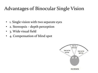
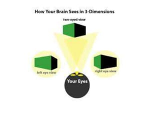
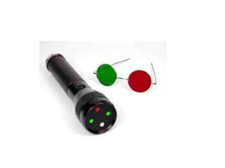
Binocular Vision
and
Stereopsis
Dr Sameeksha Agrawal
Binocular Single Vision
• It the state of simultaneous vision, which is achieved by
the coordinated use of both eyes, so that separate and
slightly dissimilar images arising in each eye are
appreciated as a single image by the process of fusion
Requirement of binocular single vision
1. Clear visual axis
2. Sensory fusion – the ability of retino-cortical element to
function in association of each other so that two disssimilar
object from either eye is fused to single image
3. Motor fusion - the precise co-ordination of the two eyes in
all direction of gazes, so that corresponding retino-cortical
element are placed in a position
Advantages of Binocular Single Vision
• 1. Single vision with two separate eyes
• 2. Stereopsis – depth perception
• 3. Wide visual field
• 4. Compensation of blind spot
Sensory aspect of BSV
The objects in space are localized by us in two ways –
A. Relative localization - relative to one another
B. Egocentric localization - in relation to ourselves.
Definition –Retinal element
• It is a retino-cerebral apparatus engaged in elaborating a
sensation in response to excitation of a unit area of retinal
surface.
• The retinal area, when stimulated, identifies the
brightness, colour and form and also the direction of the
object in visual space.
• This direction is relative to the visual direction of the fovea
Visual axis
• It is an imaginary line which connects the object to its
image formed on retinal surface
• If the line connects to the fovea – Principal visual axis
• If the principal visual axis from two eyes bisect each other
at the object point – binocular fixation
Common subjective visual direction
• It consists of all object points that stimulates
simultaneously to corresponding retinal elements in two
eyes ; example two foveas
• The common subjective visual direction for fovea forms a
plane which coincides with the median plane of head
Retinal correspondence
• Retinal elements of the two eyes that share a common
subjective visual direction are called corresponding retinal
points.
• Rest are non corresponding points or disparate points
• Existence of corresponding retinal elements with their
common relative subjective visual direction is the essence
of binocular vision
Relative position of two object
Retinal correspondence
Two types –
• Normal retinal correspondence – two foveas have common
subjective visual direction
• Abnormal retinal correspondence – the fovea of one eye
shares subjective visual direction with extra foveal point.
• Phenomenon helps to have some stereopsis
• Normal foveal fixation taken over as soon as other eye is
closed.
Sensory fusion
• Sensory fusion is the cortical process of blending the
images from each eye into a single binocular stereoscopic
image
• The striate cortex has binocular cells which are stimulated
by corresponding retinal area in each eye
• In humans, approximately 70% of the cells in the striate
cortex are binocular cells
Motor fusion
• Motor fusion is the mechanism that allows fine-tuning of
eye position to maintain eye alignment
• The correctional eye movements that maintain binocular
foveal alignment provided by motor fusion are termed
fusional vergence movements
• Unlike sensory fusion, motor fusion is the exclusive
function of the extrafoveal retinal periphery.
Diplopia
• The simultaneous stimulation of non-corresponding or
disparate retinal elements by an object point causes this
point to be localized in two different subjective visual
directions
True vertical diplopia
Retinal rivalry
• Simultaneous excitation of corresponding retinal areas by
dissimilar objects leads to confusion
• Suppression of one image occurs to prevent confusion
• The constant foveal suppression of one eye with cessation
of rivalry leads to complete sensory dominance of the
other eye
CONFUSION
Suppression
• It is a neuro-physiological active inhibitory mechanism in
which when corresponding retinal areas are stimulated by
dissimilar stimuli or
• When non-corresponding retinal areas are stimulated by
similar stimuli
Horopter
• Psychophysical experiments indicate that the locus of
points, which project to corresponding retinal points of
each eye takes the shape of an ellipse
• This ellipsoid plane is called Horopter
• Objects located in front of or behind the empirical
horopter will project to noncorresponding retinal points
Panum’s fusional area
• The finite area in front of and behind the horopter line
where objects stimulate noncorresponding retinal points,
yet are still fusible into a single binocular image, is called
Panum’s fusional area
HOROPTER AND PANUM’S FUSIONAL AREA
Panum’s fusional area
• Stimulation of noncorresponding retinal points within
Panum’s fusional area will produce three-dimensional
vision.
• This ability for the brain to determine that images are
falling on retinal points that are not exactly corresponding
produces stereoscopic vision
• The fovea has high spatial resolution
• As we move to the peripheral fields, the receptive field size
enlarges and the spatial resolution decreases
• The retinal architecture of high central resolution versus
low peripheral resolution explains the excellent
stereoacuity from central fields and progressively poorer
stereoacuity from peripheral binocular retinal fields
Panum’s fusional area
• The object outside the Panum’s area produces disparate
images that can’t be fused
• This causes diplopia – physiological diplopia
• Object closer to the panum’s area produces crossed
diplopia and outside the panum’s area produces uncrossed
diplopia.
Stereopsis
• It is the ability to fuse images that stimulate horizontally
disparate retinal elements within Panum’s fusional area
resulting in binocular appreciation of visual object in
depth
• An object confined to the horopter is seen as flat
• The sensory fusion of these horizontally disparate unequal
retinal images results in a three dimensional percept
Stereopsis
• Interpupillary distance also influences stereoacuity.
• Farther apart the two eyes, the greater the angle of visual
disparity and the greater the stereoscopic potential
• Closer an object is to the eyes, the greater the angle of
disparity; therefore, the better the stereoscopic view
Grades of Binocular Vision
• Grade I - Simultaneous macular perception
• Grade II – fusion of the images
• Grade III – stereopsis ; it involves a perceptual synthesis at
higher level
Before any test is undertaken it is essential to
assess the :
• Visual acuity
• Fixation in the squinting eye
• Direction and size of deviation
Test for retinal correspondence
• Bagolini’s striated glass test
• Red filter test
• Synaptophore test
• Worth 4 dot test
Test for suppression
• 1. Worth's four dot test
• 2. Synaptophore
• 3. Friend test
• 4. Amsler Grid
• 5. 4 Δ prism base out test
• 6. Red filter test
• 7. Bagolini’s striated glasses
Stereoacuity testing
• Stereoscopic perception can be created from two-
dimensional figures by presenting each eye with similar
figures that are horizontally offset
• Bitemporal retinal stimulation within Panum’s fusional
area gives the stereoscopic perception of an image coming
toward the observer
Stereoacuity testing
• Stereoacuity can be quantified by measuring the amount
of image disparity
• Minimum stereoscopic resolution is a disparity of
approximately 30 to 40 s of arc.
• Stereoscopic resolution depends upon visual acuity, as
poor vision in one or both eyes will decrease stereoacuity.
Contour stereoacuity tests
• It uses stereoscopic figures with a continuous contoured
edge; example Titmus test
• Measures disparities from 3000s arc (the big fly) to 40 s arc
(ninth circle)
• They have the disadvantage of having monocular clues
There is a set of monocular or experimental clues that play an
important role in our estimation of relative distance of visual
objects
• Apparent size
• Interposition
• Shading
• Geometric perspective – physically parallel lines converge
toward a vanishing point at the horizon, e.g. railroad
tracks.
Mono ocular clues
• Relative velocity – the image velocity of a moving target in
the distance is lower than the image velocity of the same
moving target when it is nearby
• Motion parallax – translocation of the head cause the
images of near objects to move opposite the head and the
images of far objects to move with the head, assuming the
fixation point is at an intermediate distance
Monocular clues
Monocular clues
• Patients with monocular vision can identify which figure is
supposed to be stereoscopic, because that figure will be
horizontally off center.
• “Image jump” patients alternate fixation between the two
horizontally displaced figures and identify the figure that
jumps back and forth
Monocular clues
Monocular clue
Monocular clues
• The patient has true stereoacuity is to retest with the
Titmus test book turned 90°
• The monocular clues still work. If the patient again
identifies the stereoscopic target, they are using
monocular clues
Random dot stereograms
• It consist of two fields of randomly scattered dots or
specks, with one field of dots projected to each eye
separately through a haploscopic device
• Each field of random dots is identical except for a group of
dots that is displaced nasally
• Random dot tests have almost no monocular clues, and a
positive response indicates true stereopsis
Thank You

Binocular vision and stereopsis

  • 1.
  • 2.
    Binocular Single Vision •It the state of simultaneous vision, which is achieved by the coordinated use of both eyes, so that separate and slightly dissimilar images arising in each eye are appreciated as a single image by the process of fusion
  • 3.
    Requirement of binocularsingle vision 1. Clear visual axis 2. Sensory fusion – the ability of retino-cortical element to function in association of each other so that two disssimilar object from either eye is fused to single image 3. Motor fusion - the precise co-ordination of the two eyes in all direction of gazes, so that corresponding retino-cortical element are placed in a position
  • 4.
    Advantages of BinocularSingle Vision • 1. Single vision with two separate eyes • 2. Stereopsis – depth perception • 3. Wide visual field • 4. Compensation of blind spot
  • 5.
    Sensory aspect ofBSV The objects in space are localized by us in two ways – A. Relative localization - relative to one another B. Egocentric localization - in relation to ourselves.
  • 6.
    Definition –Retinal element •It is a retino-cerebral apparatus engaged in elaborating a sensation in response to excitation of a unit area of retinal surface. • The retinal area, when stimulated, identifies the brightness, colour and form and also the direction of the object in visual space. • This direction is relative to the visual direction of the fovea
  • 7.
    Visual axis • Itis an imaginary line which connects the object to its image formed on retinal surface • If the line connects to the fovea – Principal visual axis • If the principal visual axis from two eyes bisect each other at the object point – binocular fixation
  • 9.
    Common subjective visualdirection • It consists of all object points that stimulates simultaneously to corresponding retinal elements in two eyes ; example two foveas • The common subjective visual direction for fovea forms a plane which coincides with the median plane of head
  • 10.
    Retinal correspondence • Retinalelements of the two eyes that share a common subjective visual direction are called corresponding retinal points. • Rest are non corresponding points or disparate points • Existence of corresponding retinal elements with their common relative subjective visual direction is the essence of binocular vision
  • 11.
  • 12.
    Retinal correspondence Two types– • Normal retinal correspondence – two foveas have common subjective visual direction • Abnormal retinal correspondence – the fovea of one eye shares subjective visual direction with extra foveal point. • Phenomenon helps to have some stereopsis • Normal foveal fixation taken over as soon as other eye is closed.
  • 13.
    Sensory fusion • Sensoryfusion is the cortical process of blending the images from each eye into a single binocular stereoscopic image • The striate cortex has binocular cells which are stimulated by corresponding retinal area in each eye • In humans, approximately 70% of the cells in the striate cortex are binocular cells
  • 14.
    Motor fusion • Motorfusion is the mechanism that allows fine-tuning of eye position to maintain eye alignment • The correctional eye movements that maintain binocular foveal alignment provided by motor fusion are termed fusional vergence movements • Unlike sensory fusion, motor fusion is the exclusive function of the extrafoveal retinal periphery.
  • 15.
    Diplopia • The simultaneousstimulation of non-corresponding or disparate retinal elements by an object point causes this point to be localized in two different subjective visual directions
  • 16.
  • 17.
    Retinal rivalry • Simultaneousexcitation of corresponding retinal areas by dissimilar objects leads to confusion • Suppression of one image occurs to prevent confusion • The constant foveal suppression of one eye with cessation of rivalry leads to complete sensory dominance of the other eye
  • 18.
  • 19.
    Suppression • It isa neuro-physiological active inhibitory mechanism in which when corresponding retinal areas are stimulated by dissimilar stimuli or • When non-corresponding retinal areas are stimulated by similar stimuli
  • 20.
    Horopter • Psychophysical experimentsindicate that the locus of points, which project to corresponding retinal points of each eye takes the shape of an ellipse • This ellipsoid plane is called Horopter • Objects located in front of or behind the empirical horopter will project to noncorresponding retinal points
  • 21.
    Panum’s fusional area •The finite area in front of and behind the horopter line where objects stimulate noncorresponding retinal points, yet are still fusible into a single binocular image, is called Panum’s fusional area
  • 22.
  • 23.
    Panum’s fusional area •Stimulation of noncorresponding retinal points within Panum’s fusional area will produce three-dimensional vision. • This ability for the brain to determine that images are falling on retinal points that are not exactly corresponding produces stereoscopic vision
  • 25.
    • The foveahas high spatial resolution • As we move to the peripheral fields, the receptive field size enlarges and the spatial resolution decreases • The retinal architecture of high central resolution versus low peripheral resolution explains the excellent stereoacuity from central fields and progressively poorer stereoacuity from peripheral binocular retinal fields Panum’s fusional area
  • 26.
    Panum’s fusional area •The object outside the Panum’s area produces disparate images that can’t be fused • This causes diplopia – physiological diplopia • Object closer to the panum’s area produces crossed diplopia and outside the panum’s area produces uncrossed diplopia.
  • 27.
    Stereopsis • It isthe ability to fuse images that stimulate horizontally disparate retinal elements within Panum’s fusional area resulting in binocular appreciation of visual object in depth • An object confined to the horopter is seen as flat • The sensory fusion of these horizontally disparate unequal retinal images results in a three dimensional percept
  • 28.
    Stereopsis • Interpupillary distancealso influences stereoacuity. • Farther apart the two eyes, the greater the angle of visual disparity and the greater the stereoscopic potential • Closer an object is to the eyes, the greater the angle of disparity; therefore, the better the stereoscopic view
  • 29.
    Grades of BinocularVision • Grade I - Simultaneous macular perception • Grade II – fusion of the images • Grade III – stereopsis ; it involves a perceptual synthesis at higher level
  • 30.
    Before any testis undertaken it is essential to assess the : • Visual acuity • Fixation in the squinting eye • Direction and size of deviation
  • 31.
    Test for retinalcorrespondence • Bagolini’s striated glass test • Red filter test • Synaptophore test • Worth 4 dot test
  • 33.
    Test for suppression •1. Worth's four dot test • 2. Synaptophore • 3. Friend test • 4. Amsler Grid • 5. 4 Δ prism base out test • 6. Red filter test • 7. Bagolini’s striated glasses
  • 34.
    Stereoacuity testing • Stereoscopicperception can be created from two- dimensional figures by presenting each eye with similar figures that are horizontally offset • Bitemporal retinal stimulation within Panum’s fusional area gives the stereoscopic perception of an image coming toward the observer
  • 35.
    Stereoacuity testing • Stereoacuitycan be quantified by measuring the amount of image disparity • Minimum stereoscopic resolution is a disparity of approximately 30 to 40 s of arc. • Stereoscopic resolution depends upon visual acuity, as poor vision in one or both eyes will decrease stereoacuity.
  • 36.
    Contour stereoacuity tests •It uses stereoscopic figures with a continuous contoured edge; example Titmus test • Measures disparities from 3000s arc (the big fly) to 40 s arc (ninth circle) • They have the disadvantage of having monocular clues
  • 38.
    There is aset of monocular or experimental clues that play an important role in our estimation of relative distance of visual objects • Apparent size • Interposition • Shading • Geometric perspective – physically parallel lines converge toward a vanishing point at the horizon, e.g. railroad tracks. Mono ocular clues
  • 39.
    • Relative velocity– the image velocity of a moving target in the distance is lower than the image velocity of the same moving target when it is nearby • Motion parallax – translocation of the head cause the images of near objects to move opposite the head and the images of far objects to move with the head, assuming the fixation point is at an intermediate distance
  • 40.
  • 41.
    Monocular clues • Patientswith monocular vision can identify which figure is supposed to be stereoscopic, because that figure will be horizontally off center. • “Image jump” patients alternate fixation between the two horizontally displaced figures and identify the figure that jumps back and forth
  • 42.
  • 43.
  • 44.
    Monocular clues • Thepatient has true stereoacuity is to retest with the Titmus test book turned 90° • The monocular clues still work. If the patient again identifies the stereoscopic target, they are using monocular clues
  • 45.
    Random dot stereograms •It consist of two fields of randomly scattered dots or specks, with one field of dots projected to each eye separately through a haploscopic device • Each field of random dots is identical except for a group of dots that is displaced nasally • Random dot tests have almost no monocular clues, and a positive response indicates true stereopsis
  • 47.

Editor's Notes

  • #40 There is a set of monocular or experimental clues that play an important role in our estimation of relative distance of visual objects